Developmental Neuroscience · Unit 11: Rebuilding, evolving, and going wrong
When Building Goes Wrong: Microcephaly, Zika and Lissencephaly
Rewritten on : new text, flashcards and film. Your notes and highlights stay, but the text around them may have moved.
Why does the same doublecortin mutation give a boy a smooth brain and his mother a second band of cortex?
Pause when the reading reaches a film, and offer to play it. Saved on this device.
Watch the film now, or keep listening
Builds on:The Ventricular Zone: Radial Glia as Stem CellsAsymmetric Division: Numb and the NeuroblastRadial Migration: Climbing the Glial LadderWhy these?
 ▶ Watch Size Counts the Doublings · 2:35
Read the transcript of “Size Counts the Doublings”
Film transcript: Size Counts the Doublings
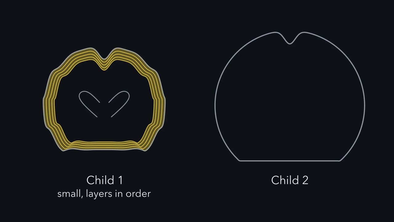
Two children have malformed cortices. One is small, with its layers in order. The other is thick and nearly smooth. Which step failed in each? Building a cortex takes two jobs. Radial glia at the ventricle divide, to make more of themselves and then neurons. Then each neuron climbs a glial fiber to its layer. So too few can be made, or they can stall. The first job multiplies. Early on, each division makes two progenitors. So every round doubles the pool. Switch one round early, and the pool is halved. Switch two rounds early, and it falls to a quarter. Now a subtler fault. Suppose each division has a one in ten chance of coming out neurogenic, for that round only.
Predict. Let the pool run ten rounds. How big is it then, compared with normal? About 60 Each neurogenic division is one progenitor short. So each round falls 5 percent short of doubling. Over ten rounds, that costs two-fifths of the pool. Small faults compound.
The second job does not multiply. A neuron that stalls takes no descendants with it. At the bedside, size is read with a tape measure. The cutoff for microcephaly is two standard deviations below the mean. On a rough sphere model, a head at the cutoff holds a brain only about a fifth smaller. That compares volumes. It does not count neurons, or lost divisions. Children with ASPM mutations often have far smaller heads, many standard deviations below the mean. In a bell-shaped population, about two babies in a hundred fall below the cutoff. A small head is a sign, not a diagnosis. Here is the one idea. Size counts the doublings. Layering records the journey.
Small with orderly layers points to production. Near-normal size with disordered layers points to migration. It is a first question, not a diagnosis. So when a cortex is malformed as it is built, ask first which job failed.
From the film
- Size counts the doublings, and layering records the journey.
- A small cortex with orderly layers points first to a problem in making neurons, and near-normal size with disordered layers points to migration.
- Because the progenitor pool doubles, a small fault that recurs in each round can compound into a large loss.
- This size-and-layering split is a first question, not a diagnosis.
Two children can both have a malformed cerebral cortex for opposite reasons. In one, the cortex is small, but its layers are in their usual order. In the other, the cortex is thick and its surface nearly smooth, and many of its neurons sit in the wrong place.
The first child has primary microcephaly. The second has lissencephaly, a word that means “smooth brain”. Each points to one step of building a cortex that failed, and to genes needed for that step. The Zika epidemic of 2015 and 2016 showed with unusual clarity that an infection can strike the same cells as a gene.
Number or place
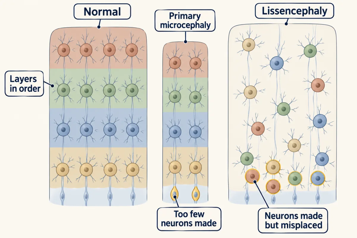
Radial glia, the progenitor cells that line the fluid-filled ventricle, divide to make more of themselves and then to make neurons (that lesson). Each newborn neuron then moves outward along a radial glial fiber to its layer, later-born neurons passing the earlier ones (that lesson). Two jobs, so two broad kinds of failure.
- Too few neurons made. Progenitors stop dividing early or die, and the cortex is small, though its plan can look normal. This is microcephaly.
- Neurons made but misplaced. Neurons stall on the way to their layer. This gives lissencephaly, or a heterotopia: a cluster of neurons sitting where they do not belong.
The first job has a property the second lacks: it multiplies. Early in development, progenitors divide symmetrically, each division making two progenitors (that lesson). Later they switch to divisions that make neurons. Each round of symmetric division doubles the pool:
progenitors at the switch = starting progenitors × 2n, where n is the number of symmetric rounds
Start from 100 progenitors.
| Symmetric rounds before the switch | Progenitors at the switch | Compared with normal |
|---|---|---|
| 7 (normal, in this model) | 12,800 | 1 |
| 6 (switch one round early) | 6,400 | ½ |
| 5 (switch two rounds early) | 3,200 | ¼ |
Switching one round early halves the pool, and if every progenitor present at the switch then makes the same number of neurons, it halves the neurons too. Real defects need not act on every progenitor at once, so try a second model, with a small, steady bias.
Predict. Suppose each division has a one-in-ten chance of being neurogenic for that round only: it gives one progenitor and one neuron instead of two progenitors, and that progenitor can divide symmetrically again next round. (A permanent switch would cost more.) Let the pool run ten rounds, longer than the table’s seven. Is the pool then about 90 percent of normal, about 60 percent, or about 10 percent?
Check your prediction
About 60 percent. Each neurogenic division is one progenitor short, so on average a division gives 1.9 progenitors instead of 2, 5 percent short of doubling. Each round the pool reaches only 0.95 of what it should, and over ten rounds that is 0.9510, about 0.60; simulations that draw each division at random give this on average, with single runs scattered around it. A fault in one division in ten has cost two-fifths of the pool, and the loss grows with every further round. (Those divisions also made a few neurons early, so total neuron output falls a little less than the pool.)
The second job does not multiply: a neuron that stalls is one neuron in the wrong place, and it takes no descendants with it.
The one idea to carry through this lesson. Size counts the doublings; layering records the journey. A cortex’s size reflects how many neurons its progenitors made and how many survived, and because the progenitor pool doubles, a small fault that recurs in each round compounds into a large loss. Its layering reports whether those neurons arrived. So a small cortex with orderly layers points first to a problem in making neurons (or in their early loss), and a cortex of near-normal size with disordered layers points to migration.
What the rule covers.
- Which malformations. Those that arise while neurons are being made and placed, from early gestation through much of fetal life. It does not cover destruction of tissue already built, which can overlap with building (much of congenital Zika syndrome, or injury from too little oxygen), or disorders of folding and wiring.
- Which measure. “Size” means cortical volume, which tracks neuron number only roughly, since neuron density, glia and white matter also vary. Head circumference is the bedside stand-in, and a rough one.
- A first question, not a diagnosis. Some genes act at both steps, and Zika also destroys tissue already built; these cases are collected in Where the sort breaks.
The rule picks up where the neurotrophic hypothesis left off: survival can never raise neuron number above the number made. This lesson is about where the number made is set: survival factors trim the cohort, and the divisions of the progenitor pool decide how large that cohort can be.
Too few: a tape measure and a spindle
Read the transcript of “Too Few: the Spindle and a Virus”
Film transcript: Too Few: the Spindle and a Virus
Why would a protein of the cell's spindle shrink the brain more than other organs? Primary microcephaly is recessive. The brain is small, and its layers are usually broadly in order. Most of its genes, such as ASPM, make proteins of the spindle pole. Radial glial progenitors are tall and thin, with a small patch facing the ventricle. In early divisions, the cleavage plane affects how that patch is shared. Tilt the spindle, and one daughter can get it all. That can change what the daughters become. But a daughter without the patch can still divide, as a progenitor away from the ventricle. In mouse embryos, knocking down Aspm tilted the division plane, and shifted divisions away from making progenitors.
In mouse progenitors, delayed mitosis can also trigger a death that depends on p53, a sensor of cell stress. And if a fault recurs round after round, it compounds. Predict. A child with no working ASPM has a markedly small brain, but mice lacking the gene have only a mild reduction. Ferrets have a folded cortex, rich in outer radial glia. Those are progenitors that have left the ventricle but keep dividing. Knock the gene out in a ferret. Less shrinkage, the same, or more?
Much more. The authors traced it to radial glia leaving the ventricle too early. A second reading, still a hypothesis, is that more rounds mean more compounding. Now Zika. How do you show a virus causes a birth defect, without testing it in pregnant women? Epidemiology. Microcephaly rose after the virus, in time and place. That shows association, not cause or mechanism. Pathology. Virus was found inside a damaged fetal brain. On its own, that cannot show the virus did the damage. The laboratory. The virus infected human cortical progenitors most readily, killing some and slowing the rest. A dish cannot show the risk in a pregnancy. Predict. The first study used an old Ugandan strain, kept for decades in the lab.
What would a skeptic say? That it might not act like the epidemic virus. Later work with epidemic strains agreed. In 2016, a review against formal criteria concluded that Zika causes microcephaly. Two cautions. Most infections in pregnancy do not produce microcephaly. And the virus also damages tissue already built.
So a spindle-pole gene, or a virus that finds the same cells, can leave too few neurons.
From the film
- Primary microcephaly genes such as ASPM mostly act at the spindle pole of dividing progenitors, leaving a small cortex with layers broadly in order.
- In mouse embryos, knocking down Aspm tilted the division plane and shifted divisions away from making progenitors.
- Evidence that Zika virus causes microcephaly came from epidemiology, pathology and laboratory models, and each kind of evidence has limits.
- Most Zika infections in pregnancy do not produce microcephaly, and the virus can also damage tissue already built.
What a tape measure can tell you
Microcephaly is defined at the bedside, not under a microscope. It means a head circumference well below the average for the child’s age and sex, measured from just above the eyebrows and ears around to the most prominent point at the back of the head. The World Health Organization uses a cutoff of more than two standard deviations (SD) below the mean, and calls more than three severe. For a family, this number is often the first sign that something is wrong.
How much smaller is the brain at those cutoffs? Less than you might expect. Make a rough model: treat the head as a sphere, so that head circumference grows as the cube root of brain volume, and ignore the scalp, skull and fluid that the tape also measures. Take a term newborn’s head as about 34.5 cm around, with an SD of about 1.2 cm.
| Head circumference, in this model | Brain volume compared with average | Volume halvings (not counted cell divisions) |
|---|---|---|
| 2 SD below the mean (the WHO cutoff), 32.1 cm | about 0.80 | about a third of one |
| 3 SD below (severe), 30.9 cm | about 0.72 | about half of one |
| volume halved, 27.4 cm (about 6 SD below) | 0.50 | one |
So on this model a head at the cutoff holds a brain only about one-fifth smaller than average. Children with ASPM mutations often have far smaller heads, many SD below the mean for their age. Still, the model only compares volumes; it counts neither neurons nor lost divisions.
The cutoff also catches some healthy babies. If head sizes follow a bell curve, about 2 in 100 lie more than 2 SD below the mean. A small head is a sign, not a diagnosis, and it has many possible causes: genes; congenital infections, among them cytomegalovirus (CMV), rubella, toxoplasmosis and Zika; alcohol; and injury around birth. This lesson follows one group of genes and one virus, which act on the same cells.
Primary microcephaly: genes at the spindle pole
Primary microcephaly (MCPH) is present at birth and inherited as a recessive trait: a child is affected when both copies of a gene are broken. That is why it is found most often in families in which the parents are related. The brain is small, and the cerebral cortex is reduced most. In most forms its layers are broadly normal, though its folds are often simpler. Some genes, including WDR62, can also disturb the structure of the cortex. Intellectual disability is common and varies in degree.
More than twenty MCPH genes are now known. In many studied populations the commonest cause is ASPM, which Woods and colleagues linked to the disease in 2002. Its name, abnormal spindle-like, microcephaly associated, comes from the fly gene abnormal spindle, which flies need to organize the spindle in dividing neural cells. Others include MCPH1 (microcephalin), CDK5RAP2 and WDR62. Most of their proteins sit at the centrosome or the spindle pole. The centrosome is the cell’s main microtubule-organizing center. At mitosis the two centrosomes form the poles of the spindle that pulls the chromosomes apart.
Why the brain?
Why would a spindle protein matter more for the brain than for other organs? One leading idea has two parts.
Radial glia are unusual dividers. A radial glial cell is tall and thin, and only a small patch of its surface faces the ventricle. In early divisions, the angle of the cleavage plane affects how that apical patch is shared. A tilted spindle can hand most or all of it to one daughter, and that can change what the daughters become. The patch does not decide fate on its own: a daughter without it can still divide, as a progenitor away from the ventricle. An experiment in mouse embryos tested the spindle idea: knocking down Aspm tilted the plane of progenitor division and shifted divisions away from the symmetric, progenitor-making kind (Fish and colleagues, 2006). A defect in how long mitosis takes can act another way: in mouse progenitors, delayed mitosis can trigger a death that depends on p53, a protein that senses stress and can halt or kill the cell.
The cortex grows through many rounds. If a fault tips progenitors toward the switch or toward death round after round, it compounds, as in the prediction above. Either history leaves fewer neurons, so a small cortex alone cannot say which happened. The neurotrophic lesson’s question, never made or made and lost, applies here too.
The brain is the most exposed organ, not the only one. Some of the same centrosome genes, such as CENPJ and CEP152, when mutated differently, shrink the whole body as well as the brain, in Seckel syndrome.
Predict. A child with no working ASPM has a markedly small brain, but mice lacking Aspm have only a mild reduction. Ferrets have a folded cortex, rich in outer radial glia: progenitors that have left the ventricle but keep dividing. Knock out Aspm in a ferret. Should its cortex shrink less than a mouse’s, about as much, or more?
Check your prediction
More, and by a lot. In ferrets an Aspm knockout reduced the cortex much more than in mice (Johnson and colleagues, 2018), which the authors attributed to ventricular radial glia being displaced from the ventricle too early.
A second, compatible reading is that a cortex that grows through more rounds of division, and through more kinds of progenitor, is more exposed to a small defect in each division. Take the same fault as before, one division in ten coming out neurogenic. After 5 rounds the pool is about 77 percent of normal, after 10 rounds about 60 percent, and after 15 rounds about 46 percent. These round counts are illustrative, not measured values for either species, and the reading remains a hypothesis. That lesson takes up how the human cortex got large.
Zika: how do you show that a virus causes a birth defect?
You cannot infect pregnant women to find out.
Zika is a flavivirus, a relative of dengue, spread mainly by Aedes mosquitoes. It was first isolated in 1947, from a monkey in the Zika Forest of Uganda, and for decades it was known for mild illness. In 2015 it spread through Brazil. Late that year, doctors in the country’s northeast reported many more newborns with microcephaly than usual.
The case was built from three kinds of evidence pointing the same way.
- Epidemiology. The rise in microcephaly followed the virus in time and place. A look back at a 2013 to 2014 outbreak in French Polynesia found an increase in microcephaly there too, with timing that fit infection in the first trimester.
- Pathology. In 2016 Mlakar and colleagues reported Zika virus RNA and virus particles in the brain of a fetus whose mother had been infected in Brazil during pregnancy. The brain was small, with calcifications.
- Laboratory models. In March 2016 the laboratories of Guo-li Ming, Hongjun Song and Hengli Tang reported that Zika infects human cortical neural progenitors grown from stem cells far more readily than it infects the stem cells or immature neurons, that it kills some of the progenitors and slows the growth of the rest. Stevens Rehen’s group in Rio de Janeiro reported that the virus stunts human neurospheres and brain organoids, tissue grown in three dimensions from stem cells (that lesson).
| Evidence | What it shows | What it cannot show |
|---|---|---|
| Epidemiology | association in time and place | cause on its own (other factors, or closer checking of newborns, can raise counts), or mechanism |
| Pathology | virus inside a damaged fetal brain | on its own, that the virus did the damage, or how often it happens |
| Cultured progenitors and organoids | which human cells the virus can infect and harm | the risk in a pregnancy |
Predict. The March 2016 progenitor study used MR766, a strain isolated in Uganda decades earlier and passaged in the laboratory ever since, not the virus spreading through Brazil. What would a skeptic say about the result, and what would answer the objection?
Check your prediction
An old laboratory strain might not behave like the epidemic virus, so its effect on progenitors might not explain the epidemic. The answer is to repeat the work with epidemic strains, and later work with them, in organoids and in pregnant mice, agreed.
In April 2016 Sonja Rasmussen and colleagues at the US Centers for Disease Control and Prevention weighed this evidence against formal criteria for calling an agent a cause of birth defects. They concluded that Zika virus causes microcephaly and other serious brain defects. The pattern in affected infants, now called congenital Zika syndrome, includes severe microcephaly, calcium deposits in the brain, damage to the eye and stiff joints.
Two cautions. Much of the mechanism comes from cultured cells, organoids and mice, so it shows what the virus can do to human progenitors, not what it does in each fetus. And most infections in pregnancy do not produce microcephaly; the risk appears highest when infection comes early.
In the wrong place: lissencephaly and the double cortex
Read the transcript of “The Nucleus Left Behind”
Film transcript: The Nucleus Left Behind
What if the neurons are made, but never arrive? In classic lissencephaly, the cortex is thick and nearly smooth, with about four crude layers. Yet in isolated lissencephaly, the head is usually near normal in size at birth. Children with Miller-Dieker syndrome lack a piece of one chromosome 17, including LIS1. Losing only one copy of LIS1 still causes lissencephaly. Half a dose is too little. A mould gave the clue. Its relative of LIS1 helps space nuclei, as do parts of dynein, a microtubule motor. In mammalian cells, LIS1 binds dynein and helps it work. A migrating neuron moves in two steps. First the leading process extends, and the centrosome moves into it.
Then dynein pulls the nucleus up toward the centrosome. Predict. Knock down LIS1 in the embryonic rat cortex. Does the process fail, the nucleus, or something earlier? Most cells never started, and stayed many-branched. In the few that did, the process kept growing, but the nucleus stayed behind.
The knockdown also blocked progenitor divisions at the ventricle. So LIS1 has one foot in too few. A second gene makes doublecortin. It is not a motor. It binds microtubules, and is thought mainly to steady the tracks. Now the puzzle. A boy and his mother share a mutation that breaks doublecortin. He has a smooth brain; she has a second band.
The clue: a male has one X. Each of a female's cells silences one of her two X chromosomes at random. Predict. Explain both brains. His one X carries the mutation, so no neuron makes working doublecortin. Most stall, and the cortex is smooth.
Her cortex is a mosaic. Neurons with the normal X active arrive. Those with the mutant X active stall partway, and gather into a band beneath the cortex. In embryonic rats, neurons with doublecortin silenced stalled, and formed such a band. Some neighbours were disturbed too, and the sorting has not been traced neuron by neuron in humans. So read a cortex two ways. Size counts the doublings. Layering records the journey.
From the film
- In classic lissencephaly the neurons are made but fail to travel, and the head is usually near normal in size at birth.
- Losing one copy of LIS1 is enough to cause lissencephaly, and LIS1 binds the dynein motor that helps pull the nucleus forward.
- Doublecortin is not a motor but binds and steadies microtubules, the tracks the nucleus travels on.
- Because of X inactivation, the same DCX mutation can give a boy lissencephaly and his mother a band of misplaced neurons.
Lissencephaly is the other category. A normal human cortex is folded and has six layers. In classic lissencephaly it is thick, has few or no folds, and is organized into about four crude layers. Affected children usually have severe intellectual disability and epilepsy. It is classed as a failure of migration: the neurons were made, but they failed to travel.
The rule predicts a near-normal size, and in isolated lissencephaly the head is usually near normal in size at birth, though head growth often slows afterwards, so the rule fits best early. The geometry allows it. Volume is area times thickness. A smooth cortex about four to five times as thick as normal (roughly 10 to 15 mm, against 2.5 to 3 mm) needs only a quarter to a fifth of the surface area to hold the same volume of gray matter. This is only a plausibility check, since the density of neurons in the malformed cortex is not normal.
The first two genes for classic lissencephaly to be found through affected children were LIS1 and DCX, the gene for doublecortin.
LIS1: half a dose is not enough
Children with Miller-Dieker syndrome have a severe lissencephaly with distinctive facial features, and most are missing a small piece of one chromosome 17, in a region called 17p13.3. In 1993 Orly Reiner, David Ledbetter, William Dobyns and colleagues isolated a gene from that region and named it LIS1.
The deletion removes one copy of LIS1, and the other chromosome 17 is normal. Children who have lost or broken only LIS1, on one copy, still have lissencephaly, called isolated lissencephaly. So half the usual amount of LIS1 is too little for neurons to migrate, a state called haploinsufficiency. (Miller-Dieker is more severe: its larger deletion also removes neighbouring genes, including the one for 14-3-3ε, that are thought to add to the severity.)
The sequence did not say what LIS1 does. The clue came from a mould. Aspergillus nidulans grows as long filaments that must space their nuclei along their length, and mutants called nud, for nuclear distribution, leave the nuclei bunched. In 1995 one of them, nudF, turned out to encode a protein 42 percent identical to human LIS1. Other nud genes encode parts of cytoplasmic dynein, a motor that walks along microtubules toward their minus ends. In mammalian cells LIS1 binds dynein, together with the partner proteins NDE1 and NDEL1, and helps the motor hold on to microtubules and assemble into working complexes. A gene found through children with smooth brains had led to a motor that moves nuclei.
Why would a nucleus motor matter for migration? A migrating neuron moves in two steps. First it extends a leading process along the radial glial fiber, and its centrosome moves forward into the process. Then dynein, pulling along microtubules that run back from the centrosome to the nucleus, draws the nucleus up toward it, with help from the motor protein myosin.
Predict. Knock down LIS1 in the embryonic cortex, in progenitors and the neurons they are about to make. Which should fail: extending the leading process, moving the nucleus, or something earlier?
Check your prediction
Tsai and colleagues did this in 2005, in the cortex of embryonic rats. Most new cells never started: they piled up near the ventricle in a many-branched, multipolar state instead of becoming bipolar migrating neurons. In the few that did become bipolar, the leading process kept extending, but the cell body and nucleus barely moved after it. In those cells the nucleus was the part that failed, and a nucleus that cannot be moved leaves its neuron short of its layer.
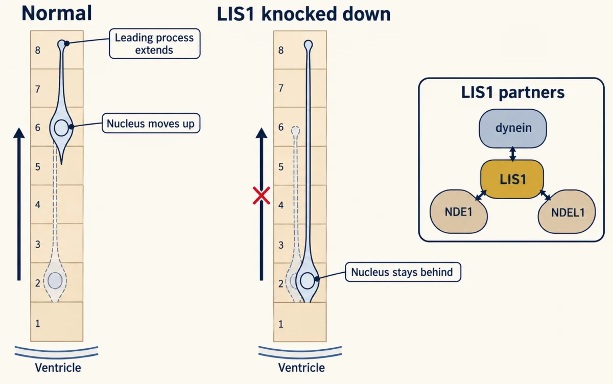
The same experiment showed something the “wrong place” label hides. Before a progenitor divides, dynein also carries its nucleus down to the ventricular surface. With LIS1 knocked down, progenitor nuclei stopped their up-and-down movement in the ventricular zone, and divisions at the ventricle were blocked. So LIS1 loss can also reduce how many neurons are made: LIS1 belongs mainly in the “wrong place” column, with one foot in “too few”. The mechanics of nuclear movement are in that lesson.
Doublecortin: one mutation, two brains
Some families showed a striking pattern. Affected boys had lissencephaly, while their mothers or sisters had a milder disorder, subcortical band heterotopia, or “double cortex”: a band of gray matter lying in the white matter beneath a nearly normal cortex. That pointed to a gene on the X chromosome. In 1998 two groups reported it in the same issue of Cell: Joseph Gleeson, Christopher Walsh and colleagues in Boston, and Vincent des Portes, Jamel Chelly and colleagues in Paris. The gene, DCX, encodes doublecortin, a protein that binds microtubules and stabilizes them in migrating neurons. It is not a motor; it is thought mainly to steady the tracks the nucleus travels on.
A male has one X chromosome. A female has two, and early in development each of her cells silences one of them at random, a process called X inactivation. The choice is inherited by that cell’s descendants, so her cortex is a mosaic of neurons with one X or the other active.
Predict. A boy with lissencephaly and his mother with band heterotopia carry the same loss-of-function DCX mutation. Using X inactivation, explain both brains.
Check your prediction
He has one X. If it carries the mutation, no neuron makes working doublecortin, and most neurons stall: lissencephaly.
In her mosaic cortex, neurons with the normal X active migrate to the cortex. Neurons with the mutant X active stall partway and gather into a band beneath the cortex. On this reading each neuron follows its own active X, so doublecortin acts mainly within the migrating cell. An experiment in rats supports it: when DCX was silenced in some cortical neurons of embryonic rats, those neurons stalled and formed such a band.
One caveat. In those rat embryos some neighbouring neurons, with DCX intact, were also disturbed, and the sorting has not been shown neuron by neuron in human tissue.
Where the sort breaks
Real cases do not always sort cleanly: some genes and infections affect both jobs. Here are those exceptions in one place.
| Case | Where the rule puts it | What else it does |
|---|---|---|
| LIS1 | wrong place: migrating neurons stall | also blocks progenitor divisions at the ventricle |
| WDR62 | too few: a primary microcephaly gene | can also disturb the structure of the cortex |
| Zika | too few: it attacks progenitors, so fewer neurons are made | also infects young neurons and other brain cells; affected brains often show destruction, such as calcifications and a partly collapsed skull, and sometimes abnormal folding or misplaced gray matter |
| Microlissencephaly (for example, mutations in both copies of NDE1) | too few: the brain is small | the cortex is also smooth |
Congenital Zika syndrome is partly a failure of building and partly damage to what was built, and a cortex made and then damaged is small because tissue was lost, not because too few neurons were made.
Putting it together
| Condition | Head size | Layering | Step that failed | Cells hit | Key evidence | What it does not establish |
|---|---|---|---|---|---|---|
| Primary microcephaly (ASPM, WDR62, CDK5RAP2) | small | broadly normal; WDR62 can disturb it | production | dividing radial glia (spindle pole) | homozygosity mapping; Aspm knockdown tilts the division plane | one mechanism for every MCPH gene |
| Zika, early pregnancy | small, sometimes with a partly collapsed skull | can be disrupted | production, plus destruction | progenitors, also young neurons | epidemiology, fetal pathology, cultured progenitors and organoids | the risk or mechanism in each pregnancy |
| LIS1 haploinsufficiency | near-normal at birth | about four crude layers, thick | migration (plus division) | migrating neurons and progenitors | Miller-Dieker deletions and isolated lissencephaly; rat knockdown stalls the nucleus | that dose loss acts only in neurons |
| DCX, male | near-normal at birth | lissencephaly | migration | migrating neurons | X-linked families; microtubule binding | normal neuron number, from head size alone |
| DCX, female | near-normal at birth | a band beneath a nearly normal cortex | migration, in neurons with the mutant X active | migrating neurons (a mosaic) | the sex difference; rat knockdown forms a band | a neuron-by-neuron proof in human tissue |
Read each row with the guiding idea, then check the exceptions before you name the step.
Design problem
You study two mouse lines. In the first, cortical progenitors switch early from symmetric proliferative divisions to neuron-producing divisions. In the second, neurons are born on schedule but cannot move their nuclei along radial glia. Predict the brain size and the layering of the cortex in each line, and say which human condition each resembles.
Saved in this browser only.
One way to do it
In the first line the pool of founder progenitors stops doubling too soon. With fewer founders there are fewer neurons, and since the pool grows exponentially, a switch even one or two rounds early cuts neuron number by a half or more, if each founder goes on to make its usual number of neurons. The neurons that are made still migrate normally and arrive inside-out, so the cortex is small with roughly normal layers. That resembles primary microcephaly.
In the second line, if the progenitors are untouched, close to the normal number of neurons is made. But neurons whose nuclei cannot follow their leading process stall in the ventricular zone, the intermediate zone or partway through the cortical plate. The cortex has a near-normal neuron count but disordered layers, with many neurons left near the ventricle. That resembles classic lissencephaly. Real LIS1 loss is messier, because LIS1 also acts in dividing progenitors.
Going deeper
The Zika emergency
Brazil declared a national public health emergency in November 2015, and the World Health Organization declared a Public Health Emergency of International Concern on 1 February 2016.
Two maps
The two lissencephaly genes leave different maps. Lissencephaly from LIS1 is usually worse toward the back of the brain, and from doublecortin (DCX) toward the front.
More genes of migration
Other genes of migration include CDK5. Mutations in both copies of RELN, the human gene for Reelin, cause lissencephaly with a small cerebellum, a human counterpart of the reeler mouse (that lesson). The word “classic” in the main text matters: a gene for cobblestone lissencephaly, fukutin, was also reported in 1998, and cobblestone malformations are a different, pial-surface defect.
Open questions
Why many mouse models of MCPH genes have milder microcephaly than patients is not settled, nor whether the mechanism is the same for every gene. For Zika, the entry receptor on human progenitors and the relative weight of direct killing, innate immune signaling and placental damage in the human fetus are still debated.
Your notes
Select any text in the lesson to highlight it or add a note to it. Your highlights and notes are saved in this browser and collected in your notebook.
This browser is not letting the site save anything (a private window or strict privacy settings), so highlights and notes will be lost when you leave the page.
How we measure it
Homozygosity mapping
In families where the parents are related, a child with a recessive disease carries two identical copies of the chromosome stretch around the broken gene. Finding the stretches that all affected children share narrows the search to a small region, where candidate genes are then sequenced. This is how ASPM and other microcephaly genes were found.
In utero electroporation with RNA interference
DNA encoding a short RNA that silences one gene, plus a fluorescent marker, is injected into the ventricle of an embryo in the uterus and driven into nearby progenitors by electric pulses. Days later, the position of the glowing cells shows whether they migrated. Live imaging of brain slices shows which step stalled, as in the LIS1 knockdown experiments.
Stem-cell-derived progenitors and organoids
Human induced pluripotent stem cells are guided into cortical neural progenitors, grown flat or as three-dimensional organoids that form layered tissue. Exposing them to a virus or a drug shows which human cell types it acts on. Organoids usually lack blood vessels, immune cells and a placenta, so they show what can happen, not what happens in each pregnancy.
Brain MRI and head circumference
Head circumference measured with a tape is the clinical test for microcephaly. Magnetic resonance imaging then shows the brain's structure: a small brain with a normal-looking cortex, a thick smooth cortex, or a band of gray matter in the white matter. The pattern points toward the step that failed and the genes to test.
Practice questions on this lesson (3) → MCAT-style, original, each with an explanation
Where this lesson fits
Before thisReview first
- The Ventricular Zone: Radial Glia as Stem CellsThis course · Lesson 13
Radial glia divide to make more progenitors and then neurons, and this lesson asks which of those divisions fails.
- Asymmetric Division: Numb and the NeuroblastThis course · Lesson 15
You need how spindle orientation sets symmetric versus neuron-making divisions to see how ASPM loss shrinks the progenitor pool.
- Radial Migration: Climbing the Glial LadderThis course · Lesson 17
You need the two-step nuclear movement along the glial fiber to see why LIS1 and doublecortin loss stall neurons.
After thisStudy next
- What Makes the Human Brain BigThis course · Lesson 50
It takes progenitor number and outer radial glia further to explain why the human cortex is so large.
- Brains in a Dish: Stem Cells and OrganoidsThis course · Lesson 51
It extends the organoid models used in the Zika work into a tool for studying human brain disease.
Elsewhere on the siteFilms, simulations and method films in other courses
- How One Set Becomes TwoFilm · Biology
Spindle poles and chromosome separation in dividing cells are the machinery that microcephaly genes disturb.
- Grip, Swing, Let GoFilm · Cell Biology
It shows how dynein walks toward the microtubule minus end, the motor LIS1 regulates.
- Tight Ends, Loose EndsFilm · Cell Biology
It explains how microtubules grow and are stabilized, the track that doublecortin steadies.
Suggested by an AI model from the lessons themselves, then checked. How we use AI
Finish line
Test yourself
8 questions
Practice and MCAT-style, with explained answers.
Next · Lesson 49
Disorders of the Synapse: Built, Then Kept
Switch a silenced gene back on in adult mice that already have symptoms, and many of the symptoms fade. If a circuit was built without a protein, is it wrong for good?
Continue →← Lesson 47 · Two Places the Adult Brain Still Makes Neurons