Cell Biology · Unit 4: Division and cell loss
Checkpoints and Irreversible Transitions
Rewritten on : new text, figures, flashcards and film. Your notes and highlights stay, but the text around them may have moved.
A yeast cell missing one gene keeps dividing after irradiation and dies. Hold it back with a drug, and it can repair the damage. What was the gene for?
Pause when the reading reaches a film, and offer to play it. Saved on this device.
Watch the film now, or keep listening
Builds on:Cyclin-CDK Control of the Cell CycleProtein Quality Control and Organelle TurnoverWhy these?
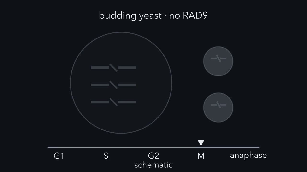 ▶ Watch A Brake Wired to a Sensor · 5:03
Read the transcript of “A Brake Wired to a Sensor”
Film transcript: A Brake Wired to a Sensor
This yeast cell lacks a gene called RAD9. Irradiate it, and it does not pause in G2. It keeps dividing for several generations, and dies. Hold it back with a drug for a few hours, and it repairs the damage. What was the gene for?
So repair could still happen. RAD9 was needed for waiting. Ted Weinert and Leland Hartwell found this in 1988. A year later, they named such controls checkpoints. A checkpoint makes one cell-cycle event wait for another. Without it, the cell divides on time, not on completion. Now run the logic the other way. What result would have said RAD9 is a repair gene after all?
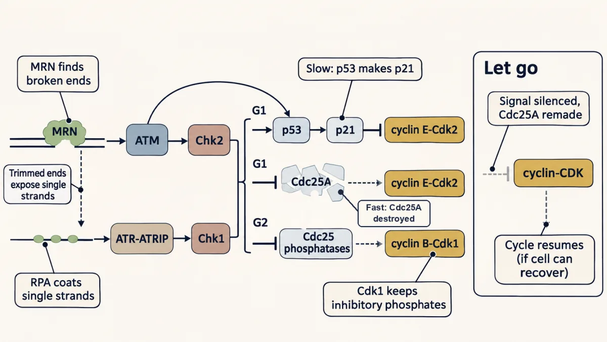
If the held cells had still failed to repair, that would point to repair, though not prove it. But more time rescued them. First, the sensor. Broken DNA ends call in a kinase, ATM. Single strands at a stalled replication fork call in ATR. ATM mainly switches on Chk2, and ATR, Chk1. In G2, these messengers inhibit the Cdc25 phosphatases. So Cdk1 keeps its inhibitory phosphates and stays off. The cell waits. In this branch, nothing was destroyed; the brake is a shift in a phosphate balance. Once the signal is silenced, a cell able to recover moves on. Here is the whole idea. A checkpoint is a brake wired to a sensor. While the sensor sees an unfinished job, the brake holds back the next transition.
When the cell finally moves, the move is hard to undo. The same wiring guards anaphase. Each pair of sister chromatids should attach to opposite spindle poles, through structures called kinetochores. In flat rat-kangaroo cells, Conly Rieder's group found one chromosome attached to only one pole. They destroyed its unattached kinetochore with a laser. Does anaphase still wait?
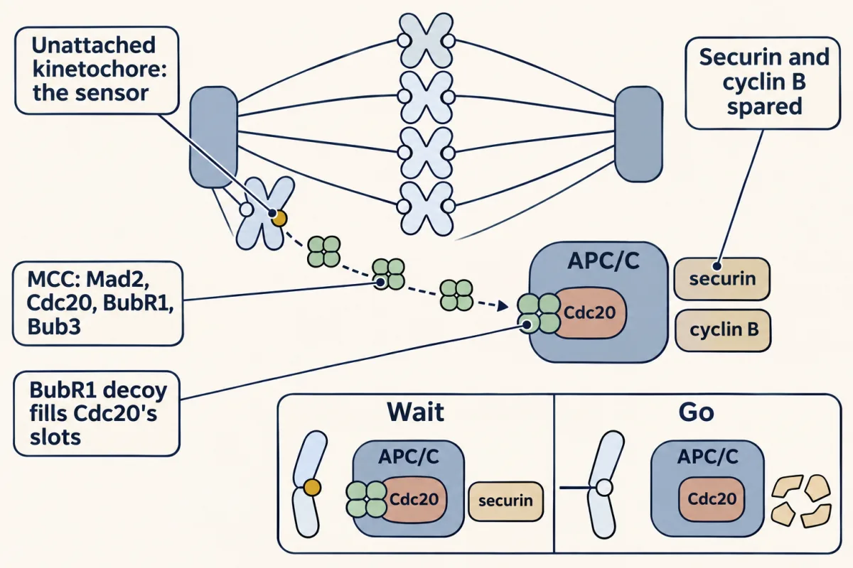
No. Anaphase followed about twenty minutes later, though that chromosome never reached the middle. The wait signal came from the unattached kinetochore. Unattached kinetochores promote assembly of the mitotic checkpoint complex. It holds back the APC/C, or anaphase-promoting complex, a ligase that marks proteins for destruction. The complex is a decoy key. It fills the slots on Cdc20 that would grip a real target. Securin and cyclin B are kept out. When the last kinetochore attaches, the decoys stop coming and come apart. Then the APC/C tags securin and cyclin B, and the proteasome destroys them. Securin had held separase, a protease, inactive; in vertebrates, so had Cdk1.
Freed separase cuts the cohesin holding sister chromatids together, and anaphase begins. Losing cyclin B is a major way Cdk1 turns off. Why does it not run backwards? A phosphate can be put back within seconds to minutes, because the protein is still there. A destroyed protein must be made again. Potapova and colleagues forced cells out of mitosis with a Cdk1 inhibitor.
Cyclin B was kept. After washout, many went back into mitosis. Cells whose cyclin B was destroyed did not. Entry into mitosis also rests on feedback. Active Cdk1 switches on its activator and switches off its inhibitor. Once the rise starts, it feeds itself. Once on, this switch tends to stay on. That may be why the damage brake normally acts in G2, before the switch flips. The brake can leak, though. A cell held in mitosis can eventually slip out, because cyclin B is still slowly destroyed. So at each guarded step, a cell asks one question: is the job finished? Until it is, a sensor holds the brake. Then destruction or feedback carries it forward, and the step is hard to take back.
From the film
- A checkpoint is a brake wired to a sensor: while the sensor sees an unfinished job, the brake holds back the next step. Without it, a cell divides on time, not on completion.
- RAD9 was needed for waiting, not for repair: held in G2 with a drug, irradiated rad9 cells repaired their DNA.
- In G2, Chk1 and Chk2 (switched on by ATR and ATM) inhibit Cdc25, so Cdk1 keeps its inhibitory phosphates and the cell waits.
- One unattached kinetochore is enough to hold anaphase: it builds the MCC, a decoy key that fills Cdc20's slots and keeps APC/C from tagging securin and cyclin B.
- The step that follows is hard to undo, because destroyed securin and cyclin B must be made again and the Cdk1 feedback switch resists reversal.
The film above opens with a budding-yeast cell that lacks RAD9: irradiated, it does not wait in G2, yet held back with a drug it repairs itself.
So the defect in rad9 cells was not in repair. RAD9 was needed for waiting. That result, from Ted Weinert and Leland Hartwell in 1988, opened a question you can ask of every step in the cycle: what makes one event hold off until another is done, and once the cell moves on, what stops it going back?
The idea: a checkpoint makes one event wait for another
In 1989 Hartwell and Weinert gave these controls a name. A checkpoint is a control mechanism that enforces dependency between cell-cycle events.
The film draws it as a brake wired to a job bar. The job is what the cell has to finish (copying DNA, repairing it, attaching chromosomes to the spindle), the engine is the enzyme that drives the next step, and the checkpoint is the wire and brake between them. Snip the wire and the cell can still divide, but on time instead of on completion.
The rad9 experiment is the test for such a wire. After irradiation, wild-type haploid yeast arrested or delayed in G2, the gap between DNA synthesis and mitosis; rad9 cells did not. When a microtubule poison held irradiated rad9 cells in G2 for several hours, repair could occur efficiently.
Predict. Run the logic the other way. What result would have told Weinert and Hartwell that RAD9 is a repair gene after all?
Check your prediction
More time rescues: a waiting defect. No rescue: perhaps a repair defect. Had the held cells still failed to repair, that would have pointed to repair, though not proved it (the block might have been too short, or the damage beyond repair). Repair itself belongs to that lesson.
Every checkpoint in this lesson is built the same way.
The one idea to carry through this lesson. A checkpoint is a brake wired to a sensor. While the sensor sees an unfinished job (a broken end, a stretch of single-stranded DNA, an unattached kinetochore), it restrains the regulators of the next transition: particular cyclin-CDKs, or APC/C-Cdc20-dependent destruction of securin and cyclin B.
- Wait. In a cell still able to resume cycling, resolving the problem and silencing the signal permit recovery, which can also need new protein. Some damage responses end in lasting withdrawal from the cycle.
- Move. The step the cell then takes is hard to undo. Depending on the step, it destroys proteins (securin and cyclin B at anaphase, Sic1 at yeast G1/S), which must be made again, or flips a positive-feedback switch (Cdk1 at entry into mitosis), which resists reversal.
The rule is about timing on the core engines of the cycle, not about repair, though the same kinases also act on replication forks. The brake is not perfect or global: the spindle checkpoint still lets cyclin A2 be destroyed, and a cell held in mitosis can eventually slip out, because cyclin B is still slowly destroyed.
The map: three checkpoints, two engines
Every name in the table is explained in the sections below.
| Checkpoint (where the cell waits) | What it senses | Signals through | Transition output restrained | Released when |
|---|---|---|---|---|
| DNA damage (at G1/S and at G2/M) | broken ends; single-stranded DNA | ATM and Chk2; ATR and Chk1; p53 and p21 | cyclin E-Cdk2 in G1; cyclin B-Cdk1 in G2 | damage signals are resolved, signalling is silenced, and the cell can still recover |
| DNA replication (at G2/M) | single-stranded DNA at stalled forks | ATR and Chk1 | cyclin B-Cdk1 | stalled forks restart and replication is finished |
| Spindle assembly (metaphase to anaphase) | an unattached kinetochore | the mitotic checkpoint complex (MCC) | APC/C-Cdc20 targeting of securin and cyclin B | the last kinetochore attaches |
The two engines, from the previous lesson:
- Cyclin-CDK. A CDK works only with its cyclin, and inhibitory phosphates can still hold it off. For Cdk1, the kinases Wee1 and Myt1 add those phosphates and Cdc25 phosphatases remove them. The cell enters mitosis only when the Cdc25 side wins.
- APC/C. This ubiquitin ligase marks proteins for destruction. To pick its mitotic targets it needs the activator Cdc20.
The table also predicts what happens when a brake is lost. A yeast rad9, mad or bub mutant often grows normally when nothing goes wrong. It fails when the event it should wait for is slow or broken, and then it moves on anyway. The cost shows up as broken or missing chromosomes in the next generation. That holds for the classic yeast mutants, not for every checkpoint protein: in budding yeast the kinases Mec1 and Rad53 are essential even without damage, and in animals some checkpoint proteins, such as Mad2, are needed in every cycle (Going deeper).
Damage and replication: kinases that keep a CDK off
Both DNA checkpoints run the same circuit in four steps. Something finds the damaged DNA, a large kinase is switched on there, it switches on a smaller messenger kinase, and the messenger’s targets keep a cyclin-CDK off. (How a phosphate works as a switch is covered in that lesson.)
Step 1: find the trouble. The cell recognizes two kinds of abnormal DNA.
- A double-strand break leaves two loose ends. The MRN complex (Mre11, Rad50 and Nbs1) binds the ends and recruits the kinase ATM.
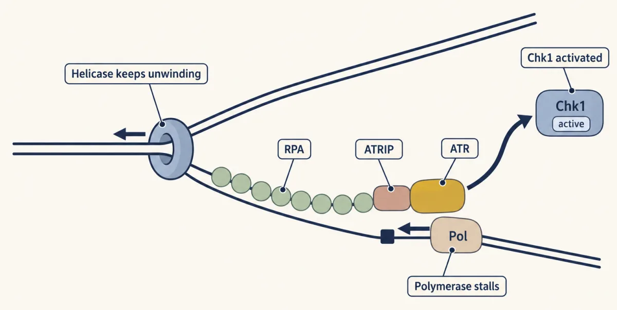
- Single-stranded DNA is coated by RPA, the single-strand binding protein. RPA helps bring in the kinase ATR with its partner ATRIP. Single strands appear at a stalled replication fork, when the polymerase stops but the helicase keeps unwinding. They also appear at a break once its ends have been trimmed back.
Step 2: pass the signal on. ATM and ATR are the upstream kinases. Each switches on a smaller messenger kinase: ATM mainly Chk2, ATR mainly Chk1. ATM also phosphorylates p53 directly. Reaching the DNA is not enough to switch ATR on: the protein TopBP1 strongly stimulates its kinase activity, and ETAA1 can do so too.
| What the cell finds | Found by | Upstream kinase | Messenger kinase |
|---|---|---|---|
| Broken ends | MRN | ATM | Chk2 |
| RPA-coated single strands | RPA and ATRIP | ATR | Chk1 |
A break in S or G2 uses both rows. ATM promotes trimming of the broken ends, which the Mre11 nuclease starts. Trimming exposes single strands, RPA coats them, and ATR joins in.
Step 3: hold the CDK off. Which CDK gets held depends on where the cell is in the cycle. Start with G2.
Predict. Chk1 and Chk2 inhibit the Cdc25 phosphatases. What happens to Cdk1, and to the cell?
Check your prediction
Cdk1 keeps its inhibitory phosphates and stays off, so the cell waits in G2. Nothing was destroyed or had to be made anew, so the brake lifts once the signal stops. The full damage response also destroys Cdc25A and makes p21 (below).
- In G2, Cdk1 stays off, as predicted. Several Cdc25 phosphatases share this job: mouse cells lacking Cdc25C still entered mitosis on time and responded normally to damage, probably because Cdc25A and Cdc25B covered for it. In human cells, p53 and p21 were needed to keep the G2 arrest going after radiation.
- In G1, fast: Cdk2 stays off. The checkpoint kinases trigger rapid ubiquitin-dependent destruction of Cdc25A, the phosphatase that activates Cdk2. Cdk2 keeps its inhibitory phosphate, and S phase does not start. This step needs no new gene expression and, in the classic experiment, no p53 (Going deeper).
- In G1, slow: p53 and p21 keep Cdk2 off. p53 is a transcription factor (that lesson). Activated after damage, it induces the gene for p21, a CDK inhibitor; in biochemical assays p21 blocks cyclin E-Cdk2 from phosphorylating Rb, so S phase stays off. In isogenic human colon cancer lines, Waldman, Kinzler and Vogelstein found that losing p21 abolished the G1 arrest after DNA damage altogether, a result in those cells, not every cell type.
Step 4: let go. As the breaks are resolved, or replication is completed, checkpoint signalling can be silenced. In a cell still able to recover, phosphatases and the remaking of destroyed proteins such as Cdc25A then restore CDK activity. That is the wait half of the rule.
Now place Rad9 from the opening. Later work showed that it is an adaptor at step 2: it helps the kinase Mec1 (yeast’s ATR) switch on Rad53, yeast’s messenger kinase. It also limits how far broken DNA ends are trimmed. So Rad9 does more than make the cell wait, but the 1988 test isolated its role in waiting.
The spindle checkpoint: one kinetochore says wait
For the chromosomes to separate correctly, every pair of sister chromatids should be attached to opposite spindle poles before anaphase. The attachment point is the kinetochore, a protein structure on each chromatid’s centromere that grips spindle microtubules. How it grips is left to the mitosis lesson.
The genes came from yeast. In 1991 Rong Li and Andrew Murray, and in the same issue of Cell Hoyt, Totis and Roberts, isolated budding-yeast mutants that kept going when a drug destroyed their microtubules. These were named mad (mitotic arrest deficient) and bub (budding uninhibited by benzimidazole).
Where does the signal come from? The film shows Conly Rieder’s laser experiment. PtK1 cells are flat, so chromosomes can be seen one by one; the group waited for a cell with a single chromosome attached to only one pole and destroyed its unattached kinetochore.
Predict. Every other chromosome is lined up at the spindle’s equator, and this one never will be. Does anaphase wait?
Check your prediction
No. Anaphase followed about 20 minutes later, although that chromosome never moved to the equator. The cell was not checking alignment. It was listening to a wait signal from an unattached kinetochore, and removing the kinetochore removed the signal.
The signal is a protein complex. Unattached kinetochores promote assembly of the mitotic checkpoint complex (MCC): Mad2, Cdc20, BubR1 and Bub3. The MCC holds APC/C-Cdc20 back. The film’s decoy key is BubR1: it carries sequences that look like a substrate’s destruction signal, and it also blocks the E2 enzyme UbcH10. Once proper attachments silence the checkpoint, kinetochore-driven MCC production subsides, and continued disassembly of the MCC lets APC/C-Cdc20 target securin and cyclin B efficiently.
Predict. Inject an antibody that blocks Mad2 into a cell in prometaphase, while some kinetochores are still unattached. Does anaphase come earlier or later?
Check your prediction
Earlier. Without Mad2 no MCC forms, APC/C-Cdc20 is free, and the wait is gone. Injecting an anti-Mad2 antibody into PtK1 cells or primary human keratinocytes in prophase or prometaphase brought on premature anaphase. Errors that escape this checkpoint produce the aneuploidy discussed in that lesson.
The block also leaks. Brito and Rieder found that PtK and human RPE1 cells held in nocodazole can slip out of mitosis while the checkpoint is still active, because cyclin B is slowly destroyed by a proteasome-dependent route.
One-way doors: destruction and feedback
Now the cell moves on, and the move is hard to undo.
Anaphase: APC/C-Cdc20 destroys securin and cyclin B
Freed APC/C-Cdc20 ubiquitinates securin and cyclin B. APC/C is the ubiquitin ligase; the proteasome does the destroying (in HeLa cells, the proteasome inhibitor MG132 blocked securin breakdown). Ubiquitin chemistry is in that lesson.
- Securin binds and inactivates separase, a protease. In vertebrate cells cyclin B-Cdk1 also inhibits separase, and Hagting and colleagues found that both securin and cyclin B1 have to be degraded before sister chromatids can separate. Freed separase cuts Scc1, the cohesin subunit that holds sister chromatids together, and anaphase begins.
Why it does not run backwards
The film contrasts the two kinds of brake: a phosphate is quickly put back (seconds to minutes, depending on the substrate), a destroyed protein must be made again. But is destruction what actually stops a cell from going back?
Predict. Potapova and colleagues forced cultured Xenopus and human cells out of mitosis with a Cdk1 inhibitor, while keeping cyclin B intact. Then they washed the inhibitor out. Did the cells stay out of mitosis?
Check your prediction
No. Many cells returned to mitosis: the cyclin B was still there, so Cdk1 switched back on once the drug was gone. Cells whose cyclin B had been destroyed before washout did not return, because Cdk1 had no partner until new cyclin was made. Destruction is what made exit stick. A later study found that after mitotic exit, inhibitory phosphorylation of Cdk1 can also hold it inactive in G1 even when cyclin B is preserved, a second lock.
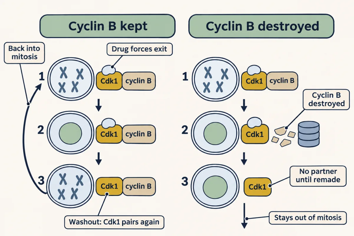
Feedback: the Cdk1 switch
The film shows Cdk1 switching on Cdc25 and switching off Wee1, so entry into mitosis rests on positive feedback.
A switch like this is bistable: over a middle range of cyclin B, it can sit stably off or on, depending on where it came from. If cyclin B is the only input varied and other regulators are held fixed, the switch turns on above an upper threshold and off below a lower one. That gap gives it memory: wobbles that stay short of the thresholds do not flip it. Changing Cdk1 inhibition or other regulators can still turn it off without lowering cyclin B. The table is a cartoon of that fixed-regulator case, with feedback strong enough to give a switch.
| Total cyclin B | Cdk1 activity, without the feedback | Cdk1 activity, with the feedback |
|---|---|---|
| Low | low | off |
| Middle | in between, following cyclin B up and down | off or on, depending on history |
| High | high | on |
This suggests why the damage brake normally acts in G2, before the switch flips: a cut in Cdc25 activity that keeps an off switch off may not turn an on switch off, because the on state is held up by its own feedback.
That is the move half of the rule.
G1/S: SCF destroys Sic1
The same logic runs at the budding-yeast G1/S transition through a second ligase, SCF. Sic1 is a CDK inhibitor that holds the S-phase cyclin-CDK off. Once phosphorylated, Sic1 is gripped by Cdc4 (the SCF subunit that recognizes it, assembled with Skp1) and ubiquitinated. When the SCF subunit Hrt1 was inactivated, Sic1 stayed stable and cells arrested at G1/S. In human cells, commitment to a new cycle also rests on a feedback switch. How destruction and feedback turn a gradual input into an all-or-none switch is taught in that lesson.
Each of these one-way steps is a destruction, and a destruction has a speed. The second film picks up here: how fast does securin go once the brake lifts, and how do you read that from one living cell?
Read the transcript of “Timing Securin's Destruction”
Film transcript: Timing Securin's Destruction
How fast can one living cell destroy a protein? Chapter 1 ended with the brake lifting. The APC/C tagged securin, and the proteasome destroyed it. Now, how can you read that speed? Tag securin with a fluorescent protein, and film a single cell. The glow tracks how much securin is left. In human HeLa cells, it starts to fall at metaphase, once the checkpoint is satisfied. Here are made-up numbers, for practice. At metaphase, 100 percent. Five minutes later, 71.
Ten minutes, 50. Fifteen minutes, 35. Treat this as exponential loss, with no new securin made. At 10 minutes, half is gone. That time is the half-life. For exponential loss, a half-life removes half of what remains. The loss is a steady fraction, not a steady amount. Here is a test for you. Ten minutes took the glow from 100 down to 50. What will it read at 20 minutes?
25 percent, not zero. Two half-lives halve it twice. The first 10 minutes lost 50 points; the next 10 lost only 25. The curve has a formula. f of t equals f zero times e to the minus k t. The rate constant k is ln 2 divided by the half-life. Here, k is about 0.069 per minute. Each securin the proteasome destroys is gone for good. While none is made, the curve can only fall. That fall is the one-way step from chapter 1, measured in minutes. So read this exponential destruction curve by its half-life. Every half-life halves what is left. From that one number you get the rate, and predict when any level is reached.
From the film
- A fluorescent tag on securin lets you time its destruction in one living cell; in HeLa cells it starts to fall at metaphase, once the checkpoint is satisfied.
- If securin is destroyed at a constant fractional rate and none is made, each half-life halves what is left.
- The loss is a steady fraction, not a steady amount: in the made-up data, 100 falls to 50 in 10 minutes and to 25 in 20.
- The rate constant k is ln 2 divided by the half-life; a 10-minute half-life gives k of about 0.069 per minute.
A worked problem: timing securin destruction
The following data are made up for practice, in the style of the live-imaging work by Hagting and colleagues, who showed in HeLa cells that securin destruction begins at metaphase once the checkpoint is satisfied. Suppose HeLa cells express fluorescent securin, and fluorescence (as percent of its metaphase value) is measured at 0, 5, 10, 15 and 20 minutes after metaphase: 100, 71, 50, 35, 25. Assume fluorescence is proportional to securin and synthesis is negligible. The film read the half-life; the steps below go past it.
In words The amount left equals the starting amount times e to the minus k t. The half-life is the natural log of 2, divided by k.
- F(t)“F of t”
- the amount left at time t, here fluorescence as percent of the metaphase value%
- F0“F zero”
- the amount at time zero%
- k“k”
- the rate constant: the fraction destroyed per minute, for small stepsper minute
- ln 2“natural log of 2”
- about 0.693, the power you raise e to in order to get 2
Sanity check After one half-life, 50% is left; after two, 25%; after three, 12.5%. Each half-life halves what remains, whatever the starting amount.
Try one number
Rate constant: at 10 minutes F = 50, so ln(100/50) = 0.693 = k × 10, and k = 0.0693 per minute.
Half-life: 0.693 / 0.0693 = 10 minutes.
Check another point: 100 × e−0.0693 × 15 = 100 × e−1.04 = 35. It fits the data.
Need a refresher? e and ln
- Rate constant. k = 0.0693 per minute; half-life 10 minutes (worked in the card).
- Time to 10 percent. t = ln 10 / k = 2.303 / 0.0693 = 33 minutes.
- Anaphase. In this made-up cell, a quarter of the securin is still there at 20 minutes. The data alone do not say when anaphase begins. If we assume separation waits until securin is below 10 percent, anaphase comes after about 33 minutes (step 2).
- Re-imposing the checkpoint. Suppose a spindle poison such as nocodazole is added at 10 minutes. Predict the fluorescence over the next 20 minutes before you open the answer.
Check your prediction
It should stay near 50, if the checkpoint switches back on quickly. The spindle is disrupted, kinetochores are unattached again, and the checkpoint holds APC/C-Cdc20 back, so destruction stops. A slow restart or leftover destruction would let it drift lower (with no stop at all it would reach 12.5 by 30 minutes). That fits Hagting and colleagues’ finding that re-imposing the checkpoint rapidly stops securin destruction (they used taxol).
- A D-box mutant. The destruction box (D-box) is a short securin sequence that APC/C recognizes. A D-box mutant securin would not fall at metaphase; in HeLa cells such a mutant inhibited sister chromatid separation.
securin, this model dashed grey: no re-imposition orange: assumed anaphase, when securin falls below 10%; dotted: checkpoint back on lesson’s made-up data (half-life 10 min)
- Rate constant
- Securin at the cursor
- At 20 min
- Time to 10 percent
Rule to keep: with a half-life h, k = ln 2 / h and F = F0 × e−kt; time to 10 percent is ln 10 / k; and once the checkpoint is back on, destruction stops and the level stays where it was: stopping destruction holds the level; it never raises it. In this sketch the stop is idealized as instant and complete (the lesson: “rapidly stops”, “near 50”). The half-life and T sliders are this sketch’s own, not the lesson’s; only 10 min and T = 10 are the lesson’s. The data are made up, as in the lesson.
Design problem
Knocking down protein Z with RNAi changes how long mitosis takes. Design experiments to decide whether Z is a spindle-checkpoint component, is needed for kinetochores to attach to microtubules, or both.
Saved in this browser only.
One way to do it
Film single cells and time nuclear envelope breakdown to anaphase in four conditions: control, Z-depleted, Mad2-depleted, and Z plus Mad2 depleted. Repeat each in nocodazole.
A checkpoint component should behave like Mad2. Mitosis may get shorter, as it did after anti-Mad2 injection, and Z-depleted cells cannot stay arrested in nocodazole.
A pure attachment protein does the opposite. Unattached kinetochores keep signalling, so mitosis gets longer. That delay should collapse if Mad2 is depleted as well, because the delay needs the checkpoint. Nocodazole arrest should be intact.
Score lagging chromosomes in anaphase in each condition. Stain for Mad2 or BubR1 at kinetochores too: some kinetochore proteins both grip microtubules and recruit checkpoint proteins, and a Z that does both could cause attachment errors and a weak arrest at once. Rescue with an RNAi-resistant Z to show the effect is Z’s. One caveat: a strong checkpoint can still look weak if cells slip out of mitosis by slow cyclin B loss, so measure how long cells stay arrested, not only whether they eventually leave.
Going deeper
Optional: the experiments behind the main text.
When checkpoint proteins are needed in every cycle
Checkpoints are not only emergency brakes. Mouse embryo cells lacking Mad2 grow normally at first, but once the epiblast starts dividing rapidly they missegregate chromosomes and die, even without spindle damage. Reducing Mad2 or BubR1 in human cancer cells killed them within six divisions.
In a 2018 study of cultured human cells, Saldivar and colleagues reported that the kinase ATR, activated by a protein called ETAA1 during ordinary replication, holds back a CDK1-driven switch on the transcription factor FOXM1 until S phase ends; when ATR was inhibited, cells entered mitosis early with under-replicated DNA.
In budding yeast, MEC1 and RAD53 are essential. Their lethality is suppressed by deleting SML1, an inhibitor of ribonucleotide reductase, so their essential job is mainly supplying DNA building blocks (dNTPs) rather than making the cell wait.
Who showed what in the DNA checkpoints
- ATM. In a purified system, Lee and Paull showed that MRN and DNA together activated ATM to phosphorylate p53 and the kinase Chk2. In mammalian cultured cells, Chk2 activation after damage depended on ATM.
- ATR. Zou and Elledge found that in human cells RPA was needed both for ATR to reach damage sites and for ATR to activate Chk1, and that RPA stimulates ATRIP to bind single strands in vitro.
- Cdc25A. In human cells, ultraviolet light or radiation triggered its destruction. After ultraviolet light this needed Chk1 but not p53; after ionizing radiation the ATM-Chk2 route also drives it (Falck and colleagues, 2001).
- At the fork. In replisomes rebuilt from purified budding-yeast proteins, the yeast checkpoint kinase Rad53 restrained excess unwinding by the CMG helicase after DNA synthesis was blocked, and only kinase-active Rad53 did so. That is a yeast reconstitution; how closely human ATR and Chk1 match it is still being worked out.
Cdc25C, serine 216 and 14-3-3
The classic example of a Cdc25 target is Cdc25C: human Chk1 phosphorylates it on serine 216, and Chk2 can do so too. A Cdc25C mutant that cannot be phosphorylated there no longer binds 14-3-3, a small adaptor protein; 14-3-3 binding is thought to keep Cdc25C away from Cdk1.
Spindle checkpoint: timing and structure
The numbers and structure behind the spindle section: in Rieder’s laser experiment, anaphase followed 20 plus or minus 2.5 minutes after the kinetochore was destroyed. The decoy-key picture comes from the cryo-EM structure of human APC/C bound to the mitotic checkpoint complex (MCC), in which BubR1 sequences that mimic a substrate’s degron (the short sequence that marks it for recognition) sit in Cdc20’s degron-binding sites.
Commitment switches in data
For the human G1/S commitment switch named in the main text, Cappell and colleagues found that EMI1 is first a substrate of the ligase APC/C-Cdh1 and then becomes its inhibitor, and that this double-negative loop is bistable, a one-way switch.
For mitotic entry, frog egg extracts show the gap between the on and off thresholds directly: Sha and colleagues (2003) found that entering mitosis takes a higher cyclin B concentration than staying in it.
Securin’s backup signal
The D-box is not securin’s only handle. The D-box mutant from the worked problem escapes destruction at metaphase, but Hagting and colleagues found it was degraded later, in anaphase, through a second APC/C recognition sequence, the KEN box.
Your notes
Select any text in the lesson to highlight it or add a note to it. Your highlights and notes are saved in this browser and collected in your notebook.
This browser is not letting the site save anything (a private window or strict privacy settings), so highlights and notes will be lost when you leave the page.
How we measure it
Imposed-delay rescue
Damage a checkpoint mutant, then supply the missing delay artificially, for example with a drug that blocks division. If the cells now survive or repair, the gene is needed for the delay. Weinert and Hartwell used this test on irradiated rad9 yeast.
Laser microsurgery with video microscopy
A focused laser destroys one chosen structure, such as a single kinetochore, in a living cell while the cell is filmed. Timing the next event shows whether the destroyed structure was the source of a signal. Rieder's group used it in PtK1 cells.
Antibody microinjection
An antibody against one protein is injected into a living cell at a chosen stage to block that protein acutely, avoiding the slow protein loss of a knockout. Injecting anti-Mad2 into mitotic PtK1 cells and human keratinocytes caused premature anaphase.
Live imaging of a fluorescent reporter
A protein such as securin is tagged with a fluorescent protein and filmed in single cells, so its destruction can be timed against visible events like metaphase and anaphase. Drugs added during filming test what the destruction depends on.
Practice questions on this lesson (3) → MCAT-style, original, each with an explanation
Where this lesson fits
Before thisReview first
- Cyclin-CDK Control of the Cell CycleThis course · Lesson 17
You need the cyclin-CDK engine and its inhibitory phosphates from there to see what each checkpoint holds back.
- Protein Quality Control and Organelle TurnoverThis course · Lesson 9
Regulated destruction of proteins and the ideas of flux and turnover from there are used when reading securin degradation.
After thisStudy next
- Mitosis and Chromosome SegregationThis course · Lesson 19
It uses the spindle checkpoint and APC/C-Cdc20 securin destruction to explain how sister chromatids separate at anaphase.
- Cell Death and ClearanceThis course · Lesson 21
Cells with damage that cannot be repaired may withdraw or die, so it picks up where the DNA damage response ends.
- Cell Identity, Cell State and DiseaseThis course · Lesson 25
It shows how losing checkpoint control and the damage response lets mutations build up in cancer.
Elsewhere on the siteFilms, simulations and method films in other courses
- Mitosis stepper and phase counterSimulation · Biology
This is not a real sim id; ignore.
Suggested by an AI model from the lessons themselves, then checked. How we use AI
Finish line
Test yourself
8 questions
Practice and MCAT-style, with explained answers.
Next · Lesson 19
Mitosis and Chromosome Segregation
How does a dividing cell give each daughter exactly one sister of every chromosome, and what happens when one attachment goes wrong?
Continue →