Cell Biology · Unit 4: Division and cell loss
New to this? Review it at high school level: The Cell Cycle and Mitosis
Cytokinesis and Organelle Inheritance
Rewritten on : new text, flashcards and film. Your notes and highlights stay, but the text around them may have moved.
Push the spindle off-centre in a sea urchin embryo cell, and the signal for the furrow follows it before the furrow does.
Pause when the reading reaches a film, and offer to play it. Saved on this device.
Watch the film now, or keep listening
Builds on:Mitosis and Chromosome SegregationCytoskeletal Polymers and DynamicsVesicle Budding, Targeting and FusionWhy these?
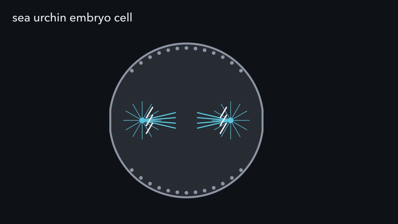 ▶ Watch The Band Before the Furrow · 3:26
Read the transcript of “The Band Before the Furrow”
Film transcript: The Band Before the Furrow
This sea urchin embryo cell is in anaphase: its two sets of chromosomes are moving apart. Now it must cut itself in two, right between them. How does its surface know where the middle is? The cut happens at the cell surface. There the surface pinches in, making a groove called the furrow. But the thing that marks the middle sits deep inside. It is the spindle, a bundle of microtubules with a star-shaped aster at each end. There are three simple answers. One: the spindle pulls the surface in directly, by force. Two: an actin ring gathers at the middle on its own. Three: the spindle only sends a signal. The signal marks the surface, and the ring is built under that mark.
To tell them apart, researchers watched a switch protein called RhoA. RhoA is on when it holds GTP, and off when it holds GDP. They added a glowing probe that sticks only to switched-on RhoA. A band of active RhoA, shown here in amber, lights up just under the membrane. One to four minutes later, the furrow starts underneath it. The same order was seen in frog embryos. The mark comes before the cut. Next, they blocked actin assembly with a drug, so no ring could be built. In sea urchin embryos, the amber band still formed. So the signal does not need the ring. Answer two is out. Now the decisive test. This cell has already begun to furrow. A blunt needle pushes the spindle to one side.
What moves first: the furrow, the amber band, or nothing? Over a few minutes, active RhoA gathers over the spindle's new middle. Only then does the furrow follow it there. The band leads, and the furrow follows. That order argues against the spindle simply dragging the surface, and it supports answer three: signal first. Here is the rule for these embryo cells. The cut goes where the anaphase microtubules place a band of active RhoA. Move the microtubules, and the band moves first. The furrow follows. Under the band, active RhoA turns on formins and promotes myosin II. Formins build long actin filaments.
Myosin II, a motor, pulls those filaments past each other. In its simplest picture, the result is a ring that tightens like a drawstring. Now the two daughters are joined only by a thin bridge. Cutting that bridge is a separate job. One question stays open: how do microtubules deep inside switch on a protein at the membrane? But where the cut went was settled earlier, where the microtubules placed the band.
From the film
- In the sea urchin and frog embryo cells studied, a band of active RhoA appears just under the membrane before the furrow.
- In sea urchin embryo cells the band still forms when actin cannot assemble, so the signal does not need the ring.
- When a needle pushes the spindle aside in these cells, active RhoA gathers over its new middle first and the furrow follows.
- Under the band, active RhoA turns on formins and promotes myosin II, which build and tighten the ring.
- In the embryo cells studied, the cut goes where the anaphase microtubules place a band of active RhoA.
Mitosis has sorted the chromosomes into two sets, and anaphase, the stage in which they move apart to the two poles, is under way. The cell has three jobs left: place the cut, make the cut, and give each daughter a working share of what it contains. The first film followed the first job and ended on its rule.
The one idea to carry through this lesson. In the embryo cells studied, the cut goes where the anaphase microtubules place a band of active RhoA. Move the microtubules, and the band moves first; the furrow follows. Other animal cells can add cues of their own (Going deeper).
The first film left a question open: how do microtubules deep inside the cell switch on a protein at the membrane? The first section answers it. The next two follow the second job, making the cut, in an animal cell and then in a plant cell boxed in by a wall. The second film takes the third job, sharing the organelles. Microtubules keep turning up: the furrow leaves a bridge packed with them, a plant guides its new wall along them, and the spindle carries some of what the daughters inherit.
From microtubules to the band
The first film showed that microtubules place the band, not how. Start with which microtubules.
Predict. Raymond Rappaport reshaped a fertilized sand dollar egg into a ring, like a doughnut. Its first division cut the ring on one side only, leaving one horseshoe-shaped cell with two nuclei. At the next division that cell held two spindles side by side, each with an aster at both ends. In the gap between them, an aster of one spindle faced an aster of the other, with no spindle joining them. Does a furrow form in that gap?
Check your prediction
Yes. Furrows formed across each spindle and also in the gap between the facing asters (Rappaport, 1961). That is why the one idea says microtubules, not the spindle: in this egg, two facing asters were enough to mark a furrow site.
Now the how. RhoA is a small GTPase, a switch whose on-off cycle belongs to GTPase switches and integrins and Rho GTPases. Two proteins relay the microtubules’ position to it.
- Ect2 is the enzyme that switches RhoA on. It is a guanine-nucleotide exchange factor (GEF): it makes RhoA let go of its GDP, and GTP, which is plentiful in the cell, takes its place. On its own, Ect2 stays folded in an inactive form.
- Centralspindlin is a protein complex that gathers where microtubules from the two poles overlap. The most prominent overlap is the spindle midzone, the dense bundle of overlapping microtubules between the separating chromosomes (see mitosis).
At anaphase, centralspindlin binds Ect2 and helps activate it. In large echinoderm embryo cells the two collect on overlapping microtubules at the equator, including where astral microtubules from the two poles meet, with or without a midzone between them (Su and colleagues, 2014): the molecular side of Rappaport’s result. In cultured human cells, ECT2 must also associate with the plasma membrane during anaphase to switch RhoA on at the equator; how it reaches the cortex in echinoderm embryos is less clear. The band reflects this microtubule cue, but it is not a simple print of where microtubules overlap, and how a sharply bounded band forms is still debated (Going deeper).
Predict. In cultured human cells such as HeLa cells, Ect2 is depleted and the cells go through mitosis. Their chromosomes separate normally. What do you expect to find once division should have finished?
Check your prediction
In the cultured human cells used in these experiments, many cells fail to concentrate RhoA, actin filaments and myosin at the equator, their furrows fail, and they are left with two nuclei even though the chromosomes were separated. One cell with doubled DNA is a tetraploid cell. The next section shows a second route to the same result.
Making the cut
The one idea says where the furrow forms, not when the daughters finally part. The ring of formin-made actin filaments and nonmuscle myosin II pulls the furrow in (see molecular motors and cytoskeleton dynamics). The first film drew it as a drawstring, the simplest case; in fuller view, how the filaments are arranged in the ring differs between systems, and no one geometry is settled for all cells.
Ingression does not finish the job. It leaves the two daughters joined by a thin intercellular bridge packed with spindle microtubules. At its centre sits the midbody, a dense plug of overlapping microtubules and proteins left from the spindle midzone. Abscission, the final cut, is a separate, later step. In the several animal-cell systems covered by a 2022 review, ESCRT-III, a set of proteins that polymerize into spirals on membranes, assembles in the bridge, constricts it and cuts it. Chromatin left in the bridge can delay the cut.
If the cut fails, the bridge can regress and leave one cell with two nuclei and doubled DNA. That is the second route to the tetraploid cell of the Ect2 prediction, and one source of the tetraploid cells discussed in chromosome changes.
Plants build a wall from the inside out
In a plant, the first film’s question has a different answer. Microtubule arrays still place and build the division, but not through a RhoA band, and a plant cell cannot pinch, because it sits inside a rigid wall. In the prevalent mode of somatic plant cytokinesis there is no furrow at all. The new membrane starts in the middle of the cell and grows outward.
The builder is the phragmoplast, an array of microtubules and actin filaments that forms between the two daughter nuclei. It guides vesicles from the Golgi to the centre of the division plane. There they fuse with one another to form the cell plate, a flat compartment of new membrane filled with wall material. The plate expands sideways toward the parental cell surface, and cytokinesis is complete when it joins the old wall.
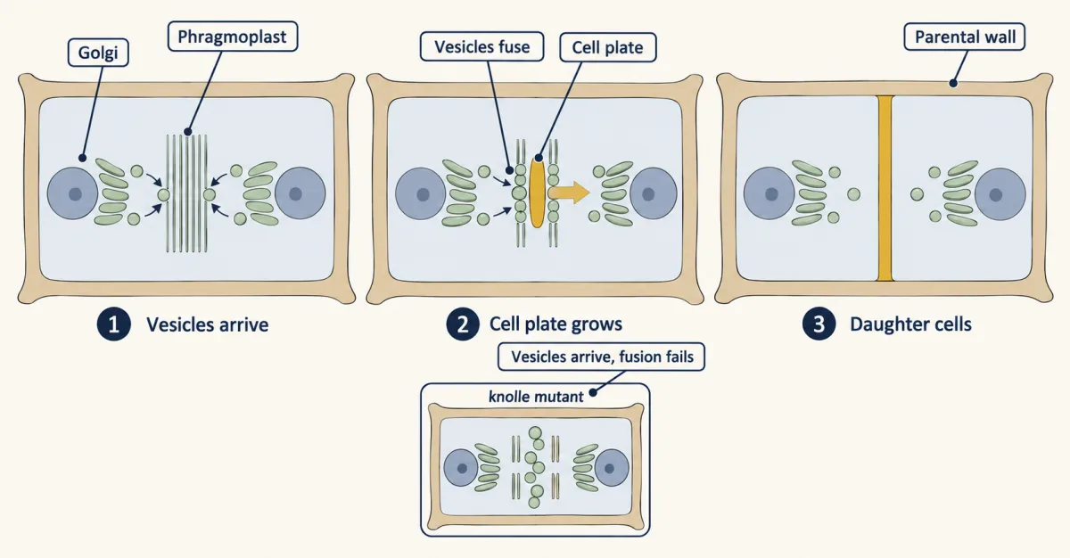
Fusion needs machinery. KNOLLE, an Arabidopsis protein related to syntaxins, the target-membrane SNAREs you met in vesicle traffic, sits at the division plane.
Predict. Arabidopsis embryos lacking KNOLLE have enlarged cells, incomplete walls and several nuclei. A plate could fail because vesicles never reach the plane, or because they arrive and cannot fuse. What would electron micrographs of the plane show in each case?
Check your prediction
A block in delivery along the phragmoplast, or in Golgi production, would leave few vesicles in the plane; a fusion block would leave a crowd. Serial-section electron microscopy of knolle embryos showed bands of vesicles in the plane and walls left incomplete. The vesicles had arrived; their fusion appeared to fail, as fits a syntaxin.
Where the plate meets the old wall is often decided earlier. Before mitosis, many plant cells build a preprophase band, a cortical ring of microtubules that marks the future division plane and then disappears. It often helps orient the division but is not indispensable everywhere; Going deeper compares two tissues.
Animal or plant, the cut falls between the two sets of chromosomes, and everything else in the cytoplasm lands on one side or the other. The second film picks up here: does the cell count its organelles out, or is chance good enough?
Read the transcript of “Chance or Control”
Film transcript: Chance or Control
In chapter 1, the microtubules placed the band, and the cut fell between the two sets of chromosomes. But the cut splits everything else too. Does the cell count its organelles out, or is chance good enough? Start with the simplest model: no control at all. Each copy of an organelle goes to either daughter by a coin toss, on its own.
With a hundred copies, the count in one daughter averages fifty, give or take about five. Most daughters get forty to sixty. The chance that either daughter gets none is about 1 .6 times 10 to the minus 30. In practice, that never happens.
Now shrink the stock to just two copies, tossed the same way. How often does one daughter end up with none? Half the time. Both copies can land left, or both right; each happens one time in four. Either daughter can be the empty one, so together they make one in two. The chance of an empty daughter is two times one half to the power n. Each added copy halves the risk. At ten copies it is about one in five hundred. At twenty-one copies it drops below one in a million.
So chance is safe only for many independent units. But some organelles are one connected structure, like a mammalian cell's Golgi ribbon, or a tubular mitochondrial network. To the coin, each is a single copy. Such a structure must be broken up first, or moved actively. In PtK1 cells, a rat-kangaroo kidney line, the Golgi ribbon comes apart during mitosis. Its membranes cluster around the spindle poles. After division, the ribbon reforms. Mitochondria in HeLa cells, a human cell line, broke into fragments early in mitosis. Without Drp1, a protein that drives mitochondrial fission, they stayed tubular. That is one cell line, so treat it as a strong lead.
Some things are not left to chance. The two centrosomes sit at the spindle poles, and the spindle carries one to each pole. So microtubules return: they place the cut, and carry part of the inheritance. One caution. Moving an organelle is not the same as changing its share. Each claim needs its own test.
So the cell need not count every unit. Chance can share many small units, often with some control alongside. Chance is safe only for many units; one connected unit needs control.
From the film
- In the simplest no-control model, each copy of an organelle goes to either daughter independently, like a coin toss.
- Under that model, the chance of an empty daughter is 2 × 0.5^n, so each added copy halves the risk.
- Chance is safe only for many independent units: with two copies, one daughter is empty half the time.
- A connected structure, such as the Golgi ribbon or a tubular mitochondrial network, counts as one unit and is broken up first or moved actively.
- In PtK1 cells the Golgi ribbon comes apart in mitosis and reforms after division; the spindle carries one centrosome to each pole.
Sharing the contents: chance or control
The second film ran the coin-toss model in pictures; here are its numbers, to check by hand. A cell does not count its organelles out one by one, so when is chance good enough? To tell when a cell must step in, first work out what pure chance would give.
Worked problem: partition by chance
The numbers are illustrative. Suppose each unit of an organelle goes to either daughter independently with probability 0.5, like a coin toss. The count in one daughter then follows a binomial distribution. This is one no-control null model, under the stated assumptions: independent units, equal chance of entering either daughter.
mean = n × 0.5; standard deviation = square root of (n × 0.5 × 0.5); P(one daughter gets none) = 2 × 0.5n
Case 1: n = 100 units. Mean = 50. Standard deviation = square root of 25 = 5. About 95% of divisions fall within two standard deviations, so most daughters get 40 to 60 units. P(one daughter gets none) = 2 × 0.5100, about 1.6 × 10⁻³⁰. In practice that never happens.
Case 2: n = 10. P = 2 × 0.510 = 2/1024, about 0.002, or 0.2%. One division in five hundred leaves a daughter with none.
Case 3: n = 2. P = 2 × 0.25 = 0.5. Half of all divisions leave one daughter with none.
Check: case 2 by counting. All ten to daughter A is one outcome of 1,024, all ten to daughter B is another, so 2 of 1,024. The factor of 2 is there because either daughter can be the empty one. Each added unit halves the risk, so it falls below one in a thousand at n = 11 and below one in a million at n = 21.
Conclusion: chance is safe only for many independent units. A single connected structure, such as the Golgi ribbon of a mammalian cell or a tubular mitochondrial network, is one unit, not many. It must be broken up first or segregated actively, as the two centrosomes, the microtubule-organizing centres at the spindle poles, are when the spindle carries one to each pole. The sampler below lets you test chance against the numbers yourself.
- Mean count in one daughter
- Standard deviation
- Within mean ± 2 SD (exact binomial)
- P(one daughter gets none)
- Divisions that left a daughter with none
The numbers are illustrative: independent 50:50 partition of each unit. Some structures are broken up first or segregated actively. Dot positions are a grid, not real positions.
Predict. Made-up data: an organelle has 50 units. By coin toss, the count in one daughter would have a standard deviation of about 3.5. You count 200 pairs of daughters and find a standard deviation of 1. Chance, or control?
Check your prediction
The data reject the coin-toss model. Its standard deviation is the square root of 12.5, about 3.5, and over 200 divisions the measured value would land close to that; 1 is far more even. That fits an equalizing mechanism, such as gathering units into two equal groups at the spindle poles, but the spread alone does not say which. A spread well above 3.5, as clumping would give, also breaks the model. If the daughters are pooled without bias, both this model and a symmetric equalizing mechanism have a mean of 25, so the much smaller spread is what rejects the coin-toss model, though the full distribution can test it too.
Chance contributes to sharing many organelles, often alongside active control, and some connected organelles are fragmented before division. In PtK1 cells, a rat-kangaroo kidney line, the Golgi ribbon (see the secretory pathway) disassembles during mitosis, its membranes cluster around the spindle poles, and the ribbon reforms after division. In HeLa cells, mitochondria fragmented in early mitosis, and cells depleted of Drp1, a dynamin-related GTPase that drives mitochondrial fission, kept tubular mitochondria through mitosis, as Taguchi and colleagues reported in 2007. These are results from single cell lines, so treat them as strong leads rather than general rules.
Moving an organelle and changing its shares are separate claims (Going deeper). Organelles also differ in how they divide (see organelles as environments), and in some cells division is unequal on purpose, as in asymmetric division.
Design problem
The Golgi’s mitotic membranes cluster around the spindle poles. Is the Golgi inherited by riding on the spindle? Design an experiment in cultured cells that could tell, and say what each model predicts before you look at any data.
Saved in this browser only.
One way to do it
Follow Jen-Hsuan Wei and Joachim Seemann, who used PtK1 cells in 2009. Induce asymmetric divisions that send the entire spindle, with the chromosomes, into one product (a karyoplast) and none into the other (a cytoplast).
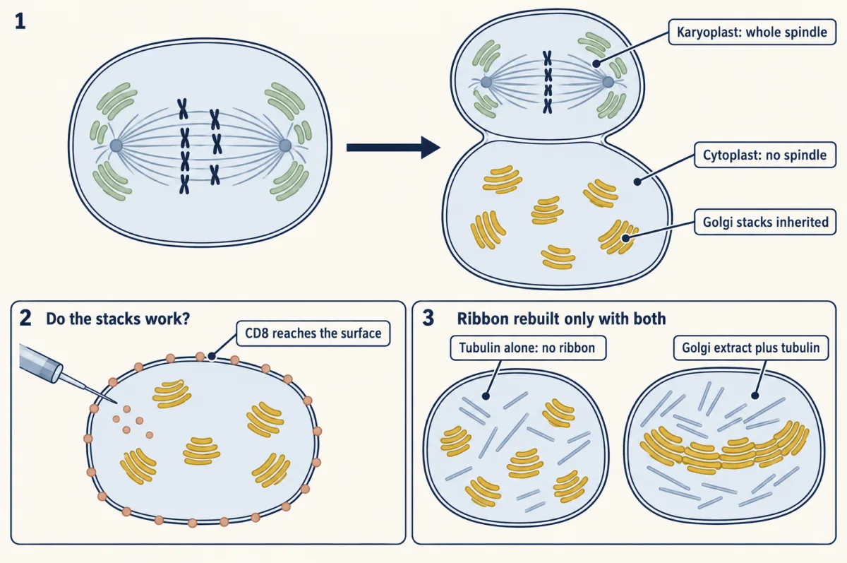
Predictions. If Golgi membranes reach daughters only on the spindle, the cytoplast should have no working Golgi. If stacks are partitioned independently, the cytoplast should have them.
Tests. Look for Golgi stacks in the cytoplast by electron microscopy and with Golgi markers. Test function: inject mRNA for a membrane reporter such as CD8 and ask whether the protein reaches the cell surface, which requires a working secretory pathway. Then ask about the ribbon separately: inject tubulin alone, Golgi extract alone, and both together, and score ribbon reassembly.
Result. The cytoplasts held scattered Golgi stacks, and CD8 reached their surface, so working stacks were inherited without the spindle. Tubulin alone did not restore a ribbon; Golgi extract plus tubulin did. A cooler division that let part of the spindle into the cytoplast also restored the ribbon, and the authors concluded that the factors needed to rebuild the ribbon travel with the spindle. So the answer to the opening question is split: working stacks do not ride on the spindle, but the ribbon’s assembly factors do.
Limits. This is an engineered division in one cell line. It does not show that no Golgi membrane ever uses the spindle, and it does not settle which Golgi components pass through the ER during mitosis.
Going deeper
The numbers behind the first film
In the 2005 work of Bement, Benink and von Dassow, the band appeared one to four minutes before the furrow in two sea urchin species and the frog Xenopus laevis; the actin drugs were cytochalasin D and latrunculin B. In green sea urchin blastomeres (early embryo cells) already furrowing, after the needle moved the spindle, the RhoA zone relocated over three to five minutes, and the furrow followed. These timings come from large embryonic cells; do not assume the same minutes in a human cell.
Keeping the band narrow
Three mechanisms may all contribute to the sharp edge, in different proportions in different cells: midzone delivery of Ect2, inhibition of cortical activity at the poles by astral microtubules, and RhoA’s own fast cycling.
A furrow placed without the spindle
Outside the embryo cells of the first film, other cues can place the cut. In Drosophila neuroblasts, which divide asymmetrically, a polarity pathway can position a furrow independently of the spindle (Cabernard, Prehoda and Doe, 2010).
Moving organelles versus sharing them
In dividing HeLa cells mitochondria gather toward the furrow, and a dominant-negative Miro-1 (a mutant form that blocks the normal Miro-1, an adaptor linking mitochondria to motors) reduced that gathering, yet the authors saw no obvious inheritance defect, possibly because the mislocalization was only partial. In other HeLa experiments, depleting the motor Myo19 sent mitochondria toward the spindle poles and disturbed how they were partitioned at division, so position can matter for shares.
The preprophase band in two tissues
After it disappears, the preprophase band leaves a cortical division zone behind. How much the band matters varies with the cell type. In Arabidopsis roots, disrupting the preprophase band reduced the precision of division orientation without abolishing the division zone. In Arabidopsis cambium stem cells, trm678 mutants that make no band kept regular division planes, while the kinesins POK1 and POK2 were needed for ordered planes. Keep each result in its tissue.
Timing the cell plate
In Arabidopsis root cells filmed live in three dimensions by lattice light sheet microscopy, untreated plates developed over about 20 to 30 minutes, and their diameter grew at about 4.09 μm per minute early on. Callose, a polysaccharide of the forming plate, helps the plate switch from collecting vesicles to maturing: blocking its deposition with Endosidin 7 disrupted that switch. The growth rate is for the early interval in this experiment, not a constant of cytokinesis.
Your notes
Select any text in the lesson to highlight it or add a note to it. Your highlights and notes are saved in this browser and collected in your notebook.
This browser is not letting the site save anything (a private window or strict privacy settings), so highlights and notes will be lost when you leave the page.
How we measure it
Live imaging with an activity biosensor
A fluorescent protein is fused to a domain that binds only the active form of an enzyme, here the Rho-binding domain of rhotekin, which binds GTP-bound RhoA. Where the probe gathers shows where the enzyme is active, not merely where the protein is, and filming it shows when.
Microneedle spindle displacement
A blunt glass needle pressed on a large embryonic cell pushes the spindle to a new position without puncturing the cell. Watching what follows the spindle, and in what order, tests whether the spindle instructs the cortex or merely sits near it.
Serial-section electron microscopy of mutants
A tissue is cut into a series of thin sections and each is imaged by electron microscopy, so that a structure can be followed in three dimensions. In knolle embryos it showed vesicles piled in the division plane beside incomplete walls, which points to a fusion defect rather than a delivery defect.
Induced asymmetric division with a secretion assay
A dividing cell is manipulated so that the whole spindle goes to one product. The other product is then examined for organelles and tested for function, for example by injecting mRNA for a membrane protein and asking whether it reaches the cell surface.
Method films · the methods library
Practice questions on this lesson (3) → MCAT-style, original, each with an explanation
Where this lesson fits
Before thisReview first
- Mitosis and Chromosome SegregationThis course · Lesson 19
You need anaphase, the spindle midzone and chromosome separation from there, since cytokinesis cuts between the two sets.
- Cytoskeletal Polymers and DynamicsThis course · Lesson 14
The actin and microtubule behaviour you learn there is what builds the ring and the phragmoplast here.
- Vesicle Budding, Targeting and FusionThis course · Lesson 11
You need SNARE-driven vesicle fusion from there to understand how KNOLLE builds the cell plate.
After thisStudy next
- Cell Death and ClearanceThis course · Lesson 21
Cells that fail division or lose organelle shares can die, and this lesson shows how clearance follows.
- Stem Cells and Tissue RenewalThis course · Lesson 24
Stem cell renewal uses division where the cut position decides what each daughter receives.
Elsewhere on the siteFilms, simulations and method films in other courses
- How One Set Becomes TwoFilm · Biology
It shows how one cell's chromosomes are copied and separated, the step that cytokinesis follows.
- One Cell, One Shipping LineFilm · Biology
It shows how the Golgi and other organelles work as a system, which the cell must hand on to each daughter.
- A Water Balloon in a BoxFilm · Life Science
It shows the plant cell wall that the phragmoplast builds a new section of.
Suggested by an AI model from the lessons themselves, then checked. How we use AI
Finish line
Test yourself
9 questions
Practice and MCAT-style, with explained answers.
Next · Lesson 21
Cell Death and Clearance
In a developing worm, the same 131 cells die on schedule, yet in healthy tissue you rarely see a corpse. How does a cell die without leaving a mess?
Continue →