Cell Biology · Skim version
Mitosis and Chromosome Segregation
Read the full lesson → Skim the whole course →
The guiding idea
How does a dividing cell give each daughter exactly one sister of every chromosome, and what happens when one attachment goes wrong?
How does a dividing cell give each daughter exactly one copy of every chromosome?
From the film: What the Cell Waits For
- A sister pair gripped from opposite poles is under tension, seen as stretch between the kinetochores.
- Slack grips, like a syntelic pair, tend to be loosened by Aurora B and remade; taut grips tend to stay.
- The checkpoint keeps the cell waiting while even one kinetochore is empty; it does not read alignment or correctness.
- When none is empty, the brake on the APC/C comes off, separase is freed, and its cut of cohesin cannot be rejoined.
- A merotelic kinetochore is attached, so it sends no wait signal and its chromatid can lag in anaphase.
From the film: Two Motions Pull the Sisters Apart
- In anaphase A each sister moves toward its pole; in anaphase B the poles themselves move apart.
- In the mammalian cells Gorbsky and colleagues studied, k-fibres shortened mainly by losing subunits at the kinetochore.
- In fruit-fly embryos and frog egg-extract spindles, poleward lattice flux can contribute much more.
- Depending on the animal cell, midzone motors, cortical dynein or both can help drive anaphase B.
- In the film's symmetric example, sister separation is twice the anaphase A rate plus the anaphase B rate.
-
The picture: one duplicated chromosome, five phases
-
Grip: the spindle and the kinetochore
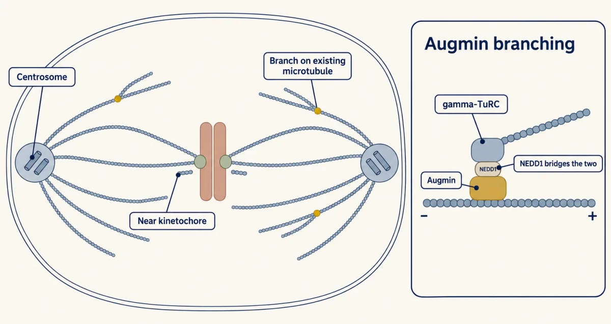
Spindle microtubules start not only at the poles but also near chromatin and kinetochores and as branches off existing microtubules, which speeds capture; the shares differ between cell types. AI-generated illustration -
Test and correct: how tension helps stabilize biorientation
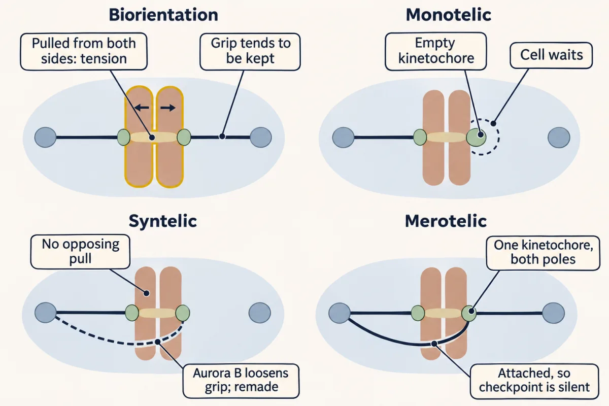
Pulled from both sides, a bioriented pair is under tension and its grip tends to be kept. An empty kinetochore makes the cell wait, a syntelic grip lacks opposing pull and is loosened by Aurora B and remade, but a merotelic kinetochore is attached, so the checkpoint stays silent. AI-generated illustration -
Wait: the checkpoint reads emptiness
-
Cut: securin, separase and cohesin
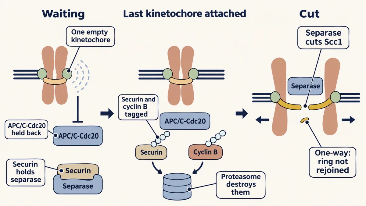
While even one kinetochore is empty, its wait signal holds APC/C-Cdc20 back. Once none is, APC/C-Cdc20 tags securin and cyclin B for the proteasome, freed separase cuts the cohesin kleisin Scc1, and the cut ring cannot be rejoined. AI-generated illustration 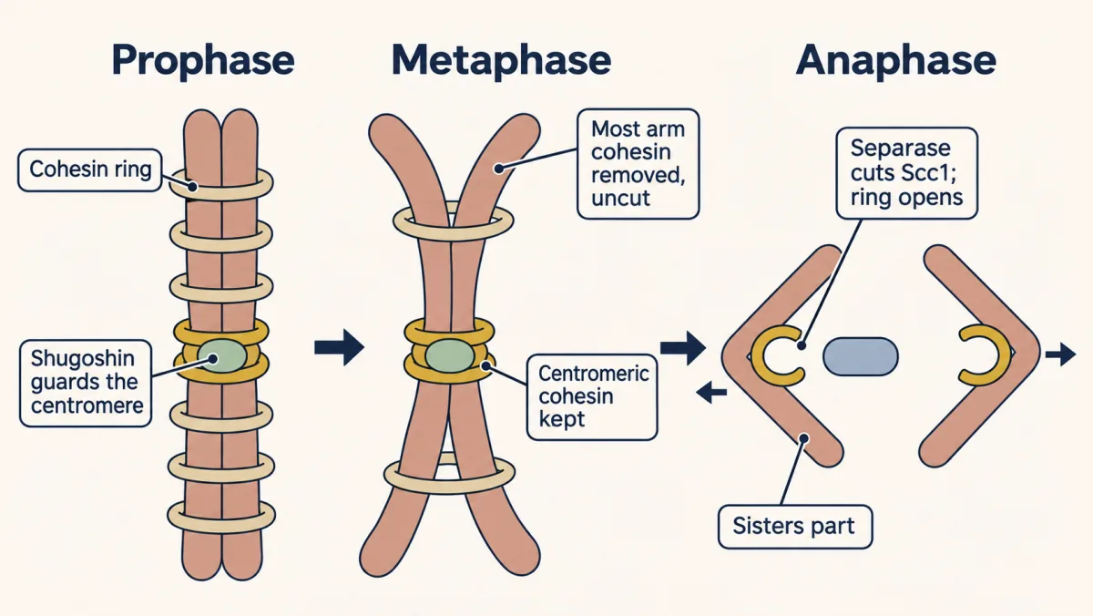
In vertebrates most arm cohesin leaves in prophase and prometaphase without Scc1 cleavage, while shugoshin protects the centromere. The sisters are held mainly at the centromere, and separase cuts that cohesin along with the small amount left on the arms. AI-generated illustration -
Move: anaphase A and B, read from a movie
-
The rule’s blind spot: merotely, laggards and micronuclei
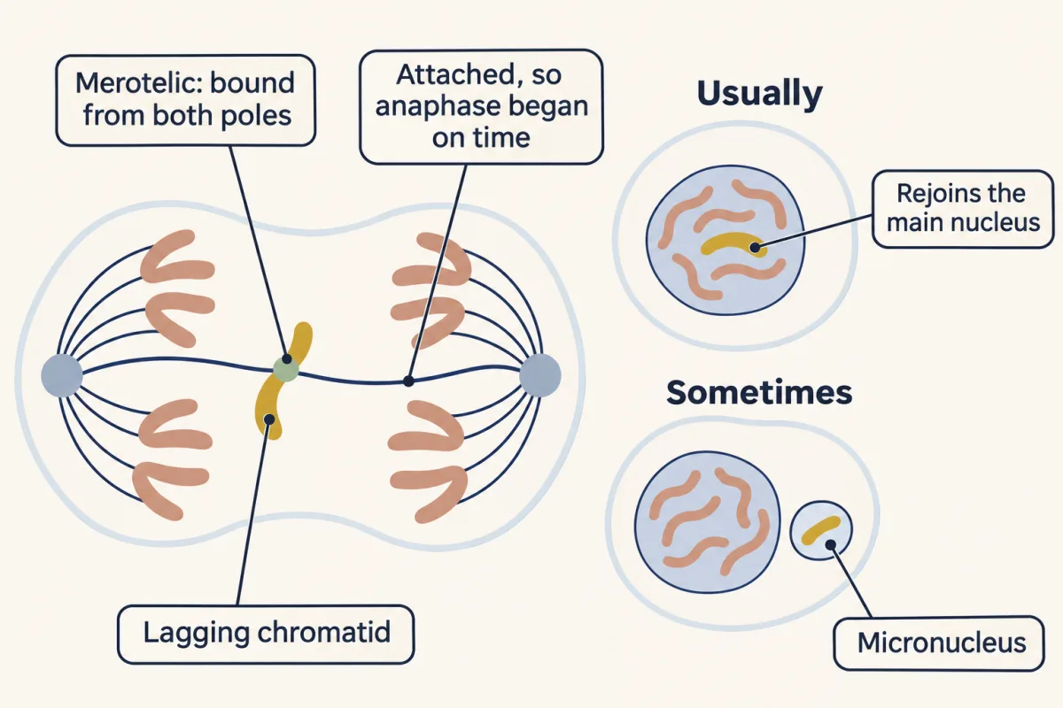
A merotelic kinetochore is attached, so the checkpoint lets anaphase start, and its chromatid lags between the poles. In human RPE1 and U2OS cells most transient laggards rejoined the main nucleus; a minority ended up in micronuclei. AI-generated illustration -
Going deeper
Where this lesson fits
Before thisReview first
- Cyclin-CDK Control of the Cell CycleThis course · Lesson 17
CDK1-cyclin B drives the cell into mitosis, and its destruction at anaphase is used here.
- Cytoskeletal Polymers and DynamicsThis course · Lesson 14
Spindle microtubules grow and shrink by dynamic instability, which the kinetochore grip depends on.
- Checkpoints and Irreversible TransitionsThis course · Lesson 18
This lesson uses the idea of a wait signal that holds the cell until the last kinetochore is attached.
After thisStudy next
- Cytokinesis and Organelle InheritanceThis course · Lesson 20
The cell splits after sisters separate, and the spindle position from this lesson sets where the furrow forms.
- Cell Identity, Cell State and DiseaseThis course · Lesson 25
Merotelic errors and lagging chromosomes feed into chromosomal instability and disease states.
- Cell Death and ClearanceThis course · Lesson 21
A cell with persistent mitotic errors can be removed by death, which this lesson's error rates lead into.
Elsewhere on the siteFilms, simulations and method films in other courses
- Mitosis stepper and phase counterSimulation · Biology
Its mitosis stepper and phase counter practise counting chromosomes and chromatids at each phase.
- How One Set Becomes TwoFilm · Biology
It shows how a copied chromosome is separated so each nucleus gets one set.
- One of Each Pair, by Coin TossFilm · Genetics and Molecular Biology
It separates sister chromatids from homologs by counting centromeres, which you need when naming what segregates.
- When Chromosomes Miss Their TurnFilm · Genetics and Molecular Biology
It shows how a missed separation at anaphase leaves a cell with an extra or missing chromosome.
Suggested by an AI model from the lessons themselves, then checked. How we use AI