Genetics and Molecular Biology · Unit 1: Inheritance
New to this? Review it at high school level: Beyond Dominant and Recessive
Chromosome Number and Structure: Aneuploidy and Rearrangements
Rewritten on : new text, flashcards and film. Your notes and highlights stay, but the text around them may have moved.
How can one missed separation give a cell an extra chromosome, and why can a balanced rearrangement still matter?
Pause when the reading reaches a film, and offer to play it. Saved on this device.
Watch the film now, or keep listening
Builds on:Chromosomes and Meiosis: The Physical Basis of Mendel's LawsSex Linkage, Dosage Compensation and Non-Mendelian InheritanceReading Pedigrees: Human InheritanceWhy these?
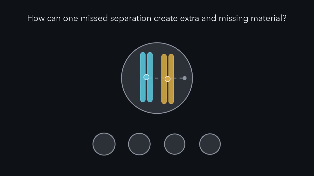 ▶ Watch When Chromosomes Miss Their Turn · 3:35
Read the transcript of “When Chromosomes Miss Their Turn”
Film transcript: When Chromosomes Miss Their Turn
How can one missed separation give one gamete extra chromosome material and another too little? Watch one replicated chromosome pair cross both meiotic divisions. Each homologue has joined sister chromatids. Blue marks one homologue, and gold marks the other. The centromeres remain visible as small circles. At meiosis I, homologues normally move apart. At meiosis II, sisters normally separate. The first division sorts homologues; the second sorts copies within each homologue. Nondisjunction means chromosomes fail to part during a division. The resulting gamete may carry an extra chromosome or none of that chromosome. This is aneuploidy, a gain or loss of individual chromosomes rather than a whole set.
First, let both homologues travel together at meiosis I. One daughter cell receives blue and gold. The other receives neither. Their sister chromatids remain joined, so the first division has already created a dose imbalance. Now suppose meiosis II runs normally. Can separating the sisters repair the chromosome dose, or is the final pattern already fixed?
The later division cannot replace a missing homologue. Normal sister separation produces matching outcomes within each branch. The products from one branch carry both homologues. The products from the other carry neither. The counts appear on screen. Reset to normal meiosis I. Blue and gold separate into different daughter cells. During meiosis II, blue sisters fail to part in only one cell. Gold sisters still separate normally in the other cell. This error makes three dose classes instead of two. One product has extra material, one is missing it, and the remaining products are normal. The stage of the missed separation sets the product pattern.
It also leaves a fingerprint that DNA markers in the child can read. A normal gamete contributes one copy at fertilization. A gamete with extra material can therefore make a trisomic zygote. A gamete missing that chromosome can make a monosomic zygote. That starting dose is copied as the zygote divides. Extra or missing chromosome material changes the copies of many genes together. That can alter development and survival. Most aneuploid human embryos do not survive pregnancy. Trisomy 21 is among the few autosomal trisomies seen regularly at birth. The division error explains the product pattern.
DNA dose explains why the pattern matters biologically. Keep those questions separate: where did separation fail, and what chromosome material became extra or missing? The same count can hide different routes to imbalance.
From the film
- Chromosome-number changes matter through DNA dose.
- Meiosis I separates homologues; meiosis II separates sister chromatids.
- A meiosis I error makes two disomic and two nullisomic gametes in the classical model.
- A meiosis II error makes one disomic, one nullisomic and two normal gametes in the classical model.
In 1959 Jérôme Lejeune, Marthe Gautier and Raymond Turpin counted 47 chromosomes, not the usual 46, in cultured cells from nine children with Down syndrome.
The extra chromosome, later numbered 21, connected chromosome content to a condition described for nearly a century. But count is only the clue: a cell can have 46 chromosomes and extra chromosome 21 material, and a rearrangement can change a gene without changing total DNA.
The one idea. Chromosome changes matter through DNA dose, gene context, and meiotic balance.
Scope: this rule covers the whole-chromosome and segment changes in this lesson. Dose asks what DNA was gained or lost; context asks what a breakpoint disrupted or joined; balance asks what chromosome combinations meiosis can pass on. It does not predict phenotype from chromosome size alone.
From one missed separation to four gametes
The first film followed one replicated pair through nondisjunction, a failure of chromosomes to part, at each meiotic division: a meiosis I error leaves two disomic products (two copies, n + 1) and two nullisomic ones (none, n − 1); a meiosis II error leaves one of each plus two normal products. With a normal egg these give trisomic (three copies) and monosomic (one copy) zygotes, both forms of aneuploidy: the gain or loss of individual chromosomes, short of a whole set.
Now scale up, keeping the film’s classical model: one error, otherwise normal divisions, all four products recovered.
A missed separation leaves one cell with two copies and another with none. Meiosis I error: two disomic and two nullisomic sperm. Meiosis II error: one disomic, one nullisomic and two normal. This is a sketch of the logic for one chromosome, with one error and all four products recovered.
Predict. Follow chromosome 21 in an invented set of 100 meiocytes (cells entering meiosis): 70 divide normally, 10 err at meiosis I and 20 err at meiosis II. How many of the 400 products are disomic?
Check your count
Meiosis I errors give 10 × 2 = 20 disomic products and meiosis II errors 20 × 1 = 20 more: 40 disomic, 40 nullisomic and 320 normal in all. With normal partners, that would give 10 percent trisomic, 10 percent monosomic and 80 percent euploid (normal-number) zygotes. These are invented teaching numbers, not measured human rates.
Which parent, which division?
An informative DNA marker has distinguishable parental alleles. Put one tightly linked to the chromosome 21 centromere: the father carries alleles 1 and 2, the mother 3 and 4. Near the centromere, homologues can carry different alleles, while sister copies usually match.
Predict. Child A has marker alleles 1, 3 and 4. Which parent supplied the extra chromosome, and which classical division failed?
Check the fingerprint
The two different maternal alleles, 3 and 4, came from both maternal homologues, so the extra chromosome is maternal and the pattern points to maternal meiosis I. By contrast, a child with 1, 3 and 3 has two copies of one maternal allele, consistent with maternal meiosis II.
One condition: a crossover between marker and centromere can make sisters differ at the marker, so a meiosis II error can resemble meiosis I, and the reverse. Markers as close to the centromere as possible reduce that risk. Human eggs also make error patterns beyond the classical two-step model (Going deeper).
Apply dose: aneuploidy in people
As the first film ended, most aneuploid human embryos are lost. Of the autosomal trisomies only 21, 18 and 13 are seen regularly at birth, and trisomy 21 is by far the commonest. Down syndrome occurs in about 1 in 700 births in widely cited United States estimates; the figure varies by population and rises strongly with maternal age. Most cases are free trisomy 21 (a separate extra chromosome); family marker studies found that most free trisomy 21 is maternal in origin and that most maternal errors arise at meiosis I.
Also in 1959, other laboratories tied an extra X to Klinefelter syndrome and a missing X to Turner syndrome. Turner syndrome occurs in females; about half have monosomy X, a 45,X karyotype, while the rest have mosaicism (a mix of cell lines), a partly lost X or a rearranged X. Most 45,X conceptions are thought to be lost during pregnancy, and many survivors are mosaic. Klinefelter syndrome is most often 47,XXY, a male with an extra X, not 47,XYY.
Predict. The X chromosome is larger and carries more genes than chromosome 21. Why is an extra X usually better tolerated than an extra 21?
Check the dose
As sex linkage and beyond explains, typical mammalian body cells largely inactivate each X beyond one, so an extra X adds much less active dose than its size suggests. Extra or missing active copies of the genes that escape X inactivation are thought to explain many remaining features of 47,XXY and 45,X. The Y also carries few genes. Better tolerated does not mean harmless, as the 45,X losses show.
Whole extra sets: polyploidy
A polyploid has complete extra chromosome sets. An autopolyploid has extra sets from one species, often after a gamete fails to reduce its chromosome number. An allopolyploid combines sets from two or more species, usually after hybridization and chromosome-set doubling.
Bread wheat is a hexaploid allopolyploid: six sets and 42 chromosomes, two from each of three ancestral grasses, so each ancestral set has a partner. Genes such as Ph1 limit pairing with close relatives from the other ancestral sets, so meiosis forms ordinary pairs.
Predict. Why do triploid seedless bananas rarely form seeds, while wheat with six sets can make balanced gametes?
Check the pairing
Three matching sets cannot split evenly at meiosis, so nearly every triploid gamete is unbalanced. Wheat’s sets each have a partner, so they form balanced pairs. Most bananas grown on a large scale are triploid.
Polyploidy is common in flowering plants and rare in animals. Plants often self-fertilize or spread without sex, so a new polyploid need not find a matching partner, and they appear to tolerate dose shifts more readily. Some fish and frogs are animal exceptions. Human triploidy, with 69 chromosomes, is almost always lost during pregnancy.
So pairing, not dose alone, decides whether gametes come out balanced. The second film shows a chromosome that breaks, rejoins, loses no DNA and still pairs awkwardly at meiosis.
Read the transcript of “Break, Rejoin, Segregate”
Film transcript: Break, Rejoin, Segregate
The first film changed whole-chromosome dose. Can a chromosome rearrangement matter when no DNA seems missing? Follow the same coloured blocks through breakage and meiosis. A chromosome is more than a counted object. Its blocks carry genes, and their order provides context. For any rearrangement, ask what material changed dose, what breakpoint changed context, and what products meiosis can make. Remove one block and the chromosome has a deletion. Its genes now have lower dose. A missing segment cannot supply its genes. Copy one block and the chromosome has a duplication. Those genes now have higher dose. An extra segment supplies another copy.
The red gauge tracks both changes. Now cut out several blocks and return them in reverse order. That is an inversion. No block is lost, so dose can stay balanced. The chromosome count also stays unchanged. During meiosis, homologous blocks bend into a loop to align. A reciprocal translocation exchanges segments between nonhomologous chromosomes. Move a teal end segment from blue to a grey chromosome, and return a grey segment.
Total dose may stay balanced even though both chromosomes have new neighbours. Balanced dose does not guarantee no biological effect. A breakpoint can interrupt a gene block. It can also place pieces from different genes together. The result depends on which sequence meets the breakpoint. These are changes in gene context, not whole-chromosome count. The carrier still faces meiosis.
Normal and translocated partners must align matching blocks. All four chromosomes form a cross-shaped group. The way this cross separates decides whether each gamete receives one copy of every block. Which separation preserves one copy of every coloured block: opposite chromosomes moving together, or neighbouring chromosomes moving together? Look at the block ledgers.
Opposite partners preserve one copy of every block. This alternate segregation makes normal or balanced gametes. Neighbouring partners give each gamete an extra copy of one segment and no copy of another. This adjacent segregation makes unbalanced gametes. That explains a common puzzle. A carrier can be healthy because their chromosome set is balanced. Yet meiosis can produce unbalanced gametes. Balance must therefore be tested at both levels. The rearrangement's consequence depends on breakpoint context and segregation, not chromosome count alone. Trace three things after every break. Ask what material was gained or lost. Ask which genes were interrupted or newly joined.
Then ask which chromosome combinations can pass through meiosis. Rearrangements matter through DNA dose, gene context, and meiotic balance.
From the film
- Rearrangements matter through DNA dose, gene context, and meiotic balance.
- Deletions and duplications directly lower or raise DNA dose.
- A balanced translocation can still make unbalanced gametes, by adjacent segregation.
- A balanced exchange can still disrupt or join genes at its breakpoints.
Broken and rejoined
The second film built these changes from blocks; here are the patients, inversion crossover products and fusions it left out.
In people, deletions and duplications show the dose part of the rule. Loss of part of chromosome 5’s short arm causes cri du chat syndrome. A small deletion at 7q11.23 that includes the elastin gene underlies Williams syndrome. In 1991 Lupski and colleagues associated a 17p duplication with Charcot-Marie-Tooth disease type 1A.
A pericentric inversion includes the centromere; a paracentric inversion does not. One crossover inside a paracentric loop produces one dicentric chromatid (two centromeres), which breaks at anaphase, and one acentric fragment (no centromere), which is lost. One crossover inside a pericentric loop gives two chromatids, each carrying a duplication and a deletion. Either way the recombinant gametes are unbalanced, so few recombinants appear among offspring.
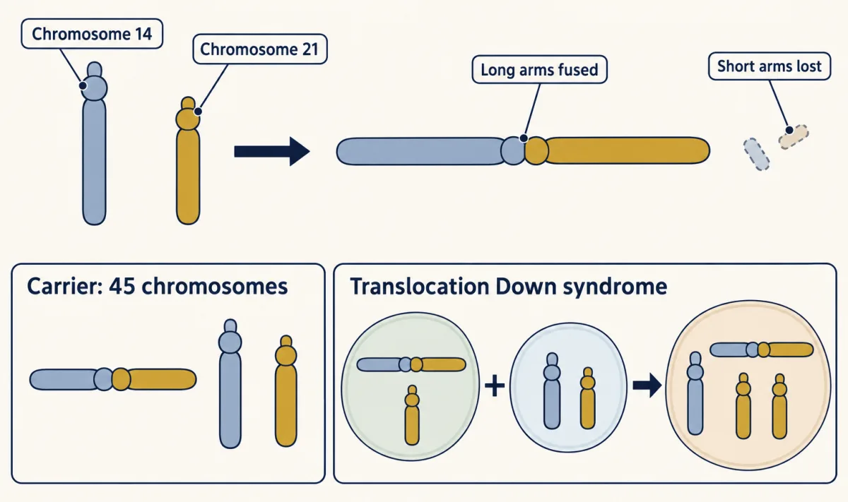
A Robertsonian translocation is a fusion, not a swap of ends: it joins the long arms of two acrocentric chromosomes, whose centromere sits near one end, and loses most of their tiny short arms. In humans the acrocentrics are 13, 14, 15, 21 and 22. Those short arms carry repeated ribosomal RNA genes and other repetitive DNA; the remaining acrocentrics retain many rRNA-gene copies. So a carrier can have 45 chromosomes and usually be unaffected. If a fusion carrying chromosome 21 material travels with a free 21, the child can have translocation Down syndrome with 46 chromosomes.
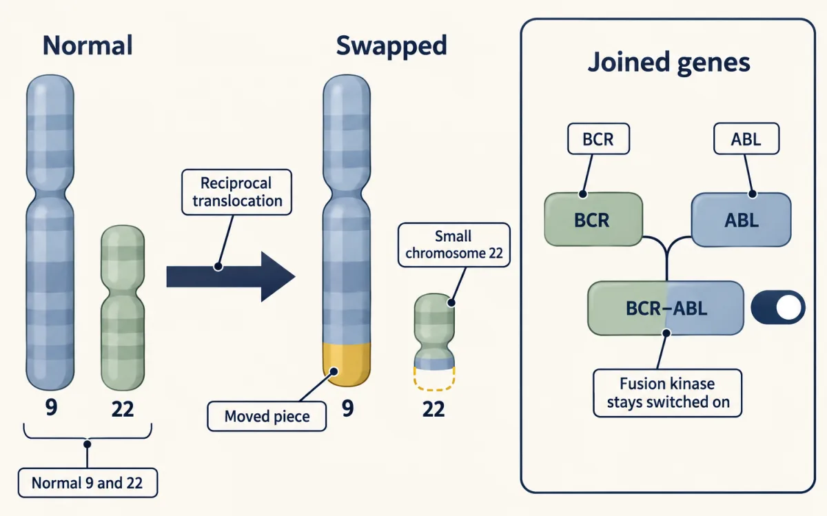
The Philadelphia chromosome is a real case of the second film’s reciprocal translocation, a swap of segments between nonhomologous chromosomes. It is the small chromosome 22 left by an acquired 9;22 exchange in blood cells. Nowell and Hungerford saw it in 1960, Rowley’s banding in 1973 suggested it was half of a swap, and in the 1980s other laboratories found the joined genes, BCR and ABL; the fusion protein is a tyrosine kinase that stays switched on (see oncogenes). Here the critical change is gene context, not simply total dose.
Choose a test by the question
First ask: dose, context or balance? Then choose the measurement.
- A G-banded karyotype counts chromosomes and compares their band patterns. It is the routine genome-wide test that directly shows balanced rearrangements, but sees only changes of roughly 5 to 10 million base pairs or larger, balanced or not. Some genome sequencing and optical genome mapping methods can also find balanced changes.
- FISH uses a fluorescent DNA probe for one chosen region, often about 100 thousand base pairs. It works in nondividing nuclei, but says nothing about regions outside its target.
- A chromosomal microarray measures segment dose across the genome. Where probes are dense, it can find gains and losses down to tens of thousands of base pairs, depending on array and region. It is blind to balanced changes, and a dose-only array can miss triploidy. In one large prenatal study, microarray found an additional clinically relevant deletion or duplication in 6.0 percent of karyotypically normal samples from pregnancies with a structural anomaly.
- Breakpoint-focused or genome sequencing can sometimes resolve junctions at or near base-pair scale, depending on method and repetitive sequence. It has shown that rearrangements that look balanced under the microscope can break genes or hide small losses (Going deeper).
Predict. A child’s banded karyotype looks normal, yet a dose-only microarray finds a small loss on one chromosome. Why could the karyotype miss it, and what kind of change could the array itself still miss?
Check the tests
The loss is below the karyotype’s routine resolution; the array reads dose far more finely where probes are dense. It would still miss a balanced rearrangement, because no dose changes, and can miss triploidy.
Design problem
A man with typical features has 45 chromosomes, including a Robertsonian 14;21 translocation, and a child with Down syndrome. Explain why he is unaffected, list his gametes, identify the zygotes expected to survive with a normal egg, and choose tests to confirm the explanation.
Saved in this browser only.
One way to do it
He has two doses of the long arms of 14 and 21, one free and one fused; the lost short arms’ rRNA genes have copies on other chromosomes.
The free 14, free 21 and fused t(14;21) pair as a group of three. In the usual 2:1 split, six gamete types result; a rare 3:0 split is omitted:
- 14 + 21: normal.
- t(14;21): balanced carrier.
- t(14;21) + 21: translocation trisomy 21 after fertilization.
- 14 alone: monosomy 21.
- t(14;21) + 14: trisomy 14.
- 21 alone: monosomy 14.
Types 4, 5 and 6 are expected to be lost early in pregnancy. If all six gametes were equally frequent and only types 1, 2 and 3 survived, one in three liveborn children would have Down syndrome. Observed risks are well below that conditional value, and published series find lower risks when the father is the carrier than when the mother is.
Karyotype the child and both parents; the expected child has 46 chromosomes, including t(14;21) and two free 21s. FISH with a chromosome 21 probe can confirm the extra dose. This is a mechanism exercise, not reproductive advice.
Going deeper
Human egg errors and age
In human eggs, sister chromatids can separate early at meiosis I. In reverse segregation, both pairs of sisters split at meiosis I. In donated human eggs, Gruhn and colleagues found a U-shaped relation between donor age and aneuploidy, with higher values among the youngest and oldest donors. That finding concerns aneuploid eggs from donors, not live-born trisomy 21 risk, which rises with maternal age.
When balanced does not mean harmless
Balance depends on resolution. Redin and colleagues sequenced apparently balanced abnormalities from 273 people with congenital anomalies. They revised 93 percent of the karyotypes, found complexity hidden from karyotyping in 21 percent of abnormalities, and judged gene disruption likely to contribute in at least a third. This selected cohort cannot say how often a healthy carrier’s rearrangement hides a small loss or disrupts a gene.
Sex determination and extra chromosome sets
Some animals, including fruit flies, set sex by counting X chromosomes during a short window of early development. An extra chromosome set can upset that count.
Your notes
Select any text in the lesson to highlight it or add a note to it. Your highlights and notes are saved in this browser and collected in your notebook.
This browser is not letting the site save anything (a private window or strict privacy settings), so highlights and notes will be lost when you leave the page.
How we measure it
G-banded karyotyping
Dividing cells are stopped at metaphase, swollen, fixed and spread on a slide, then treated with trypsin and stained with Giemsa so that each chromosome shows its own pattern of light and dark bands. Counting and comparing bands reveals extra or missing chromosomes and rearrangements large enough to see, about 5 to 10 million base pairs in routine work.
Fluorescence in situ hybridization (FISH)
A fluorescent DNA probe pairs with its matching sequence in fixed cells. Counting spots gives the copy number of that one region, even in nuclei that are not dividing. On spread metaphase chromosomes, the spots also show which chromosome carries the region. A FISH result says nothing about regions the probe does not cover.
Chromosomal microarray
Patient DNA is labelled and compared with reference DNA or reference data at thousands of mapped sites, giving the relative dose of each segment. It finds gains and losses far smaller than a karyotype can, but it cannot see a balanced rearrangement, and a dose-only array can miss triploidy unless it also reads SNP genotypes (which base a person carries at common single-base variant sites).
DNA marker typing of parent and child
Short, variable DNA sequences near a centromere are typed in a child and both parents. Which parental alleles the child carries, and in how many copies, shows which parent contributed an extra chromosome and, if no crossover separates marker and centromere, whether the error looks like one at meiosis I or meiosis II.
Practice questions on this lesson (3) → MCAT-style, original, each with an explanation
Where this lesson fits
Before thisReview first
- Chromosomes and Meiosis: The Physical Basis of Mendel's LawsThis course · Lesson 2
You need how homologues and sister chromatids separate in meiosis I and II to follow where a missed separation happens.
- Sex Linkage, Dosage Compensation and Non-Mendelian InheritanceThis course · Lesson 6
Dosage compensation of the X explains why extra or missing sex chromosomes are better tolerated than autosomal ones.
- Reading Pedigrees: Human InheritanceThis course · Lesson 5
Reading family trees helps in the design problem, where a translocation carrier's gametes and children are worked out.
After thisStudy next
- Linkage and Recombination MappingThis course · Lesson 8
Crossing over between a marker and the centromere, used here to infer the error, is the basis of recombination mapping.
- Cancer as a Genetic DiseaseThis course · Lesson 21
The Philadelphia chromosome's joined BCR and ABL genes show how a rearrangement changes gene context in tumours.
- Sequencing and GenomesThis course · Lesson 23
Sequencing resolves breakpoints and hidden losses that a karyotype misses in apparently balanced rearrangements.
Elsewhere on the siteFilms, simulations and method films in other courses
- Cell, primary and explant cultureMethod film
Karyotypes are made from cultured dividing cells arrested in metaphase, so the lesson's tests depend on it.
- Homologs First, Sister Chromatids SecondFilm · Biology
It shows how homologs and sister chromatids separate in meiosis I and II, the steps where errors arise.
- Heal the scrapeSimulation · Life Science
A hands-on look at how copied chromosomes are shared between daughter cells, the process that can go wrong.
Suggested by an AI model from the lessons themselves, then checked. How we use AI
Finish line
Test yourself
8 questions
Practice and MCAT-style, with explained answers.
Next · Lesson 8
Linkage and Recombination Mapping
Independent assortment predicts four equal test-cross classes. Why do two classes dominate?
Continue →← Lesson 6 · Sex Linkage, Dosage Compensation and Non-Mendelian Inheritance