Cell Biology · Unit 4: Division and cell loss
Cell Death and Clearance
Rewritten on : new text, flashcards and film. Your notes and highlights stay, but the text around them may have moved.
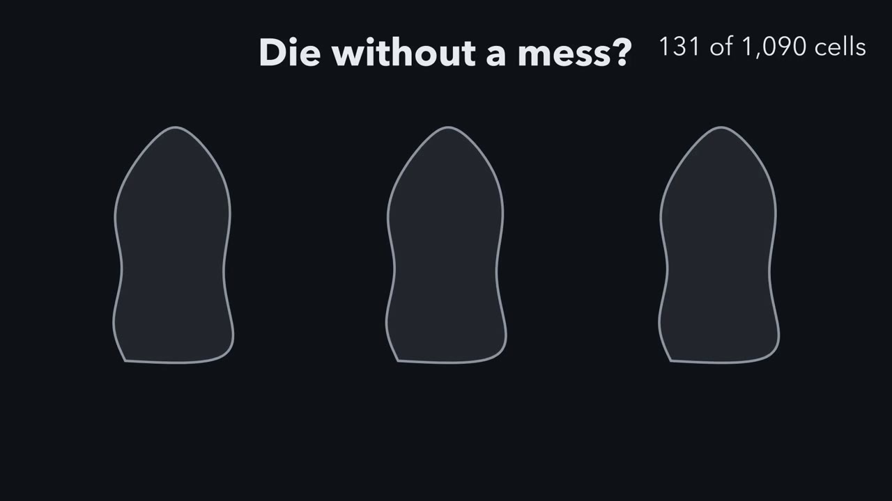
In a developing worm, the same 131 cells die on schedule, yet in healthy tissue you rarely see a corpse. How does a cell die without leaving a mess?
Pause when the reading reaches a film, and offer to play it. Saved on this device.
Watch the film now, or keep listening
Builds on:Membrane Organization and TopologyVesicle Budding, Targeting and FusionWhy these?
 ▶ Watch Sealed or Torn · 3:17
Read the transcript of “Sealed or Torn”
Film transcript: Sealed or Torn
Every one of these worms kills the same 131 cells. How does a body lose that many cells without leaving a mess? In healthy tissue, dead cells are cleared so fast that you rarely see a corpse. Follow one doomed cell. Its own enzymes, called caspases, switch on. They cut other proteins. The cell shrinks, its chromatin packs down, its nucleus breaks into pieces, and its surface bubbles. But watch the membrane. It has not broken. The cell is taking itself apart, but for now every piece stays wrapped inside a closed membrane. This tidy, self-run death is called apoptosis. While still sealed, it can send two signals. First, a channel in its membrane opens, and small molecules puff out.
They are a find-me signal. They call a phagocyte, a cell that eats other cells. Second, a lipid called phosphatidylserine normally sits mostly on the membrane's inner face. The caspases cut the membrane proteins that control which face it sits on. Now it appears on the outer face. That is the eat-me flag. Here is the idea to carry. In apoptosis, the membrane holds while the cell takes itself apart. That buys time for a phagocyte to eat it before anything leaks. The phagocyte arrives and recognises the flag. In the simplest picture, it wraps around the whole cell and swallows it. A lysosome digests it. Nothing spills. That is why you rarely see a corpse.
Now put the same kind of dying cell in a dish, where no phagocyte will come. Time passes. What happens to its membrane? Often, it fails. The membrane that held through the early stages now leaks, and the contents spill. This late leak is called secondary necrosis. In a tissue, a phagocyte usually gets there first. Some cells die the other way round. This one swells, and its membrane tears early. Its contents spill at once, including alarm signals. These can call in the immune system and cause inflammation.
This messy death is called necrosis. Tearing does not mean accident: some tearing deaths are genetic programs too. In its simplest form, the difference is timing. Apoptosis keeps the membrane intact early. Necrotic deaths generally open it early. So back to the worm. Its doomed cells are usually eaten by neighbouring cells before they spill. A membrane that holds until the meal often makes for a quieter death.
From the film
- In apoptosis, the cell's own caspases take it apart while its plasma membrane is still intact, at least at first.
- While still sealed, an apoptotic cell can send find-me signals and expose phosphatidylserine, an eat-me flag, on its outer face.
- In healthy tissue, a phagocyte usually eats the cell before it leaks, so nothing spills.
- In a dish with no phagocyte, an apoptotic cell often leaks later: secondary necrosis.
- In necrotic deaths the membrane generally opens early, and spilled alarm signals can cause inflammation.
The one idea to carry through this lesson. In apoptosis the cell’s own caspases take it apart while its plasma membrane is still intact, which gives a phagocyte time to find and eat it before anything leaks. Necrotic deaths generally open the membrane early and spill. Its limits: an uneaten apoptotic cell often leaks later (secondary necrosis), and some tearing deaths are genetic programs too.
The first film showed what happens; the rest of the page asks how we know, one piece of its idea at a time. The worm shows who takes the cell apart, clearance experiments show what happens while the membrane holds, necrotic programs show what opens it early, and two dyes tell these apart in a dish.
The worm shows the cell kills itself
The evidence that the cell’s own caspases do the killing came from a worm. In the hermaphrodite Caenorhabditis elegans, development makes 1,090 somatic cells and kills 131 of them, leaving 959 in the adult (males have a different lineage and count). In living transparent embryos every division can be followed, and the lineage and its fates, including which cells die, are highly invariant. But an invariant pattern says death is reproducible, not that a gene causes it: a cell might die because it is squeezed or starved at the same place every time. The test is genetic: find mutants in which those cells fail to die.
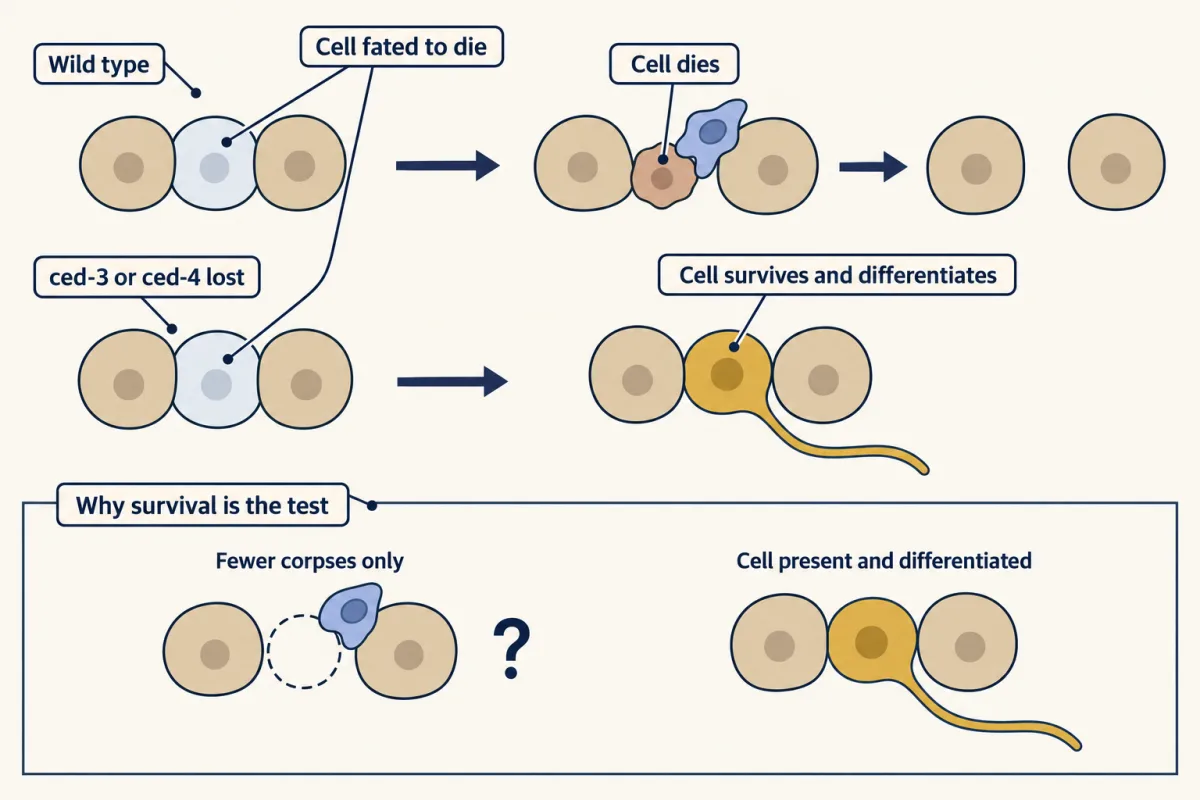
Hilary Ellis and H. Robert Horvitz did that. Their 1986 paper reported that wild-type function of two genes, ced-3 and ced-4 (ced for cell death abnormal), is needed for the deaths they examined.
Predict. Suppose all you knew was that worms lacking ced-3 show fewer cell corpses. Does that show the cells did not die? What observation would?
Check your prediction
No. Fewer corpses alone could mean corpses were cleared too fast to count (the corpse-count problem below puts numbers on this). Ellis and Horvitz saw more: cells normally fated to die survived, and some differentiated into recognizable cell types. A living, differentiated cell shows the death itself did not happen.
A third gene acted the other way. In 1992 Michael Hengartner, Ronald Ellis and Horvitz reported that an activating mutation in ced-9 prevented the normal developmental deaths, while inactivating mutations made cells that normally live die.
Geneticists order genes like these by epistasis: in a linear pathway, when two loss-of-function mutations have opposite effects, the double mutant usually shows the effect of the gene that acts later.
Predict. If ced-3, the killer, acts after ced-9, the protector, what does a worm lacking both do?
Check your prediction
It lives and keeps the cells that normally die, as the real double mutant does. The killing that follows loss of ced-9 needs ced-3, so ced-3 acts downstream, carrying out the death that ced-9 normally prevents.
Ordering of this kind placed a fourth gene, egl-1, upstream of ced-9, then ced-4, then ced-3. When the genes were cloned, CED-3 proved to be a caspase, CED-4 resembled human Apaf-1 and CED-9 resembled BCL-2 (Going deeper has a caution about how CED-9 works). In human cells this machinery runs as a cascade: in the intrinsic route BAX and BAK permeabilize the mitochondrial outer membrane, cytochrome c escapes, and caspases (proteases that cut after aspartate) are activated in turn. You do not need those names here: death receptors, BCL-2 and caspases teaches them, stress signals covers how p53 can push a damaged cell toward this death, and for the nervous system see naturally occurring death and death machinery in neurons.
Clearance: separating each step from the death
Each clearance step was shown by blocking it without stopping the death. Clearance of apoptotic cells is often called efferocytosis.
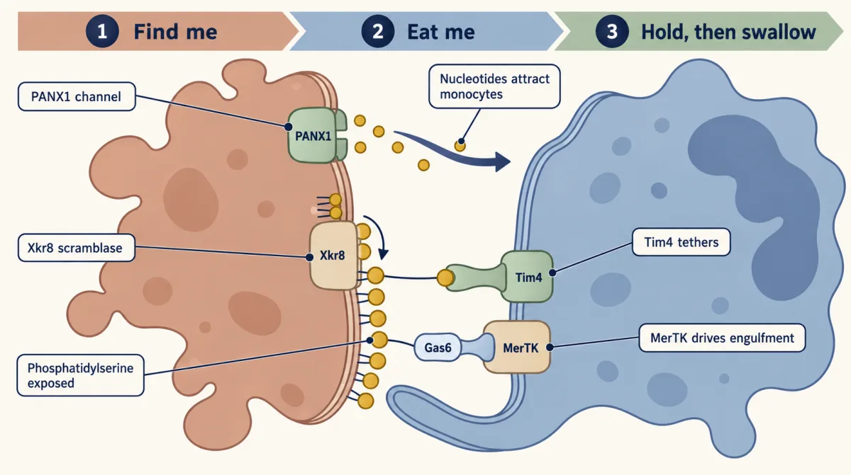
Find me. The first film’s channel is PANX1. In apoptotic Jurkat cells (a human T-cell line), the effector caspases 3 and 7, the ones that dismantle the cell, cut PANX1 and open it. ATP and UTP leave through it and attract monocytes, blood cells that can become macrophages. Knocking PANX1 down cut nucleotide release without slowing caspase activity, so the signal can be separated from the death. Nucleotides are one of several find-me signals; dying cells also release the lipids lysophosphatidylcholine and sphingosine-1-phosphate and the chemokine CX3CL1.
Eat me. Valerie Fadok, Peter Henson and colleagues reported in 1992 that macrophages recognize exposed phosphatidylserine (PS) on apoptotic lymphocytes. PS is normally kept on the inner leaflet, the inner layer of the plasma membrane (see lipids and membrane organization and topology). The first film’s caspases cut the membrane proteins that set which face PS sits on, and there are two kinds. A flippase is a pump that keeps returning PS to the inner face. A scramblase is a door that lets lipids mix between the two faces. Caspases cut and activate the scramblase Xkr8, and in mouse cells lacking Xkr8, apoptotic cells failed to expose PS and were engulfed poorly.
Predict. Caspases also cut the flippase ATP11C, and cutting switches it off. A cell carries an ATP11C that caspases cannot cut, so the pump keeps running, while Xkr8 is switched on as usual. Does PS reach the outer face during apoptosis?
Check your prediction
No. Cells carrying a caspase-resistant ATP11C did not expose PS during apoptosis. With the pump still running, the door alone is not enough. Exposure needs both steps: the scramblase on and the flippase off.
Hold, then swallow. The first film used the simplest picture, a single gulp; in resident mouse peritoneal macrophages it takes two receptors. Tim4 binds PS directly and tethers the dying cell. MerTK, a receptor tyrosine kinase (it signals by phosphorylating proteins on tyrosine), binds PS through the bridging proteins Gas6 or Protein S and drives the engulfment. The phagosome then fuses with lysosomes, which digest the cell, as in endocytosis.
Design problem
Mouse resident peritoneal macrophages are mixed with apoptotic cells. Macrophages lacking Tim4 bind fewer apoptotic cells. Macrophages lacking MerTK bind them normally but take few inside. Design an assay that separates binding from engulfment, and say what each receptor does. (Hint: you need one readout for “stuck to the outside” and another for “inside”.)
Saved in this browser only.
One way to do it
Score bound and internalized targets separately. Label targets with a pH-sensitive dye that brightens in acidic compartments, so targets inside acidified phagosomes glow and surface-bound ones do not (acidification takes time, so a target engulfed moments ago may still be dim), and count total contacts by microscopy. Or quench or strip the surface signal after incubation. Run a parallel incubation at 4 °C, where binding still occurs but uptake stops.
Controls: mask PS on the targets with annexin V, which should block both binding and uptake; use live, non-apoptotic targets, which should be ignored. Then put each receptor alone, and both together, into a cell line that does not normally eat cells, and ask which confers binding and which confers engulfment.
The expected answer from Nishi and colleagues (2014) is two steps in this macrophage population: Tim4 tethers, MerTK engulfs, and cells expressing both engulf strongly. Other mouse macrophages depend on Tim4 to different degrees (Yanagihashi and colleagues, 2017), so the result should not be generalized beyond the cells tested.
Worked problem: why you rarely see a corpse
The numbers are illustrative. At steady state, corpses on view = death rate × time each stays visible. If a tissue replaces 1% of its cells a day, 10,000 per million die daily, about 417 an hour; if each corpse is visible for 1 hour, about 417 per million (0.04%) are on view.
Predict. Clearance slows, so each corpse stays visible for 12 hours. The death rate does not change. How many corpses do you see now?
Check your prediction
About 417 × 12 = 5,000 per million, or 0.5%: twelve times as many corpses with no extra death. So a corpse count cannot tell faster clearance from death that failed, which is why the ced-3 evidence had to be living, differentiated cells.
Why it matters: inflammation. The first film’s alarm signals are spilled molecules, such as the protein HMGB1, that can call in immune cells, so apoptosis is usually the quieter death. Usually, not always: apoptosis and its clearance are not always immunologically silent; outcomes depend on context, as the lesson on TLRs and NF-κB helps explain. Phagocytes differ too: microglia also clear dying cells and pruned synapses (see microglia and complement pruning).
That quieter-or-louder contrast was taught for decades as programmed versus accidental. The second film picks up here: what a torn cell’s look can and cannot tell you about what killed it.
Read the transcript of “A Look Is Not a Cause”
Film transcript: A Look Is Not a Cause
This cell tore open. What killed it: an accident, or a program? Chapter one showed two endings: an apoptotic cell held its membrane until eaten; a necrotic cell tore early. For decades, these two looks were taught as a pair. Apoptosis, named in 1972 by Kerr, Wyllie and Currie, was the programmed death. Necrosis was the accident. That pairing has not survived. A regulated death runs on dedicated machinery.
Genes or drugs can turn that machinery up or down. Such a cell can still look necrotic. Three such deaths are defined by what they depend on. First, necroptosis. Activated RIPK3 adds phosphate to MLKL. Clusters of MLKL then move to the plasma membrane.
Second, pyroptosis. Gasdermins are cut, freeing fragments that form pores in the plasma membrane. It often, but not always, follows inflammatory caspases. Third, ferroptosis, driven by severe, iron-dependent damage to membrane lipids.
GPX4 is the main guard, reducing damaging lipid hydroperoxides. In cultured cells, drugs that trap iron block it. Here is the idea to carry. A death's look names an endpoint, not a cause. To find the cause, block the machinery and see whether the death changes. Now take dying cells in a dish, with no phagocyte. Two dyes read chapter one's two membrane events. Annexin V binds phosphatidylserine it can reach from outside. Propidium iodide, a DNA dye, is kept out by an intact membrane. A flow cytometer reads both colours on each cell and places it in one of four boxes. An apoptotic cell in a dish often becomes flagged while intact. After secondary necrosis, it is flagged and leaky.
The boxes are staining states, not verdicts. Other markers each report one event. TUNEL labels free DNA ends, whatever broke them. Cleaved caspase-3 shows the protease switched on, not that it killed. LDH, a cytosol enzyme, leaks through a torn membrane. So does LDH mean a cell died?
Researchers removed NINJ1, a small plasma membrane protein, from mouse macrophages. Then they triggered death. Will LDH pour out? Very little came out. The cells died, but they stayed swollen, like balloons that never popped. Death and the final tear are separate steps. So LDH measures the tear, not the death. So back to the torn cell. Its look tells you how it ended, not what killed it. To find the killer, block each candidate mechanism and watch what changes.
From the film
- Some deaths that tear the membrane are genetic programs, not accidents.
- In the mammalian model, necroptosis needs RIPK3 and MLKL; pyroptosis needs pore-forming gasdermins; ferroptosis is driven by iron-dependent lipid damage.
- A death's look names an endpoint, not a cause; to find the cause, block the machinery and see whether the death changes.
- Annexin V reads phosphatidylserine it can reach from outside, and propidium iodide reads a leaky membrane.
- Mouse macrophages lacking NINJ1 died but released little LDH, so LDH reports the final tear, not the death.
Necrotic deaths can be programs too
The second film named the three programs; this section adds what it left out. Kerr, Wyllie and Currie described the apoptotic look in many tissues in 1972: chromatin condenses against the nuclear envelope, and blebs can pinch off as membrane-bound pieces. Necrosis shows swollen organelles and a ruptured membrane.
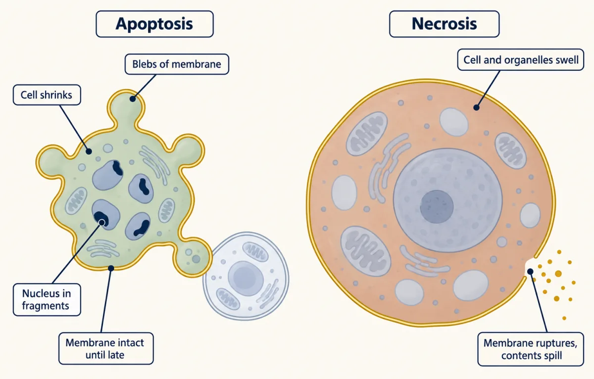
A death is regulated when it relies on dedicated molecular machinery that genetic changes or drugs can alter, and it can still end with necrotic morphology. The three regulated deaths, in fuller form:
- Necroptosis needs RIPK3 and MLKL in the mammalian model: activated RIPK3 phosphorylates its substrate MLKL, and the MLKL oligomers then move to the plasma membrane.
- Pyroptosis needs gasdermins: cutting frees the fragments that form pores, and often, but not always, follows activation of inflammatory caspases.
- Ferroptosis: the iron-dependent damage is severe lipid peroxidation (oxidative damage to membrane lipids). GPX4 holds it off by reducing lipid hydroperoxides; parallel systems such as FSP1, which works through coenzyme Q10, also do. In cultured cells it is blocked by iron chelators and lipophilic antioxidants.
The second film’s test, perturb the machinery and see the death change, is the logic of experiments that distinguish, applied to a dying cell.
Reading the flag and the leak with two dyes
The second film introduced the two dyes; here is how to read them in a dish, where no phagocyte eats the cells.
- Annexin V reads the flag. It is a calcium-dependent protein that binds phosphatidylserine (PS), as Tim4 does on a macrophage. Binding means PS is reachable from outside.
- Propidium iodide (PI) reads the leak. It is a DNA dye that an intact membrane keeps out. PI entry means the membrane is leaky, whatever made it so.
A flow cytometer reads the fluorescence of single cells streaming past a laser and places each in one of four boxes:
| PI negative: membrane intact | PI positive: membrane leaky | |
|---|---|---|
| Annexin V negative: no PS seen outside | neither signal (AnnV−PI−) | leaky, no flag seen (AnnV−PI+) |
| Annexin V positive: PS accessible to annexin V | external PS detected, membrane PI-impermeable (AnnV+PI−) | annexin-accessible PS, membrane PI-permeable (AnnV+PI+) |
The boxes are staining states, not verdicts on life or death: a cell with neither signal may already be committed to die, and some living cells expose PS too.
Predict. An apoptotic cell in a dish with no phagocyte starts in flagged, intact and, after secondary necrosis, ends in flagged, leaky. Where does a cell sit that tore open in the first minutes of a necrotic death? Can one time point tell the two apart?
Check your prediction
The torn cell lets PI in, and annexin V gets inside and finds PS on the inner face. So it goes straight to flagged, leaky. Both deaths end in the same box; one time point cannot tell them apart. The flagged, intact box fits the apoptotic order but does not prove it: necroptotic cells can also expose PS before they leak, and how long cells stay there must be measured in each system.
Worked problem: does this death need caspases?
Made-up data: 10,000 Jurkat cells per sample, counted as neither / flagged, intact / flagged, leaky / leaky only.
- Untreated: 9,300 / 400 / 200 / 100
- Drug, 6 hours: 5,800 / 2,900 / 1,000 / 300
- Drug plus a pan-caspase inhibitor (a drug that blocks many caspases at once), 6 hours: 8,600 / 600 / 500 / 300
Each row adds up to 10,000. Assume the inhibitor does not affect the cells on its own, and that the untreated sample is the baseline for every condition.
Step 1. How much of the flag needed caspases? Annexin-positive cells (both flagged boxes): 6%, 39% and 11%. The excess is 33 points with the drug and 5 with the inhibitor.
inhibitor-sensitive share of the drug-induced annexin-positive excess = (33 − 5) / 33 = 28 / 33 = 0.85, about 85%
That is a share of the PS-exposing cells, not of the death. In apoptosis, PS exposure itself needs caspases, so the inhibitor can lower the annexin signal even in cells that still die. The remaining 5 points are caspase-independent PS exposure or incomplete inhibition; these data cannot tell which.
Step 2. Is the pattern flag-first? With the drug, the excess is 2,500 flagged, intact and 800 flagged, leaky, about three to one; with the inhibitor it is 200 and 300. That shape is compatible with a flag-first death, but one snapshot cannot show cells moving between boxes, since the ratio also depends on how fast cells enter each box and how long they stay; repeated time points or single-cell tracking would test it. The leaky-only box (200 above baseline in both treated samples, unchanged by the inhibitor) means nothing until you check that its events are whole cells, not debris.
Step 3. What 6 hours cannot tell you. Under these assumptions, most of the drug-induced annexin signal is inhibitor-sensitive, which is compatible with caspase-dependent PS exposure but does not by itself establish caspase dependence or show that caspases did the killing. The inhibitor may only delay death, or reroute it: in cells that make RIPK3, blocking caspase-8 can open the necroptosis route. So follow PI uptake or viability to, say, 24 hours, with and without a RIPK3 or MLKL inhibitor, and add a direct caspase readout and a test of whether the cells can still recover and grow.
| Quadrant, and what it usually means | Expected | Observed | Share |
|---|
Annexin V and PI at one time point cannot tell late apoptosis from necrosis, and the caspase-dependent share of annexin-positive cells is the drug’s excess annexin-positive points minus those left with the inhibitor, divided by the drug’s excess; it assumes the inhibitor does not affect the cells on its own and that the untreated sample is the baseline for every condition; this is the lesson’s made-up data, dots sit at random inside each quadrant, and only the counts carry information.
The other common markers each report one event that can occur in more than one kind of death:
- TUNEL labels free DNA ends in fixed cells. In rat liver it labelled necrotic and autolytic (self-digested after death) cells as well as apoptotic ones.
- Cleaved caspase-3 on an immunoblot shows the protease was activated, not that the cell died or that caspase-3 killed it.
- LDH release into the medium reports the final rupture that lets out large molecules such as the cytosolic enzyme lactate dehydrogenase (LDH). In mouse macrophages lacking NINJ1, a small plasma membrane protein, cells died yet stayed swollen and released little LDH, showing that rupture is its own step.
So the first film’s idea holds, with its limits: an apoptotic membrane stays intact early, often until the cell is eaten. Each marker reports a state that more than one death can reach, in no fixed order, so combine markers over time, then perturb.
Going deeper
The hermaphrodite lineage counts
John Sulston, Einhard Schierenberg, John White and Nichol Thomson followed every embryonic division under a light microscope. They reported in 1983 that 113 cells die during embryogenesis in the hermaphrodite; the rest of the 131 die after hatching.
From worm genes to the human cascade
In a human cell-free system, Li and colleagues showed in 1997 that cytochrome c and dATP let Apaf-1 bind and activate caspase-9, which then cleaves and activates caspase-3.
The textbook cartoon has CED-9 simply holding CED-4 until EGL-1 releases it. That is a simplification: the genetic phenotypes stand, but the mechanism by which CED-9 protects is less settled. A 2024 study of CED-9 mutants that cannot bind CED-4 found that the binding was needed for a pro-apoptotic role of CED-9 but not for its protective role, in their assays.
Your notes
Select any text in the lesson to highlight it or add a note to it. Your highlights and notes are saved in this browser and collected in your notebook.
This browser is not letting the site save anything (a private window or strict privacy settings), so highlights and notes will be lost when you leave the page.
How we measure it
Annexin V and propidium iodide flow cytometry
Cells are stained with fluorescent annexin V, which binds exposed phosphatidylserine, and with propidium iodide, a DNA dye that enters only cells whose plasma membrane is leaky. A flow cytometer counts each cell in one of four quadrants, giving surface lipid change and membrane integrity for thousands of cells at once.
Cell-lineage tracing and forward genetic screens
Dividing cells are followed in a living transparent embryo to record which cell gives rise to which, and which die. Animals are then mutagenized and screened for ones in which the fixed deaths do not happen, identifying the genes the deaths need.
TUNEL staining
An enzyme adds labelled nucleotides to free DNA ends in fixed cells or tissue sections, so cells with fragmented DNA light up. It detects DNA breaks, which occur in apoptosis but also in necrosis and autolysis, so it needs a second readout.
LDH release with caspase-3 immunoblotting
Lactate dehydrogenase, a cytosolic enzyme, is measured in the culture medium as a readout of plasma membrane rupture. An immunoblot of the cells shows whether caspase-3 has been cut into its active fragments. Together they separate a protease program from a broken membrane.
Practice questions on this lesson (3) → MCAT-style, original, each with an explanation
Where this lesson fits
Before thisReview first
- Membrane Organization and TopologyThis course · Lesson 4
You need the two-leaflet membrane and its lipid asymmetry from there to follow how phosphatidylserine moves to the outer face.
- Vesicle Budding, Targeting and FusionThis course · Lesson 11
Engulfment and phagosome fusion with lysosomes build on how membranes bud, target and fuse.
After thisStudy next
- Stem Cells and Tissue RenewalThis course · Lesson 24
Tissue renewal balances cell loss against replacement, so the steady-state clearance idea applies directly.
- Cell Identity, Cell State and DiseaseThis course · Lesson 25
Disease states often involve altered death and survival, using the programmed-death machinery learned here.
- Designing a Cell Biology ExperimentThis course · Lesson 26
Combining ambiguous markers over time and then perturbing is the design reasoning practised there.
Suggested by an AI model from the lessons themselves, then checked. How we use AI
Finish line
Test yourself
8 questions
Practice and MCAT-style, with explained answers.
Next · Lesson 22
Adhesion and the Extracellular Matrix
Take the calcium away and a sheet of epithelial cells comes apart. What was the calcium holding together?
Continue →