Cell Biology · Unit 4: Division and cell loss
New to this? Review it at high school level: The Cell Cycle and Mitosis
Cyclin-CDK Control of the Cell Cycle
Rewritten on : new text, figures, flashcards and film. Your notes and highlights stay, but the text around them may have moved.
From yeast to humans, one kinase family drives every cell cycle, and it works only while a partner protein is present. Why would a cell destroy that partner every time it divides?
Pause when the reading reaches a film, and offer to play it. Saved on this device.
Watch the film now, or keep listening
Builds on:Experiments That Distinguish MechanismsProtein Quality Control and Organelle TurnoverThe Nuclear Envelope and Nuclear TransportWhy these?
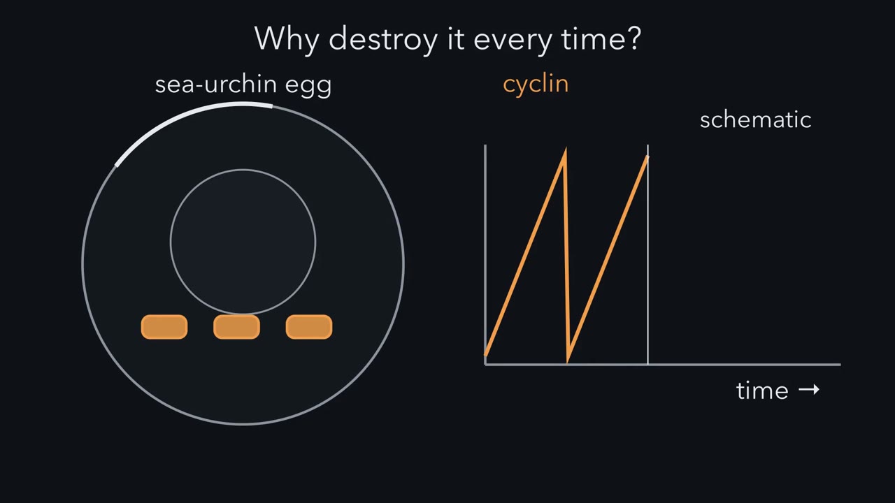 ▶ Watch One Rise, One Fall · 5:15
Read the transcript of “One Rise, One Fall”
Film transcript: One Rise, One Fall
Every time this egg divides, it destroys one of its own proteins. Then it builds it up again. Why would a cell destroy its engine's partner, every single time? In 1982, at Woods Hole, Tim Hunt's group saw this in dividing sea-urchin eggs. The protein climbed through each cycle and vanished at every division. They named it cyclin. The eggs make it all the time. It falls because it is destroyed.
Cyclin's partner is a kinase, a CDK, or cyclin-dependent kinase. Alone, the CDK is inactive. A bound cyclin is needed to switch it on. Every eukaryotic cell, from yeast to human, runs its cycle on this engine. Now add a second trace: the kinase's activity. In cycles like a frog embryo's first, it does not follow the ramp. It stays low through most of interphase. Then it can snap up just before mitosis, and crash as cyclin is destroyed. Here is the puzzle. Cyclin climbs steadily, and cyclin switches the kinase on. So why does the activity wait, and then jump?
Because a pair can be built and still be held off. The mitotic pair is CDK1 with cyclin B. All through G2, Wee1 adds an inhibitory phosphate to each new pair. Inactive pairs pile up, loaded and waiting. Then a phosphatase, Cdc25, takes the phosphate off. Once a little CDK1 escapes, it boosts Cdc25 and slows Wee1. Each active pair frees more. Activity does not creep; it snaps, all or none. Checkpoints pull the same levers. If DNA is damaged or unfinished in G2, sensor kinases switch off Cdc25.
CDK1 keeps its brake, and the cell waits. Here is the one idea. CDK activity rises and falls once per cycle. The rise carries the cell from G1, through S, into mitosis. Destroying cyclin brings the fall. And that one rise and fall copies the genome once. Now the fall. It starts with one enzyme: the APC/C, or anaphase-promoting complex. Until every chromosome grips the spindle, a checkpoint holds the APC/C back. The cell waits before anaphase. Once the checkpoint falls silent, the APC/C tags two proteins for destruction.
Securin goes, so the sister chromatids separate. Cyclin B goes, so activity falls and the cell leaves mitosis. Why not just switch the kinase off? A phosphate can be removed; that is only a pause. A destroyed protein cannot be switched back on. It must be made again. That is a main reason the big transitions run one way. Here is the test. Frog egg extracts cycle in a tube. Give them a cyclin that cannot be destroyed. What happens?
The extract went into mitosis. The kinase stayed active, and the extract did not come out. Making cyclin drives entry. Destroying it permits exit. The same rise and fall controls copying. An origin of replication must be licensed before it can fire. Licensing happens only while activity is low, in late mitosis and G one. Firing needs the rising activity of S phase. High activity also blocks new licences, in yeast, and helps block them in animal cells. So each origin can fire at most once per cycle. New licences wait for the fall, normally at mitosis. Fission-yeast cells test the rule. Without their mitotic cyclin, activity falls after S phase, with no mitosis.
Origins are licensed afresh, and the cells copy their DNA again, without dividing. So why destroy cyclin every time? Because the kinase must climb once and fall once per cycle. Destruction is what normally makes the fall one way. The cell leaves mitosis, resets its origins, and copies its genome once per cycle.
From the film
- In the sea-urchin egg record, cyclin is made steadily and destroyed at every division, making a sawtooth; a CDK is inactive alone and needs a bound cyclin to switch on.
- Wee1 holds loaded CDK1-cyclin B pairs off through G2, then Cdc25 releases them, so activity snaps on all at once.
- CDK activity rises and falls once per cycle; the rise carries the cell into mitosis.
- The APC/C destroys securin and cyclin B, so the sisters separate and activity falls; a destroyed protein must be made again, so the fall runs one way.
- Origins are licensed only when activity is low and fire only when it is high, so the genome is copied once per cycle.
In the summer of 1982, at Woods Hole, Tim Hunt’s group found a protein in dividing sea-urchin eggs that built up through each cell cycle and vanished at every division.
The first film, above, asks why a cell destroys its engine’s partner, a cyclin, every time it divides; the answer is how a cell makes its cycle run forwards only, and copy its genome exactly once. The text keeps the film’s names (the cyclin sawtooth, the brake, the rise and fall) and adds what it leaves out: the evidence, the other cyclin-CDK pairs and the harder cases.
The record: a sawtooth and a snap
The first film draws the simplest case: two schematic traces on one time axis, for a cycle with strong brake control, such as the first cycle of a frog embryo or the frog egg extracts met below. The exact waveform depends on organism and stage; later rapid frog cleavage cycles show smoother kinase oscillations.
- Cyclin. A sawtooth: it climbs steadily and drops sharply at each division, because it is destroyed.
- Kinase activity. The activity of the mitotic kinase that cyclin switches on (first measured as an activity called MPF, maturation-promoting factor) does not follow the ramp: it stays low, snaps up just before mitosis, and crashes with cyclin.
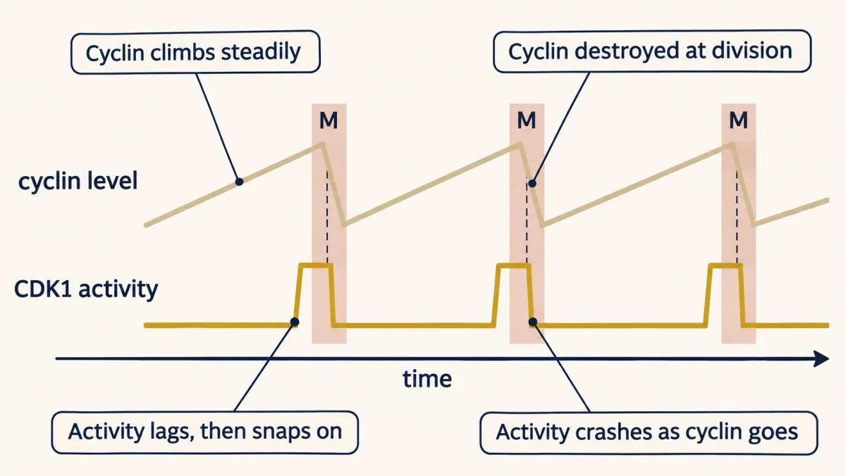
The two traces pose three puzzles.
- Why does the kinase need cyclin at all?
- If cyclin climbs steadily, why does activity lag behind and then jump?
- Why does the cell destroy cyclin to end mitosis, instead of simply switching the kinase off?
One idea answers all three.
The one idea to carry through this lesson. CDK activity rises and falls once per cycle. Rising activity carries the cell from G1 through S and into mitosis; brakes and checkpoints decide when it may rise. At the metaphase-to-anaphase transition one enzyme, the APC/C, tags two targets for destruction: securin, so the sister chromatids separate, and cyclin B, so activity falls and the cell can leave mitosis. Low activity resets the replication origins and high activity starts copying, so the genome is copied once per cycle.
This is an organizing model, not a single dial. “CDK activity” stands for several cyclin-CDK pairs that rise in turn (the table below), and phosphatases, checkpoints and protein destruction shape each step. The closest thing to a single dial is a fission-yeast cell engineered to run on one fused cyclin-CDK: S phase starts at a lower level of its activity and mitosis at a higher one. Meiosis and endocycles (cycles that copy DNA without dividing) bend the pattern, and endocycles turn out to test it (Copy once, below).
The engine: a kinase and a partner that comes and goes
The cycle has four phases. G1 is the gap between mitosis and the start of DNA replication. In S phase the genome is copied (the mechanics belong to DNA replication). G2 is the gap after copying and before mitosis. In M phase, mitosis sorts the duplicated chromosomes into two nuclei, and cytokinesis, the split of the cell itself, usually follows. Mammalian cells can also leave G1 for G0, a resting state they enter when conditions change and can leave again.
CDKs (cyclin-dependent kinases) are the catalytic subunits, and they are inactive alone. Cyclins are regulatory subunits that switch them on and steer them to targets. That answers the first puzzle. CDK levels stay fairly even while most cyclin levels rise and fall (cyclin D tracks growth signals most closely), so the rise and fall of each cyclin switches its kinase on and off. A bound cyclin is needed but not enough.
Different cyclin-CDK pairs carry the rise through different stretches of the cycle. In mammals:
| Stretch of the cycle | Cyclin | CDK |
|---|---|---|
| G1 | D | CDK4 or CDK6 |
| G1/S transition | E | CDK2 |
| S | A | CDK2 |
| G2 and M | A and B | CDK1 |
This is the usual division of labour, not a strict requirement: CDKs can stand in for one another (Going deeper).
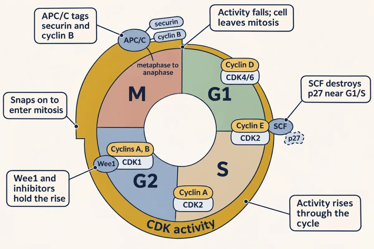
The mitotic pair has an older name. Maturation-promoting factor (MPF), the activity that makes frog oocytes mature and is also found in mitotic cells, is CDK1 (first named Cdc2, from fission yeast) bound to cyclin B.
Holding the rise back, and letting it go
Now the second puzzle: if cyclin B climbs steadily and switches CDK1 on, why does activity lag and then jump? Because a cyclin-CDK pair can be built and still be held off. To be active, CDK1 needs four things at once:
- a cyclin bound;
- an activating phosphate, which a CDK-activating kinase (CAK) adds to a loop near the active site;
- no inhibitory phosphates: Wee1 adds one near the ATP site, on tyrosine 15, and in animal cells the related kinase Myt1 adds one on threonine 14 as well as tyrosine 15;
- no inhibitor protein bound.
The names to hold on to are the brake, Wee1, and its release, Cdc25; the rest is detail.
The inhibitory phosphate is a brake. Wee1 keeps phosphorylating new CDK1-cyclin B complexes through G2, as the first film shows, so inactive complexes pile up. The phosphatase Cdc25 takes them off (the chemistry is in the phosphorylation switch).
Inhibitor proteins are a second brake. In mammals, p21 and p27 bind CDK2 and CDK1 complexes and block them without any chemistry; the structure of p27 lying across cyclin A-CDK2 is in the 3D viewer on this page. (With cyclin D-CDK4/6 they act differently; Going deeper.) One important way to remove an inhibitor is to destroy it. Around G1/S, cyclin E-CDK2 phosphorylates p27, the SCF ubiquitin ligase tags it for the proteasome (Sic1 is the yeast counterpart), and with its brake gone cyclin E-CDK2 switches fully on.
The release is a switch. Once a little CDK1 escapes its brake, it phosphorylates Cdc25, making it more active, and Wee1, making it less active. These two positive loops are the snap in the first film: each active complex frees more. A switch built this way also has memory: once mitosis starts, a small dip in cyclin will not reverse it (signaling circuits).
Predict. A fission-yeast cell is engineered to make much more Cdc25 than normal. When does it divide, and at what size?
Check your prediction
Earlier and smaller. With more of the phosphatase, the brake comes off before the cell reaches its usual size, CDK1 snaps on early, and the cell enters mitosis small. Losing Wee1 gives the same result, which is how that gene got its name: wee.
A decision in G1
Before any of this, a cell in G1 has to decide whether to divide at all. That decision is the restriction point (in yeast, Start), and a cell passes it only if it has enough nutrients and growth factors. Growth-factor signals raise cyclin D (Ras to MAPK). Cyclin D-CDK4/6 begins to phosphorylate Rb, a protein that holds the E2F transcription factors off. Freed E2F switches on genes for S phase, including cyclin E, and cyclin E-CDK2 phosphorylates Rb further. In the classical model that loop is another switch: once it flips, the cell finishes the cycle even if the growth factors are taken away (later work adds a second switch; see Going deeper). Rb’s role in cancer is in cancer genetics. Strictly, this is a commitment point, not a check that a step is finished.
Checkpoints pull the same levers
A checkpoint, in Leland Hartwell and Ted Weinert’s term, holds the cycle until an earlier step is finished or repaired. The three you need all act on the engine:
- DNA damage in G1. p53 builds up and switches on the gene for p21. p21 binds and blocks CDK2 complexes, and the cell waits in G1.
- Damaged or unfinished DNA in G2. The sensor kinases ATM and ATR, through Chk1 and Chk2, inactivate Cdc25 (and Chk1 activates Wee1), so CDK1 keeps its inhibitory phosphate and the cell waits in G2. In frog egg extracts, unreplicated DNA raised the amount of cyclin needed to enter mitosis.
- An unattached chromosome in mitosis. Unattached kinetochores, the sites where chromosomes grip the spindle, switch on the spindle assembly checkpoint. It stops the APC/C and its partner Cdc20 from destroying securin and cyclin B, so the cell waits before anaphase, often in prometaphase. Attachment normally silences the signal, but some errors escape it: a merotelic kinetochore, gripped from both poles, can silence it and enter anaphase misattached. (Some other APC/C targets, such as cyclin A, are still destroyed.)
Predict. Cells lacking p53 are irradiated in G1. What happens to them?
Check your prediction
They lose the main burst of p21, so the lasting G1 stop fails and many enter S phase with damaged DNA. Other damage responses can delay them briefly, and they can still be stopped later, at the G2 checkpoint, which acts through Cdc25 and Wee1. Losing the G1 stop is one reason p53 is the most often mutated gene in human cancers.
The fall: destruction makes it one way
Falling activity carries the cell out of mitosis. The G1 and G2 brakes and checkpoints act on the rise, and the spindle checkpoint holds back the fall. Each is a pause: a phosphate can be removed, an inhibitor let go, a checkpoint silenced. The way down is different, and it answers the third puzzle; the first film shows it as one enzyme tagging two proteins. Two ubiquitin ligases tag regulators for destruction by the proteasome (ubiquitin and proteolysis): the SCF, met above, and the APC/C, which acts from mitosis through G1. Once kinetochore checkpoint signaling has been silenced, the APC/C with Cdc20 tags securin and cyclin B. Losing securin frees separase, the enzyme that cuts cohesin, the protein ring holding sister chromatids together, so the sisters separate (in vertebrates, losing cyclin B also lifts CDK1’s own block on separase). Losing cyclin B drops CDK1 activity, phosphatases strip the mitotic phosphates, and the cell leaves mitosis. Then the APC/C with Cdh1 keeps mitotic cyclins low through G1, so activity starts the next cycle from the bottom.
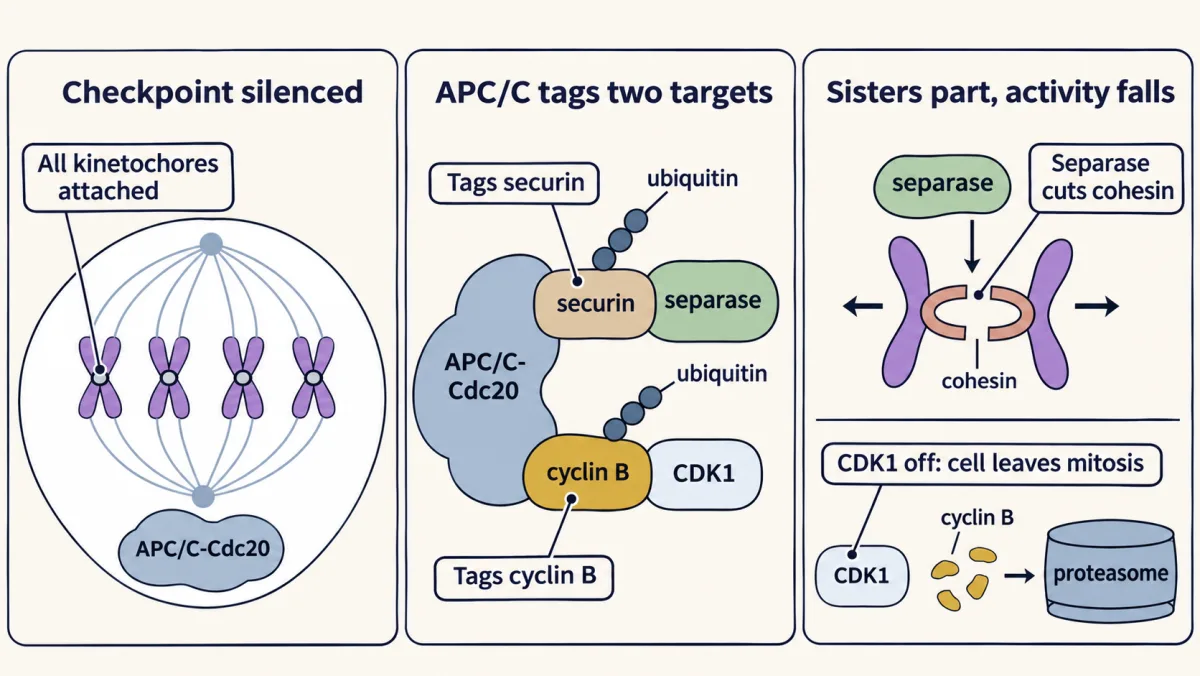
As the first film says, a destroyed protein has to be made again. Destruction, together with feedback switches, is a main reason the cycle’s big transitions run one way (at G1/S the Rb-E2F switch adds its own memory). Destroying a brake drives the cycle forward; destroying cyclin resets it.
The first film’s frog egg extract test shows that a cyclin that cannot be destroyed holds the extract in mitosis. A second test asks whether low activity alone would do.
Predict. Vertebrate cells in mitosis are blocked from destroying cyclin B. A drug that inhibits CDK1 is added briefly, and they start to leave mitosis. Then the drug is washed out. What happens?
Check your prediction
They go back toward mitosis. Cyclin B was never destroyed, so once the drug was gone CDK1 switched back on. After a short treatment, low activity on its own was only a pause; destroying cyclin is what normally keeps activity low and makes the exit one way. Longer inhibition can lock cells out of mitosis even with cyclin B present, so feedback helps too (Potapova and colleagues: Nature, 2006, and 2009).
Copy once: low activity licenses, high activity fires
Before an origin of replication can fire, it must be licensed: loaded with the helicase that will later unwind the DNA there. Licensing needs low CDK activity; firing a licensed origin needs S-phase CDK activity, together with a second kinase, DDK. High CDK activity from S phase to mitosis also blocks relicensing in yeast, and contributes in animal cells, which add further locks, such as the protein geminin and destruction of the licensing protein Cdt1 (the details belong to DNA replication). So, as the first film shows, an origin can fire at most once per cycle; many licensed origins never fire and are copied by forks from their neighbours. In an ordinary cycle the way back to low activity, and to new licences, is through mitosis, where cyclin is destroyed. The prediction below shows another way.
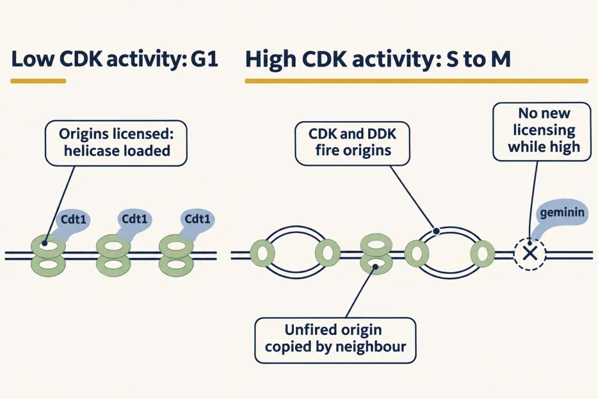
Predict. Fission-yeast cells lack their mitotic B-cyclin, Cdc13. They finish S phase, but CDK activity then falls without any mitosis. What happens to their DNA?
Check your prediction
They copy it again, as the first film shows: repeated S phases without dividing. Cells that overproduce the CDK inhibitor Rum1 do the same (Hayles and colleagues, 1994; Moreno and Nurse, 1994). Some normal cells use this on purpose: endocycles are the natural version.
Before anyone knew of CDKs, cell fusion showed that a replicated nucleus does not simply start again. In 1970 Rao and Johnson fused cultured HeLa cells taken from different phases, so that two nuclei shared one cytoplasm. As later reviews describe the results, a G1 nucleus in a cell fused with an S-phase cell started replicating early, while a G2 nucleus in the same situation did not. Two conclusions follow. S-phase cytoplasm carries an activity that can start replication, which in the rule’s terms fires the origins a G1 nucleus has licensed. And under these fusion conditions a G2 nucleus did not start another round, which fits licensing: its fired origins cannot be licensed again until activity falls, normally at mitotic exit (the Cdc13 and Rum1 cells and endocycles show it can happen without mitosis).
Fusing a mitotic cell with an interphase cell had a different outcome: the interphase nucleus condensed its chromosomes prematurely (Johnson and Rao, 1970), and how the chromosomes looked depended on the phase of that nucleus. Mitotic cytoplasm is dominant: it is full of active CDK1-cyclin B, which, with the kinases and phosphatases it controls, can condense an interphase nucleus from G1, S or G2.
Design problem
Using cell fusion, design experiments to ask whether S-phase cytoplasm can drive replication in another nucleus, and why a G2 nucleus might not respond.
Saved in this browser only.
One way to do it
Synchronise cultured mammalian cells in G1, S and G2 and confirm each population by DNA content (2C or 4C, where 1C is the DNA of one haploid genome; see Measuring the states below), using a mitotic marker such as phosphorylated histone H3 to show that the 4C cells are in G2 and not M. Pre-label one partner before fusing, for example with a nuclear dye or a long label in the S cells, so that every nucleus in a fused cell can be identified.
Fuse S with G1, and S with G2. Give a short pulse of a DNA precursor (radioactive thymidine or EdU) and score each nucleus separately. Expected result, as in Rao and Johnson’s 1970 work: G1 nuclei start replicating early, G2 nuclei do not.
Controls: same-phase fusions, where G1 with G1 and G2 with G2 test whether fusion itself starts replication and S with S tests whether fusion blocks it, plus unfused neighbours on the same slide.
Interpretation: S cytoplasm carries a replication-starting activity, and under these fusion conditions a replicated G2 nucleus does not initiate another round in S-phase cytoplasm. A third pairing, mitotic with interphase, should give premature chromosome condensation.
Limit: fusion identifies an activity and a nuclear competence, not the molecules. Naming them needs biochemistry or genetics.
How the engine was found: frogs, yeast and sea urchins
The model came from three lines of work that began apart and met in one complex. Frog oocytes gave an activity: in 1971 Yoshio Masui and Clement Markert injected cytoplasm from progesterone-treated oocytes into untreated ones, which then matured, and named the activity maturation-promoting factor (MPF). Like cell fusion, this revealed an activity, not a molecule. Yeast genetics gave the genes: budding-yeast CDC28, needed at Start, and fission-yeast cdc2, an accelerator needed both in G1 and in G2, with wee1 as its brake; CDC28 and then a human gene could each stand in for cdc2. Sea urchins gave cyclin (published in 1983), which the frog egg-extract tests in The fall confirmed in 1989. The lines met in 1988, when highly purified MPF from Xenopus eggs proved to contain the frog relative of Cdc2, and in 1990, when its other component proved to be cyclin B.
One idea from the yeast work is still how mutants are read. In 1970 Leland Hartwell, Joseph Culotti and Brian Reid described temperature-sensitive mutants of budding yeast, Saccharomyces cerevisiae, that grow at a low temperature and stop at a higher one, each with a uniform shape. They called them cdc, for cell division cycle. The bud reports the stage: none in G1, small in S, large by mitosis.
Their key idea was to separate two times. Picture a car that must fill up at one station on every lap. If the station closes just after the car has filled up, it finishes this lap and stalls on the next; if it closes just before, the car stalls later on this lap, well past the station. The station is the execution point: the last time in the cycle the gene’s function is needed. Where the car stalls is the termination point, or arrest point: where cells collect when the function fails. Shift a growing culture to the restrictive temperature and follow single cells: the fraction that finishes this cycle tells you where the execution point lies.
Reading a population needs one more tool: telling, in one frozen moment, which phase each cell is in. The first film connected the activity cycle to one round of replication, so DNA amount constrains the phase without identifying it exactly. The second film picks up here: how a frozen dish of cells is classified with added markers.
Read the transcript of “A Snapshot of the Cycle”
Film transcript: A Snapshot of the Cycle
Which phase is each cell in? Freeze this dish of thousands: every cell looks unlabelled, but each carries clues. In chapter 1, one cell's kinase rose and fell once. Its genome was copied once. So DNA doubles once per cycle, then halves at division.
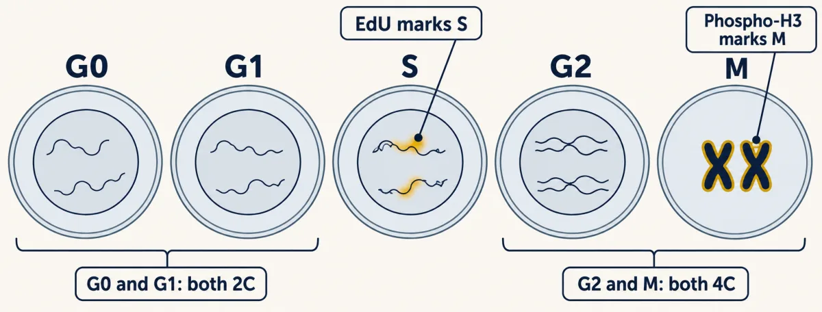
Start with DNA content. One C is one haploid genome's DNA. A diploid cell holds 2C in G1, and 4C in G2 and mitosis. The cell has not become tetraploid. It still has two homologous sets of chromosomes. By G2, each chromosome has two sister chromatids, doubling DNA without changing ploidy. C measures DNA amount, not chromosome-set number.
Stain fixed cells with propidium iodide, a dye that fluoresces when bound to nucleic acid. Add RNase, because the dye also binds RNA. A flow cytometer reads each cell's fluorescence. After RNA is removed, fluorescence is a proxy for DNA amount. Under the same staining conditions, a 4C cell gives about twice a 2C cell's signal. The comparison needs single cells measured consistently.
The dots pile up into a histogram. There is a peak at 2C and another at 4C. Cells in S spread between them, partway through copying. Read the axes before the labels. Horizontal position is fluorescence per cell, used here for DNA amount. Bar height is the number of cells. This plot is a population snapshot, not one cell moving through time. Here is one cell from the 4C peak. It has copied all its DNA. Is it still in G2, or already in mitosis?
The histogram cannot say. Mitotic cells share the 4C peak with G2. Quiescent G0 cells share the 2C peak with G1. DNA content narrows the phase; other markers can separate some look-alikes. Two more stains help. EdU is a thymidine look-alike. During a short pulse, cells copying DNA build it in, and click chemistry adds a glowing tag. It marks cells in S. EdU also catches cells at the borders. Early S cells can sit near the 2C peak, and late S cells near 4C. DNA content can blur both into neighbouring phases; EdU reveals ongoing synthesis. An antibody detects histone H3 phosphorylated on serine 10. This mark spreads across chromosomes as they condense.
It helps find mitotic cells, but it is not an exact boundary. The signal begins near centromeres in late G2, before mitosis. It spreads by prophase, remains strong through metaphase, then fades during anaphase. So a positive cell is usually mitotic, but timing keeps the call approximate.
G0 is harder: no single stain marks it cleanly. Together, these measures classify most cycling cells. But G0 versus G1 and the G2-mitosis boundary remain imperfect. So which phase is each cell in? DNA amount narrows the choices; EdU and phospho-H3 sharpen the answer. G0 remains unresolved.
How long each phase lasts is the text's next question.
From the film
- Because the genome is copied once per cycle, a diploid cell holds 2C of DNA in G1 and 4C in G2 and mitosis, with S-phase cells in between.
- Cells stained with propidium iodide and read one at a time in a flow cytometer give a histogram: a 2C peak, a 4C peak and S smeared between.
- DNA content alone cannot tell G0 from G1, or mitosis from G2.
- A short EdU pulse marks cells in S; histone H3 phosphorylation begins in late G2, spreads over condensing mitotic chromosomes and fades during anaphase.
- No single stain marks G0 cleanly.
Measuring the states
The first measurement is DNA content. One C is the DNA of one haploid genome, so a diploid cell has 2C in G1 and 4C in G2 and M. Doubling the DNA does not double the ploidy: a G2 cell is still diploid, with each chromosome made of two sister chromatids. To read DNA content, fix the cells and stain them with propidium iodide, a dye that fluoresces when bound to nucleic acid, plus ribonuclease (RNase), because the dye also binds RNA. A flow cytometer measures each cell’s fluorescence one at a time and builds a histogram: a 2C peak, a 4C peak, and S-phase cells smeared between.
The histogram has two blind spots. G0 cells sit in the 2C peak with G1, and mitotic cells sit in the 4C peak with G2. Two more stains help. EdU, a thymidine analogue, is built into DNA made during a short pulse and is then detected by click chemistry with a fluorescent tag, so it marks cells in S and separates them from the edges of the 2C and 4C peaks, where early and late S cells blur into G1 and G2. An antibody to histone H3 phosphorylated on serine 10 marks condensing mitotic chromosomes. It is close to a mitotic marker but not exact: the mark begins in late G2, spreads across chromosomes as they condense and fades during anaphase. G0 is harder, and no single stain marks it cleanly (see Going deeper).
Worked problem: fractions are not durations
The numbers here are illustrative. Suppose an asynchronous culture of a diploid human cell line doubles every 24 h, every cell is cycling, and none die. Of 10,000 cells, 5,500 fall in the 2C peak, 2,000 in the 4C peak and 2,500 between. A 30-minute EdU pulse labels 25% of cells, which agrees that the cells between the peaks are in S. And 3% are phospho-H3 positive; assume the stain marks every mitotic cell and nothing else.
Step 1. Fractions: G1 0.55, S 0.25, G2 plus M 0.20, of which M is 0.03.
Predict. G1 holds 55% of the cells, and the culture doubles every 24 h. Does G1 last 13.2 h (0.55 × 24), more, or less?
Check your prediction
Less: about 11.1 h. Multiplying by 24 h (13.2, 6.0 and 4.8 h for the three fractions) is the tempting move, and it is wrong. Each division turns one old cell into two newborn ones, so a growing culture always holds twice as many cells just born as cells about to divide. Early phases are overrepresented, so G1’s share of the cells is bigger than its share of the time.
Step 2. Line the cells up by age, newborn on the left: the line slopes down to half its height at division, and that slope gives a formula.
In words The time to pass the youngest fraction F equals minus the doubling time, times the base-two log of one minus half of F.
- t“t”
- the time from a cell's birth to the boundary that has the fraction F of all cells younger than ith
- T“T”
- the doubling time of the culture, which here is also the length of one cycleh
- F“F”
- the fraction of the population younger than the boundary; for the end of G1, the fraction in G10 to 1
- log2“log base two”
- the power you must raise 2 to in order to get the number in brackets
Sanity check At F = 0, t = 0. At F = 1 the bracket is one half, log2(0.5) = −1, and t = T: the oldest cells are just dividing. At F = 0.5, t is about 0.415 × T, less than half the cycle, because young cells are overrepresented.
Try one number
End of G1 in the worked problem: F = 0.55 and T = 24 h.
1 − F/2 = 1 − 0.275 = 0.725, and log2(0.725) = −0.464.
t = −24 × (−0.464) = 11.13 h.
Where it comes from optional
Step 1, the picture: cells of age a were born when the culture was smaller by a factor of 2a/T, so the number of cells of age a is proportional to 2−a/T. Newborns (a = 0) are twice as common as cells about to divide (a = T).
Step 2, add up the cells younger than t and divide by all cells: F = 2 × (1 − 2−t/T).
Step 3, solve for t: 2−t/T = 1 − F/2, so t = −T × log2(1 − F/2).
Need a refresher? powers and roots · log scales
Step 3. Apply it at each boundary, using the cumulative fraction of cells younger than that boundary.
- End of G1 (F = 0.55): 11.13 h, as in the example above.
- End of S (F = 0.80): t = -24 × log2(0.600) = 17.69 h, so S lasts about 6.55 h (working from unrounded values).
- Start of M (F = 0.97): t = -24 × log2(0.515) = 22.98 h. M lasts 24 - 22.98 = 1.02 h, and G2 lasts 22.98 - 17.69 = 5.29 h.
Check: 11.13 + 6.55 + 5.29 + 1.02 = 23.99 h, 24 h within rounding. The naive estimate made G1 two hours too long and G2 plus M (6.31 h) one and a half hours too short. In this model the first phase, G1, always looks longer than it is and the last, G2 plus M, shorter; a middle phase can go either way (here S looks about half an hour short). The model assumes identical phase lengths in every cell and no resting or dying cells, so treat these as estimates.
- 2C (G1)
- 5,500
- Between (S)
- 2,500
- 4C (G2 + M)
- 2,000
- EdU pulse labels (30 min, equals S)
- 25%
| Phase | Fraction | Naive: fraction × T | Lesson’s formula |
|---|
Same area, different width: young phases are tall, so they hold more cells per hour.
Defaults reproduce the worked problem.
Keep the rule: a phase’s share of the cells is not its share of the cycle, because a growing culture holds twice as many newborn cells as cells about to divide; use t = −T × log₂(1 − F/2) on the cumulative fraction. This is the lesson’s illustrative culture (every cell cycling, none dying, identical phase lengths), assuming the phospho-H3 stain marks only mitotic cells. The histogram’s peak shapes are a sketch, and slider ranges go beyond the worked problem. The gold band is a cartoon of the lesson’s one idea, not a measurement.
Going deeper
The extract thresholds
In Xenopus egg extracts with protein synthesis blocked, entry into mitosis needed about 32 to 40 nM of added nondegradable cyclin B, but an extract already in mitosis stayed there down to about 16 to 24 nM (Sha and colleagues, 2003). Two thresholds with a gap between them are the mark of a switch with memory; the numbers belong to that assay, not to every cell.
A second switch in G1
The Rb-E2F loop is not the only memory at the restriction point. Recall that the APC/C with Cdh1 keeps mitotic cyclins low through G1. In human MCF10A cells, Cappell and colleagues (2018) found a second G1 switch with memory: the switching-off of APC/C-Cdh1, held in place by a double-negative loop with EMI1 (knocking down EMI1 abolished the memory).
Beyond the usual pairings
Mouse embryos lacking CDK2, CDK3, CDK4 and CDK6 still develop to mid-gestation, with CDK1 binding all the cyclins. CDK inhibitor proteins are also less simple with cyclin D-CDK4/6 than with CDK2 and CDK1 complexes: they help that complex assemble, and tyrosine-phosphorylated p27 can leave it active.
Marker fine print
Cells out of the cycle lose Ki67, a protein made in cycling cells, but its level falls gradually through G1 and G0, so it does not mark G0 cleanly. Saldivar and colleagues combined DNA content, EdU and phospho-H3, read by image-based cytometry rather than flow, to separate phases in human RPE-1 and MCF10A cells (Science, 2018).
The three lines in full
Frogs. A frog oocyte waits in prophase of its first meiotic division until progesterone tells it to resume. Masui and Markert’s oocytes were from the leopard frog Rana pipiens, and removing the donor’s nucleus did not prevent the activity.
Fission yeast. Paul Nurse used Schizosaccharomyces pombe, a rod that divides in the middle, so length tells you its age. In 1975 he described a mutant that divided at a smaller size than normal. The mutation changed when division happened, not whether. Most later small-cell mutants mapped to a gene named wee1; one mapped to cdc2, whose other alleles block mitosis. In 1982 budding-yeast DNA carrying CDC28 rescued a fission-yeast cdc2 mutant: two distant yeasts, one job. In 1987 a human gene did the same.
Sea urchins. In work done in the summer of 1982 and published in 1983, Tim Hunt’s group followed new proteins in dividing embryos of the sea urchin Arbacia punctulata. Cyclin’s disappearance at division was a correlation, so tests came next. In Xenopus egg extracts, destroying the messenger RNA stopped the cycles in interphase and adding back cyclin mRNA restored them (Murray and Kirschner, 1989).
Yeast names and early mutants
In the 1970 paper, the cdc-3 mutant kept budding and dividing its nucleus while its cells failed to separate, so steps of the cycle can come apart. A naming warning: Hartwell’s budding-yeast cdc-2, which stopped at mitosis, is not the fission-yeast cdc2. Nurse and Yvonne Bissett concluded in 1981 that cdc2 acts both in G1 and in G2, and the 1982 CDC28 rescue was by David Beach, Barbara Durkacz and Nurse.
Who found MPF’s parts
Smith and Ecker reported a cytoplasm transfer like Masui and Markert’s in the same year, 1971. Hunt’s group saw similar proteins in another sea urchin and a clam, and clam cyclin A mRNA injected into Xenopus oocytes made them resume meiosis (Swenson, Farrell and Ruderman, 1986). The nondegradable-cyclin experiment was by Murray, Solomon and Kirschner (1989). In 1988 Lohka, Hayes and Maller purified maturation-promoting factor (MPF) from Xenopus eggs; antibodies to fission-yeast Cdc2 recognised its 32 kDa protein (Gautier and colleagues), and Dunphy and colleagues reached Cdc2 by a separate route.
Your notes
Select any text in the lesson to highlight it or add a note to it. Your highlights and notes are saved in this browser and collected in your notebook.
This browser is not letting the site save anything (a private window or strict privacy settings), so highlights and notes will be lost when you leave the page.
How we measure it
DNA-content flow cytometry with EdU and phospho-H3
Fixed cells are stained with a DNA dye (propidium iodide plus RNase, or DAPI) and passed one at a time past a laser, giving a histogram with a 2C and a 4C peak. A short EdU pulse marks cells in S phase, and an antibody to histone H3 phosphorylated on serine 10 marks mitotic cells (and some late-G2 cells), separating states that DNA content alone lumps together.
Temperature-sensitive mutants and shift experiments
A mutant protein works at a permissive temperature and fails at a restrictive one. Shifting a growing culture and following single cells shows where cells stop (the arrest or termination point) and, from which cells finish their cycle first, when the gene was needed (the execution point).
Cell fusion and cytoplasm transfer
Two cells in different phases are fused so their nuclei share one cytoplasm, or cytoplasm from one cell is injected into another. Each nucleus is scored separately for DNA synthesis or chromosome condensation. The result shows a transferable activity and whether a nucleus can respond to it, not the molecule responsible.
Cycling egg extracts
Crushed Xenopus eggs give a cytoplasm that runs through repeated cycles in a tube. Messenger RNA can be destroyed and added back, and cyclin added at known concentrations, so the cause of entry into and exit from mitosis can be tested directly.
Method films · the methods library
- Cell, primary and explant culture 3:58
- Cell-free extracts and reconstitution 4:01
- Forward genetic screens 4:07
- Immunohistochemistry and immunofluorescence 4:06
- Live-cell time-lapse imaging 4:01
- Mathematical and computational models 4:00
- Protein purification and column chromatography 4:14
- Radioactive and stable isotope tracers 4:03
Practice questions on this lesson (3) → MCAT-style, original, each with an explanation
Where this lesson fits
Before thisReview first
- Experiments That Distinguish MechanismsThis course · Lesson 3
The cell-fusion, extract and temperature-sensitive mutant experiments here use the reasoning for telling mechanisms apart taught there.
- Protein Quality Control and Organelle TurnoverThis course · Lesson 9
Regulated destruction of proteins by ubiquitin tagging and the proteasome is used here to reset the cycle, so you need that idea.
- The Nuclear Envelope and Nuclear TransportThis course · Lesson 8
Cyclin B-CDK1 and licensing factors move in and out of the nucleus, which you need to understand regulated nuclear access.
After thisStudy next
- Checkpoints and Irreversible TransitionsThis course · Lesson 18
It takes the Wee1, Cdc25, p21 and APC/C levers from here and asks how checkpoints make transitions irreversible.
- Mitosis and Chromosome SegregationThis course · Lesson 19
It uses the CDK1 rise, and the APC/C destruction of securin and cyclin B, to explain chromosome segregation.
- Cytokinesis and Organelle InheritanceThis course · Lesson 20
It builds on the fall of CDK activity at mitotic exit, which triggers furrow formation and organelle partitioning.
Elsewhere on the siteFilms, simulations and method films in other courses
- How One Set Becomes TwoFilm · Biology
It shows the copy-then-separate sequence of chromosomes that the cyclin-CDK engine times.
- Mitosis stepper and phase counterSimulation · Biology
Its phase counter shows how the proportion of cells in each phase relates to the cycle.
- Pulse-chase labellingMethod film
Following a cohort labelled in a short window is the logic behind the EdU pulse used to mark S-phase cells.
Suggested by an AI model from the lessons themselves, then checked. How we use AI
Finish line
Test yourself
8 questions
Practice and MCAT-style, with explained answers.
Next · Lesson 18
Checkpoints and Irreversible Transitions
A yeast cell missing one gene keeps dividing after irradiation and dies. Hold it back with a drug, and it can repair the damage. What was the gene for?
Continue →