Genetics and Molecular Biology · Unit 4: From gene to protein, and its control
Cancer as a Genetic Disease
Rewritten on : new text, flashcards and film. Your notes and highlights stay, but the text around them may have moved.
If cancer is a genetic disease, why are most of the DNA changes in a tumor not inherited?
Pause when the reading reaches a film, and offer to play it. Saved on this device.
Watch the film now, or keep listening
Builds on:Reading Pedigrees: Human InheritanceDNA Repair and RecombinationMutation: Where Variation Comes FromWhy these?
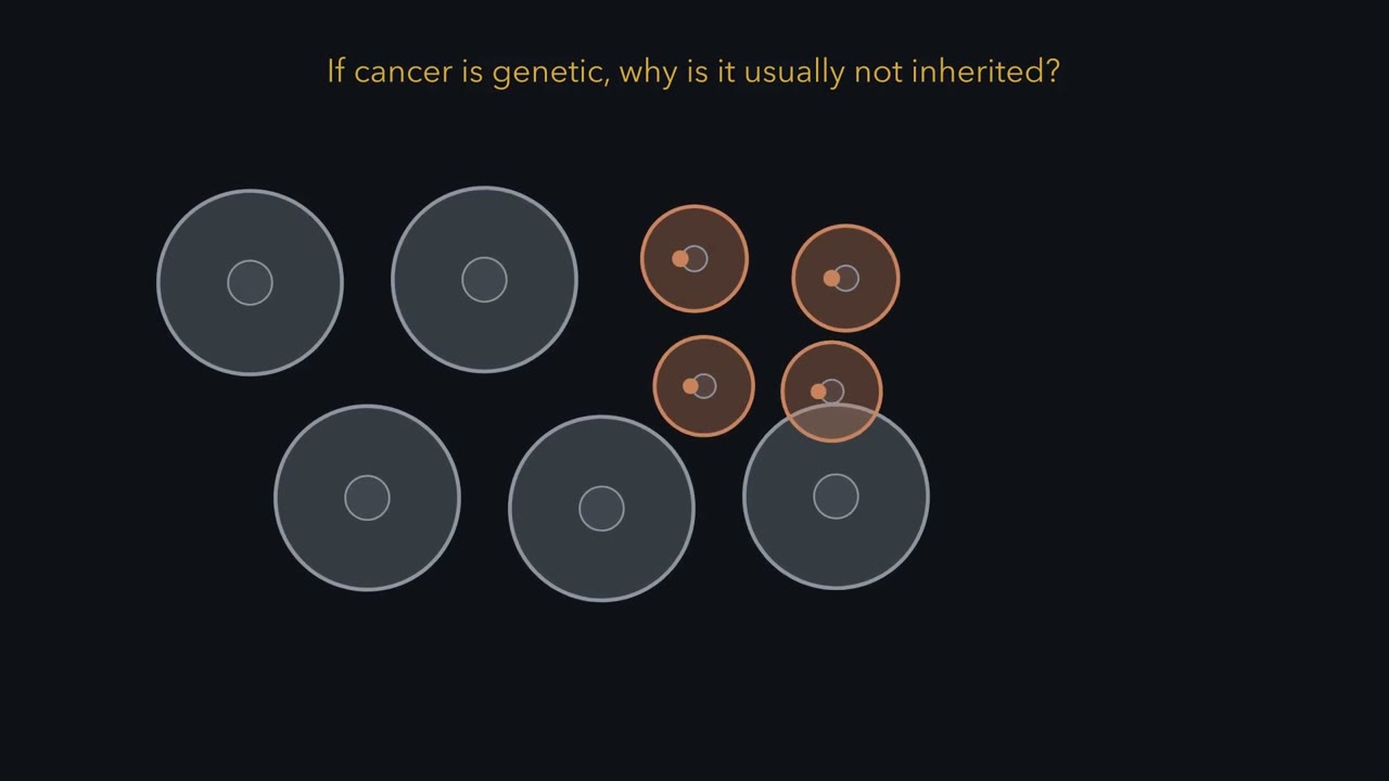 ▶ Watch One Cell, Two Routes · 3:29
Read the transcript of “One Cell, Two Routes”
Film transcript: One Cell, Two Routes
If cancer is genetic, why is it usually not inherited? Watch one pale tissue cell as a clay-colored DNA change appears. That cell divides, and the change follows its daughters. Nearby cells lack it. This is a somatic change, acquired during life, not a change carried in every cell.
The change matters only if it changes the lineage's future. A driver lets cells divide more or die less. Its branch widens while other branches continue nearby. A passenger may ride with that branch, but it did not cause the widening.
Selection acts on cells, and their DNA marks spread with them. One route changes an accelerator. A proto-oncogene normally promotes growth when signals call for it. A gain-of-function change can make an oncogene overactive.
Picture two gene copies, with one switch locked on. One altered allele is usually enough to change the cell's behavior. Extra gene copies or a translocation can also activate this route. The other route changes a brake. Here are two tumor-suppressor copies. One turns red and loses function, while the other remains green.
Predict: does the cell lose the brake now, or can the working copy still hold it? Usually, one working copy still provides enough brake. A second loss removes that remaining function. This can give the cell a growth or survival advantage, so its branch may expand. It does not by itself guarantee cancer. Tumor-suppressor changes are therefore usually recessive within one cell. Later, the text adds exceptions to these rules. RB1 makes the two-hit logic visible. A child may inherit one nonworking copy. Gold first-hit badges then appear in every cell, including every retinal cell. That inheritance raises risk, but it does not make every cell a tumor.
Each cell still has one working copy. This germline first hit can also pass to the next generation. A rare somatic second hit removes the working copy in one retinal cell. Only that cell's descendants expand. Without an inherited first hit, both hits must arise in the same body-cell lineage. This is why inherited risk can look dominant in a family while the change remains recessive within a cell. Cancer is genetic because altered DNA passes through dividing cell lineages. It is usually not inherited because most relevant changes arise somatically. The route may activate an accelerator or remove a brake. Cell lineages expand when their DNA changes improve growth or survival.
Every change a lineage carries, driver or passenger, spreads with it.
From the film
- Cell lineages expand when their DNA changes improve growth or survival. Every change a lineage carries, driver or passenger, spreads with it.
- Most cancer-associated changes arise in body cells during life, not through inheritance.
- Oncogenes usually gain function, while tumor suppressors usually lose function.
- An inherited RB1 change raises risk, but a retinal cell still needs a second hit.
The first film followed one somatic change through a dividing lineage. A somatic change arises in a body cell during life and passes to its daughters, but usually not to the person’s children. A germline change comes through the egg or sperm, is usually in every cell, and can be inherited. A driver gives its cell a selective advantage. A passenger gives none.
Two kinds of cancer gene: accelerators and brakes
The first film set the one-copy and two-copy rules; here are the molecules behind them, then the exceptions. One change alone usually does not make a malignant cancer.
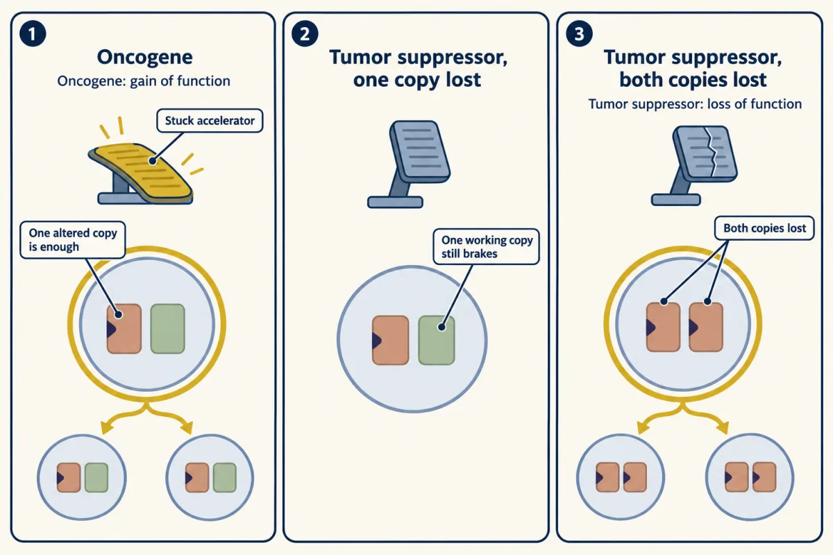
A proto-oncogene is a normal gene whose product drives growth or division; an oncogene is its altered, overactive form, a gain of function dominant within the cell.
RAS is a switch: on while it holds GTP, off once it hydrolyzes GTP to GDP. A one-base change in HRAS from a human bladder carcinoma line replaces glycine with valine at codon 12. Purified Val12 protein hydrolyzed GTP several times more slowly than normal HRAS made in E. coli. The larger effect: a GTPase-activating protein, or GAP, sped hydrolysis by normal RAS more than 200-fold but did not speed codon 12 mutants (signaling in the oncogenes and RAS-to-MAPK lessons).
Predict. If a RAS mutant ignores the GAP that normally speeds its off reaction, does it spend more or less time in the GTP-bound state?
Check your prediction
More time. Without GAP speeding its off reaction, the mutant stays GTP-bound, and on, longer.
MYC is usually activated another way: by extra copies (amplification) or by a translocation that puts it under the control of a highly active gene’s regulatory DNA. In Burkitt lymphoma the commonest change is t(8;14), which places MYC beside an immunoglobulin heavy-chain gene (see chromosome changes).
A tumor-suppressor gene encodes a brake: its product restrains division or stops a damaged cell. Losing both alleles, a change recessive within the cell, can help a cell become cancerous, usually together with other changes.
RB1 encodes RB. In its underphosphorylated form, RB binds E2F transcription factors that turn on genes for DNA replication. Returning a cloned RB gene to RB-deficient human tumor cells suppressed their tumor-like growth in a 1988 test. TP53 encodes p53, which accumulates after DNA damage and can halt the cell cycle or trigger cell death (see stress signals and death receptors and BCL-2).
The first film’s paradox, an RB1 allele dominant in a family yet recessive in a cell, rests on pedigree terms: the allele shows autosomal dominant inheritance: about half a carrier’s children inherit the risk, and affected people can appear generation after generation (see reading pedigrees). Both hold because a pedigree tracks risk, and a carrier has very many cells that each already have one hit, so a second hit in at least one is likely. Penetrance, the fraction of carriers who show the trait, is therefore high but below 100%.
p53 shows two exceptions to the copy rules. It acts as a tetramer, and many TP53 missense mutant proteins can disable wild-type subunits they join, a dominant-negative effect. In mice with one p53-null allele, many tumors kept a functional wild-type allele, so reduced dose may suffice in that model.
Predict. If mutant p53 disables a tetramer that also contains wild-type subunits, must both TP53 alleles be lost before function falls?
Check your prediction
Not necessarily. A mutant subunit can interfere with wild-type subunits in the same tetramer.
Retinoblastoma and the two-hit arithmetic
Retinoblastoma is a tumor of the developing retina. Hereditary cases tend to be bilateral (both eyes) and diagnosed earlier; nonhereditary cases usually involve one tumor in one eye. Most hereditary cases lack a family history because the mutation arose new, most often in the father’s sperm, and some carriers have a tumor in only one eye.
The first film’s two routes come from Alfred Knudson. In 1971 he analysed 48 cases with published series and concluded that two events are needed. He estimated that carriers develop about three tumors on average, not a whole retina of tumor. Knudson did not find the gene; Stephen Friend and colleagues isolated a cDNA from the chromosome 13 RB1 locus in 1986.
A simple model shows what the two hits predict. If each tumor in a carrier arises from an independent second hit, tumors per eye follow a Poisson distribution. With about three tumors per carrier and independent eyes, about 5% of carriers have no tumor, so penetrance is about 95%, and most affected carriers have bilateral disease (full calculation under Going deeper).
Predict. Under this model, is a carrier more likely to have tumors in both eyes or in one eye only?
Check your prediction
Both eyes: about 60% of carriers, against about 35% with one eye only.
In a nonhereditary case, both rare hits must fall in one lineage, so the expected number of tumors per eye is far below 1. Such tumors tend to be single, one-sided and diagnosed later.
| Inherits one mutant allele | Inherits none | |
|---|---|---|
| Mean tumors per eye (this sketch) | ||
| ≥ 1 tumor: expected | ||
| ≥ 1 tumor: observed | ||
| Tumors per person: expected / observed | ||
| Tumors in both eyes: expected / observed |
In this sketch the carrier mean grows in proportion to age and the non-carrier mean with its square (relative units, not data), both scaled to the lesson’s end-of-window figures.
How the second hit happens: loss of heterozygosity
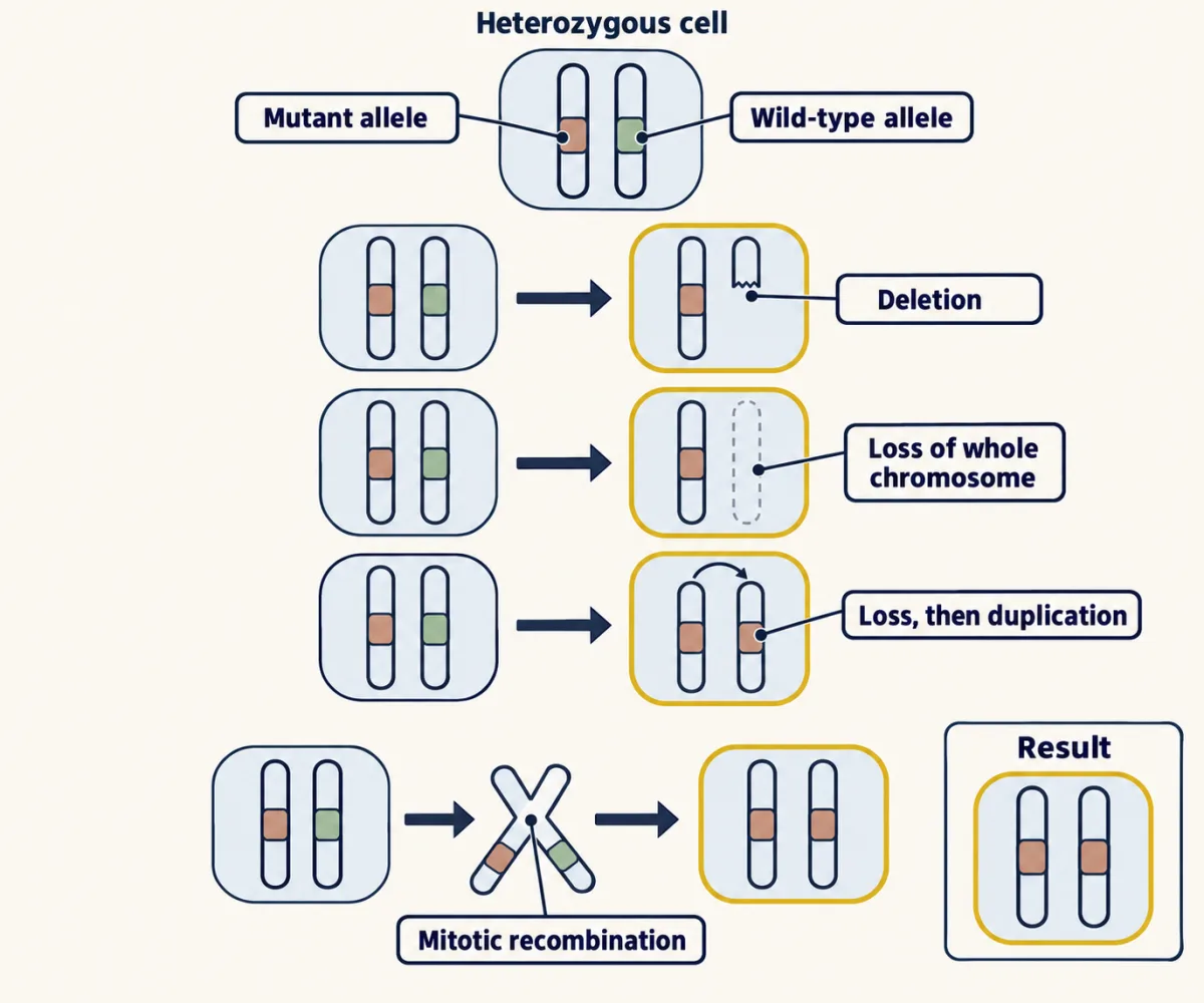
A carrier is heterozygous at RB1: one mutant allele and one wild-type allele. A second hit can be a new point mutation in the wild-type allele or silencing of its promoter by DNA methylation (see eukaryotic regulation).
Often it is larger. Loss of heterozygosity (LOH) means that a site no longer has two different allele identities. In a carrier’s tumor, only the mutant identity remains. The cell may keep one physical copy, after a deletion or chromosome loss, or two matching copies, after loss and duplication of the mutant chromosome or after mitotic recombination, a crossover between homologs in a dividing body cell that leaves a daughter cell with two copies of the mutant region.
Predict. A tumor retains two copies of chromosome 13 but shows only the mutant marker near RB1. Does that pattern require a deletion?
Check your prediction
No. Mitotic recombination or loss followed by duplication can leave two copies with one allele identity.
In 1983 Webster Cavenee, Thaddeus Dryja and colleagues tested this with DNA markers that differ between the two copies of chromosome 13. In several patients heterozygous at those markers in normal tissue, the tumor showed only one allele. The patterns fitted mitotic recombination, or nondisjunction with loss of the wild-type homolog. The second hit was often chromosomal, and LOH in tumor DNA became a way to find suppressor genes in other cancers.
Instability and clonal evolution
The same lineage logic scales from one retinal cell to competing subclones in a tumor. A tumor is usually a clone, the descendants of one cell. In 1976 Peter Nowell proposed that tumors evolve: acquired genetic variability lets more aggressive sublines be selected. Treatment can fail when a resistant subline emerges. As in bacteria under an antibiotic, mutation supplies variants and selection keeps those that divide more or die less.
Genome instability raises the supply. Mutations in DNA-repair genes tend to cause further mutations in other genes. Cells lacking mismatch repair, as in Lynch syndrome tumors, accumulate copying errors in short repeats. Loss of BRCA1 or BRCA2 impairs repair of double-strand breaks by homologous recombination (see DNA repair and recombination and mutation). Faulty chromosome segregation produces whole-chromosome gains and losses, and chromothripsis can produce many clustered rearrangements in one catastrophic event.
Predict. Does losing mismatch repair directly make every new mutation advantageous, or does it mainly change the supply of variants?
Check your prediction
It raises the supply. Selection favors lineages whose drivers improve growth or survival, while passengers carried by those lineages can hitchhike with them.
Colorectal tumors show the accumulation. RAS mutations were found far more often in larger adenomas and in carcinomas than in small adenomas, and the changes accumulated as tumors progressed (Vogelstein and colleagues, 1988). Eric Fearon and Vogelstein’s 1990 model stressed the accumulation of changes over their exact order; it is a colorectal model, not a fixed sequence for every tumor.
Back to the one idea: instability creates more candidates, and growth or survival decides which lineages expand. In whole cancer genomes from many tumor types, each with matched normal tissue, the Pan-Cancer Analysis of Whole Genomes consortium identified an average of 4.6 drivers per tumor with current knowledge. That figure depends on the rules used to call drivers. Selection also works before cancer, in clones of normal human esophagus; a selected clone is not automatically a cancer.
Reading a tumor genome
All those competing lineages end up mixed in one tumor sample. Can its reads still show which lineage carries a change?
Read the transcript of “Reading a Tumor Family Tree”
Film transcript: Reading a Tumor Family Tree
The first film followed DNA changes through one expanding lineage. How can mixed DNA reads reveal which tumor lineage carries a change? Start with that same clay-colored family tree. An early clay marker appears near its trunk. Every later branch inherits it.
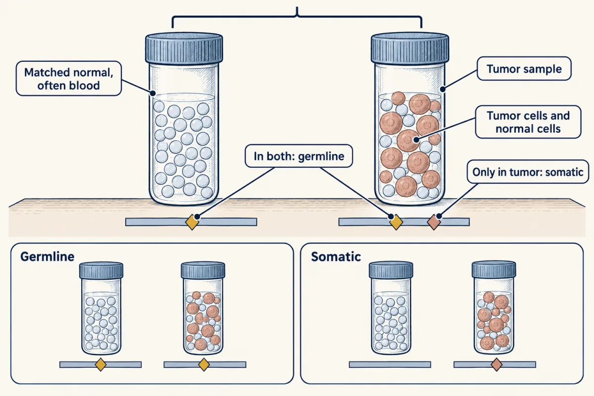
Later, one branch gains a second, square marker. Only that branch carries it, so it is a subclone. The trunk marker is clonal because every tumor branch carries it. The pattern records ancestry, even when the sample mixes all branches together. Now place a matched-normal sample beside the tumor. It comes from the same person, often from blood. A variant found only in tumor is usually somatic. A variant found in both samples is usually germline, most often inherited. The word usually matters. Here is a gold variant in both tumor and matched normal.
Predict: did it most likely enter this person's cells before the tumor lineage began, or after one tumor branch formed? Most likely, it was present before the tumor lineage began. So it is usually germline, whether inherited or newly formed in an egg or sperm. The matched normal gives the timing clue. Tumor data alone cannot reliably separate that history from a somatic change. Variant allele fraction, or VAF, is the fraction of reads carrying a variant. A clonal variant can still sit below one half.
Normal cells dilute the tumor reads. Copy gains, losses, and loss of heterozygosity also change the number of variant and nonvariant copies. Purity is the fraction of cells in the sample that are tumor cells. For a heterozygous clonal variant in a two-copy region, expected VAF is purity times one half. The shortcut also assumes normal cells lack the variant. It fails after copy changes or loss of the other allele, or when only a subclone carries it. So read the evidence in order.
Compare tumor with matched normal. Ask which lineages carry the variant. Then count mutant copies against all copies. Purity, copy number, and read depth limit the inference. The result is a family-tree estimate, not a perfect replay. A high or low fraction is not a verdict by itself. Read it with the matched normal and the sample's copy structure. Different branch histories can produce similar fractions, so the supporting clues matter. That uncertainty belongs in the interpretation. Together, those clues connect mixed sequencing reads back to the expanding cell lineages that produced them.
From the film
- Variant fractions trace DNA changes through expanding tumor cell lineages.
- A matched normal helps separate variants that are usually germline from somatic variants.
- A clonal variant is in every tumor cell; a subclonal variant is in only one branch.
- Purity and copy number shape the variant fraction measured in a tumor sample.
The second film compared the tumor with a matched normal sample, often blood, from the same person, and read each variant’s variant allele fraction (VAF), the fraction of reads carrying it, against the sample’s purity, the fraction of cells that are tumor cells (method in the sequencing lesson).
A variant in both samples is usually germline, most often inherited and rarely new in the egg or sperm, but blood is not a perfect normal. Blood-cell clones that expand with age carry somatic mutations, and those cells can also enter the tumor sample.
expected VAF (heterozygous, all tumor cells, two copies) = purity × 0.5
When any of the shortcut’s four conditions fails (heterozygous, in every tumor cell, absent from normal cells, two copies), count mutant copies over all copies in the mixed sample, as the design problem does.
Genotype now shapes diagnosis. In chronic myeloid leukemia, the 9;22 translocation makes the BCR-ABL kinase that imatinib inhibits (see drugs against a part). In a 2021 study of acute myeloid leukemia and myelodysplastic syndromes, whole-genome sequencing was fast enough to change the genetic risk category for some patients.
Design problem
A colorectal tumor sample is estimated at 60% tumor cells. Tumor and matched-normal sequencing gives: KRAS G12D at VAF 0.30 in the tumor; TP53 R175H at VAF 0.60, with the other TP53 allele lost; PIK3CA at VAF 0.08; and a BRCA2 frameshift at VAF 0.50 in normal tissue and 0.80 in the tumor. Assume two copies of each region unless the data say otherwise. Which variants are inherited, which are in every tumor cell, and which are in a subclone? Why is the matched normal needed?
Saved in this browser only.
One way to do it
In this simplified problem, reads sample DNA copies evenly.
KRAS. If every tumor cell has one mutant and one wild-type copy, and normal cells have none, the expected VAF is 0.6 × 0.5 = 0.30, as observed: a heterozygous, clonal variant.
TP53. If tumor cells retain two mutant copies, every tumor-cell read is mutant: 0.6 × 1.0 = 0.60, as observed. Had the wild-type allele been deleted instead, tumor cells would carry one copy and normal cells two, giving mutant over all copies: (0.6 × 1) / (0.6 × 1 + 0.4 × 2) = 0.43. So 0.60 fits clonal, copy-neutral LOH, such as mitotic recombination, under these assumptions.
PIK3CA. If it is heterozygous, a clonal variant would sit at 0.30; 0.08 / 0.30 = 0.27, so about 27% of tumor cells carry it: a subclone that arose later.
BRCA2. A VAF of 0.50 in normal tissue fits a heterozygous germline allele, most likely inherited; testing the parents would show whether it is new. In the tumor, 0.6 × 1.0 + 0.4 × 0.5 = 0.80 fits two mutant copies in tumor cells and one of each in normal cells: copy-neutral LOH, as for TP53. A deletion of the wild-type copy would instead give (0.6 × 1 + 0.4 × 1) / (0.6 × 1 + 0.4 × 2) = 0.71. Either route supplies a second hit in a gene already present in one-hit form.
The matched normal. Tumor data alone cannot separate an inherited BRCA2 allele from a somatic one, or set aside the many harmless inherited variants every person carries. These are inferences about one sample, not clinical advice.
Going deeper
The two-hit arithmetic in full
Worked problem. Assume that in a carrier of one mutant RB1 allele each tumor arises from an independent second hit, so the number of tumors per eye follows a Poisson distribution with mean m = 1.5 (3 per person, Knudson’s approximate figure), and the eyes are independent. What fractions of carriers have no tumor, a tumor in one eye only, and tumors in both eyes?
Step 1. The Poisson probability of zero events is e−m.
P(no tumor in one eye) = e−1.5 = 0.223
Step 2. So P(at least one tumor in an eye) = 1 − 0.223 = 0.777.
Step 3. No tumor in either eye: 0.223 × 0.223 = 0.050, which equals e−3. About 5% stay tumor free, so penetrance is about 95%.
Step 4. Tumors in both eyes: 0.777 × 0.777 = 0.604.
Step 5. A tumor in one eye only, where either eye can be the affected one: 2 × 0.223 × 0.777 = 0.347.
Check: 0.050 + 0.604 + 0.347 = 1.001, which is 1 within rounding. About one carrier in twenty has no tumor yet can pass the allele on.
If a nonhereditary case’s expected number of tumors per eye were 0.0001, a tumor in each eye would have a chance of about 1 in 100 million.
Who found what, and when
In 1982 several laboratories, among them those of Mariano Barbacid, Robert Weinberg and Michael Wigler, reported in Nature the codon 12 valine-for-glycine change described above; the slower hydrolysis was measured in 1984 and the GAP effect in 1987. Riccardo Dalla-Favera and colleagues mapped c-myc to chromosome 8 in 1982.
Cohort sizes and percentages
In a 1988 series of 172 specimens, Bert Vogelstein and colleagues found RAS mutations in 9% of adenomas smaller than 1 cm, 58% of larger adenomas and 47% of carcinomas. The Pan-Cancer Analysis of Whole Genomes consortium examined 2,658 whole cancer genomes from 38 tumor types, each with matched normal tissue. In the 2021 leukemia study, whole-genome sequencing in 117 consecutive patients took a median of five days and changed the genetic risk category for 19 of them.
Your notes
Select any text in the lesson to highlight it or add a note to it. Your highlights and notes are saved in this browser and collected in your notebook.
This browser is not letting the site save anything (a private window or strict privacy settings), so highlights and notes will be lost when you leave the page.
How we measure it
Restriction fragment length polymorphism comparison
A probe detects DNA fragments whose sizes differ between the two homologs of a chromosome. Comparing a patient's normal DNA with tumor DNA shows whether the tumor has lost one of the two alleles. Cavenee and colleagues used it to detect loss of heterozygosity on chromosome 13 in retinoblastoma.
Gene transfer and functional rescue
A cloned normal gene is introduced into tumor cells that lack it, and their behavior is compared with controls, for example by colony growth in soft agar and tumor formation in nude mice. Suppression after restoration is functional evidence that the gene is a tumor suppressor, as Huang and colleagues showed for RB in 1988.
Tumor and matched-normal sequencing
DNA from the tumor and from normal tissue of the same person is sequenced and compared. Variants in both are usually germline; variants only in the tumor are usually somatic. The fraction of reads carrying each variant, read with the tumor purity, suggests whether a mutation is in all tumor cells or only a subclone.
Karyotyping and fluorescence in situ hybridization
Banded metaphase chromosomes show gains, losses and translocations large enough to see under the microscope, such as the 9;22 translocation in chronic myeloid leukemia. Fluorescent probes then test a chosen locus, for example whether two genes have been brought together.
Practice questions on this lesson (3) → MCAT-style, original, each with an explanation
Where this lesson fits
Before thisReview first
- Reading Pedigrees: Human InheritanceThis course · Lesson 5
You need to read dominant inheritance and penetrance in a pedigree to see why an RB1 allele is dominant in a family.
- DNA Repair and RecombinationThis course · Lesson 15
Mismatch repair, BRCA and double-strand break repair explain how genome instability raises the supply of mutations here.
- Mutation: Where Variation Comes FromThis course · Lesson 14
Mutation supplying variants that selection then sorts is the logic behind tumor lineages expanding.
After thisStudy next
- Recombinant DNA, Cloning and PCRThis course · Lesson 22
PCR and cloning are tools used to detect and confirm cancer-gene variants like those found in tumors.
- Sequencing and GenomesThis course · Lesson 23
Reading variant fractions in tumor and matched-normal data uses the sequencing methods taught there.
- Population Genetics: Hardy-Weinberg and Evolutionary ForcesThis course · Lesson 25
Selection acting on variants within lineages here is taken further as allele frequency change in populations.
Elsewhere on the siteFilms, simulations and method films in other courses
- Sealed or TornFilm · Cell Biology
It shows how apoptosis, the process p53 can trigger, takes a damaged cell apart.
Suggested by an AI model from the lessons themselves, then checked. How we use AI
Finish line
Test yourself
8 questions
Practice and MCAT-style, with explained answers.
Next · Lesson 22
Recombinant DNA, Cloning and PCR
A white colony survives ampicillin and a PCR tube ends with a bright band. How do you know either contains the DNA you wanted?
Continue →