Cell Biology · Unit 3: Traffic and movement
Cytoskeletal Polymers and Dynamics
Rewritten on : new text, flashcards and film. Your notes and highlights stay, but the text around them may have moved.
A microtubule in a dish of pure tubulin grows for minutes, then collapses many times faster. Why would a polymer be built to fail? Because a polymer that switches by itself lets a cell explore space, rebuild its scaffolding and organize division.
Pause when the reading reaches a film, and offer to play it. Saved on this device.
Watch the film now, or keep listening
Builds on:Transport Across MembranesExperiments That Distinguish MechanismsWhy these?
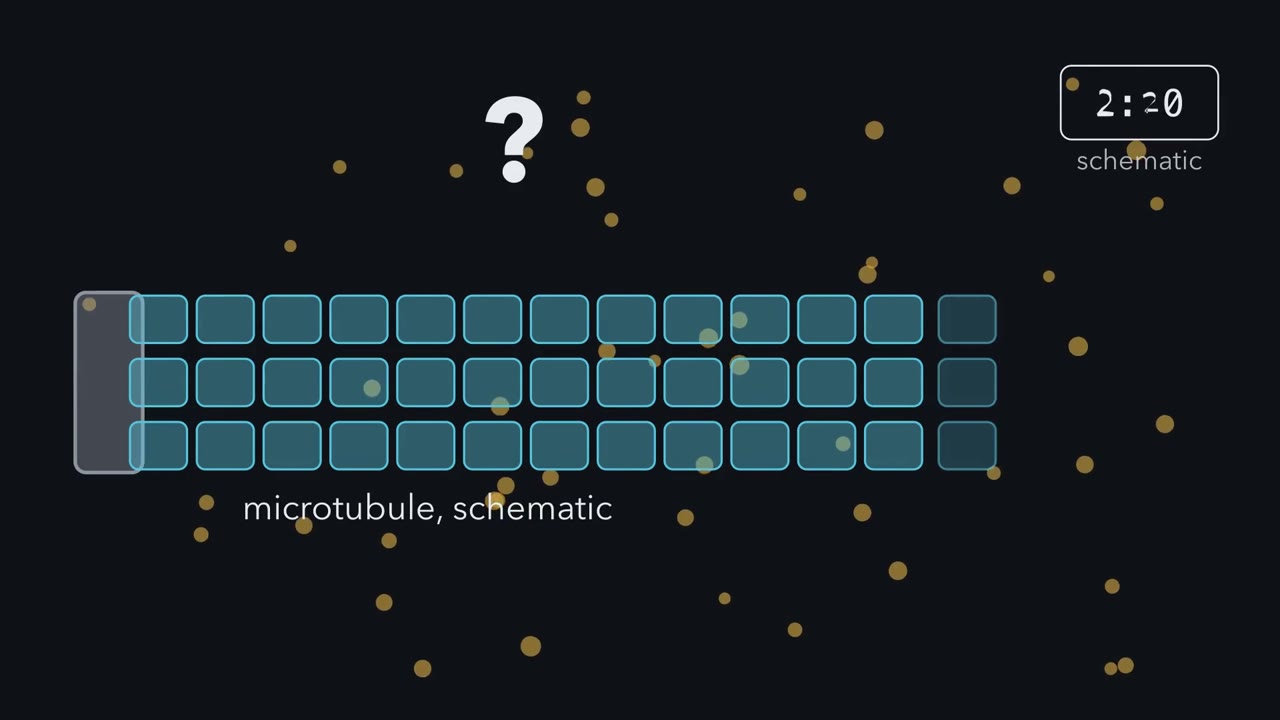 ▶ Watch Tight Ends, Loose Ends · 3:10
Read the transcript of “Tight Ends, Loose Ends”
Film transcript: Tight Ends, Loose Ends
Why would a polymer fall apart while its building blocks crowd around it? Watch one microtubule grow from a seed in purified tubulin. It lengthens slowly for minutes, then collapses many times faster. A crystal in its solution does not do this.
Start with one end, and treat it as a doorway. Subunits arrive at a rate set by how many are free. They leave at a rate that does not depend on that. At one free concentration, the two balance. That is the end's critical concentration, Above it, the end grows.
Below it, the end shrinks. If subunits never used their nucleotide, both ends would share one critical concentration. The polymer would grow until free subunits fell to that level, then simply settle. Real actin and tubulin differ. Each subunit arrives carrying fresh ATP or GTP. Some time after joining, it hydrolyses that nucleotide and releases the phosphate. Those old subunits then hold on less tightly. Here is the whole idea. Hydrolysis after joining gives one polymer two kinds of end. A tight end shows fresh subunits and has a low critical concentration. A loose end shows old subunits and has a high one. So set the free subunits between the two levels: above the tight end's, below the loose end's. What can the polymer do now?
It cannot come to rest. Actin answers in space. Its barbed end adds subunits fast and stays fresh: a tight end that grows. Its pointed end shows old subunits: a loose end that shrinks. When gain and loss match, the length holds while subunits flow through. That is treadmilling. A microtubule answers in time instead. While growing, it wears a cap of fresh GTP-tubulin, so its end stays tight. Behind the cap, GDP-tubulin is held straight only by its neighbours. The cap can be lost by chance, or when growth slows and hydrolysis catches up. Then the end turns loose. Its protofilaments peel outward, and it shortens many times faster than it grew. That switch is a catastrophe.
Growth starting again is a rescue. Exactly what commits an end to collapse is still argued, and in cells other proteins tune the rates. Hydrolysis gives tight and loose ends, so the polymer cannot simply settle. Cells use that restlessness to search their interior and rebuild.
From the film
- Each filament end has a critical concentration: above it the end grows, below it the end shrinks.
- If subunits never hydrolysed their nucleotide, both ends would share one critical concentration and the polymer would simply settle.
- Hydrolysis after joining gives a polymer tight ends, with a low critical concentration, and loose ends, with a high one.
- At treadmilling steady state, actin grows at its tight barbed end and shrinks at its loose pointed end.
- A microtubule end is tight while it keeps its GTP cap and collapses many times faster when it loses it.
The first film above opens on a microtubule that will not settle down, where a crystal would grow until crystal and solution balance and then stop.
The instability is not a defect. Ends that grow and collapse by themselves let a microtubule array search the cell interior, keep the ends that find a target, and rebuild quickly when the cell changes shape or divides; regulated actin turnover lets a cell build and take down protrusions. This lesson asks what the three filament systems are made of, what decides whether an end grows or shrinks, and why two of them spend nucleotide energy in a way that keeps them unstable.
Three polymers built for different jobs
The cytoskeleton is the set of protein filaments in the cytosol, the fluid part of the cytoplasm outside organelles. It has three classes.
| Actin filament | Microtubule | Intermediate filament | |
|---|---|---|---|
| Subunit | globular actin | α/β-tubulin heterodimer (a two-protein unit) | fibrous proteins (vimentin in fibroblasts, keratins in epithelial cells) |
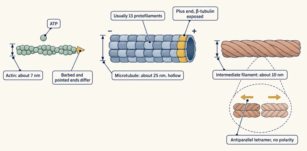
| Width | about 7 nm | about 25 nm, a hollow tube of (usually) 13 protofilaments (rows) | about 10 nm |
| Two different ends? | yes: barbed and pointed | yes: plus (β-tubulin exposed) and minus | no |
| Nucleotide | ATP | GTP | none, and no enzyme activity |
| Bending | in between | stiffest | most flexible |
Actin’s barbed and pointed ends are named after the arrowhead pattern seen when fragments of the motor protein myosin decorate a filament in the electron microscope. Actin subunits add head to tail, which is why the filament has two different ends. Tubulin dimers also stack head to tail, into rows called protofilaments, and usually (not always) 13 rows lie side by side to close the tube. Every dimer points the same way, so the tube has a plus end and a minus end. In vimentin and other cytoplasmic intermediate filaments, two rope-like coiled-coil dimers line up antiparallel (in opposite directions) and half staggered (offset) to form a tetramer. A building block that points both ways gives a filament with no polarity. These filaments are not permanent: they exchange subunits slowly, and cells take them apart by phosphorylation, the addition of phosphate tags, as with the nuclear lamins, fibrous proteins of the nuclear scaffold, at mitosis.
In comparisons of purified filaments, microtubules are the stiffest in bending, which suits them to serve as tracks and struts. Intermediate filaments are the most flexible in bending, yet a single filament can be stretched beyond two or three times its resting length without breaking. Think of a rope beside a fishing rod: the rope bends at a touch and is far harder to snap. Flexible is not weak; bending and stretching are different tests.
The one idea to carry through this lesson, the one the first film animates. Each filament end, in a given state, has a critical concentration, Cc = koff / kon. Hydrolysis after assembly gives a polymer a tight end (fresh nucleotide, low Cc) and a loose end (old nucleotide, high Cc). Between the two Ccs the polymer cannot come to rest with nothing moving. Actin separates the two kinds in space (treadmilling); a microtubule end switches between them over time (dynamic instability).
This rule is for single ends with purified proteins. It says whether a polymer can settle and which way each end moves, not the rates in a cell, where accessory proteins (profilin, formins, cofilin, capping protein, end-binding (EB) proteins and others) change them, some by an order of magnitude or more. It does not say when a capped end loses its cap. Intermediate filaments, with no nucleotide and one kind of end, are the contrast case: the rule predicts neither treadmilling nor instability.
One end: the doorway and its critical concentration
Start with a measurement. Let tubes of pure actin at a range of total concentrations settle, then measure how much is in filaments and how much is free, and plot free actin against total actin.
- Below a certain total, nothing polymerizes, so free actin rises in step with total actin.
- Above that total, free actin stays flat and every extra subunit goes into polymer, one for one.
The flat level of free actin is the critical concentration, Cc, and it is also the total where the line bends. (For real ATP-actin, whose two ends differ, free actin settles between the two ends’ values; next section.) Followed over time, one tube shows a lag, then growth, then a plateau. The lag is there because starting a filament is hard: a few subunits must come together before an end exists. So cells control where filaments start, with formins and the Arp2/3 complex for actin and γ-tubulin at the centrosome for microtubules. Rho-family GTPases decide where actin assembles (that lesson).
Why does free actin stop at one value? Treat a filament end as the film’s doorway: subunits arrive at a rate proportional to the free subunit concentration C, and leave at a rate that does not depend on C.
In words Net rate equals k on times C, minus k off. Setting it to zero gives the critical concentration, k off divided by k on.
- net rate“net rate”
- subunits gained per second at one end; negative means shrinkingper s
- kon“k on”
- joining rate per micromolar of free subunitper µM per s
- C“C”
- the free subunit concentrationµM
- koff“k off”
- how fast subunits leave the end, whatever C isper s
- Cc“C c”
- the critical concentration, where the end neither grows nor shrinksµM
Sanity check At C = Cc, joining and leaving balance. Above Cc, kon × C beats koff and the end grows; below Cc, loss wins and it shrinks.
Try one number
A growing microtubule plus end in one purified preparation: kon = 8.9 per µM per s and koff = 44 per s.
At 10 µM tubulin: 8.9 × 10 − 44 = 89 − 44 = +45 dimers per s, so the end grows.
Its critical concentration: 44 / 8.9 ≈ 4.9 µM.
Need a refresher? rates and “per” · units and prefixes
Predict. A tube holds total actin at twice the critical concentration. How much is free, and how much is polymer?
Check your prediction
For an end with one Cc: one Cc is free and the other Cc’s worth is polymer. Any free actin above Cc joins filament ends until gain and loss match again.
Two ends: why hydrolysis makes them differ
So far, one end. A filament has two, and they are built differently: the barbed end adds subunits much faster than the pointed end does (about nine times faster for ATP-actin). Does that give the two ends different critical concentrations?
Predict. Imagine actin that never hydrolysed its ATP. Could the barbed end have a lower critical concentration than the pointed end?
Check your prediction
No. Adding one subunit at the barbed end, or one at the pointed end, gives exactly the same result: the same filament, one subunit longer. The energy change is the same, so the balance point, koff/kon, must be the same. The barbed end can be faster at both adding and losing, but the ratio cannot differ. Actin holding ADP, which cannot be hydrolysed further, shows this: its two ends have critical concentrations of about 1.9 and 1.7 µM, equal within measurement error.
So a difference between the ends needs a step that spends energy, and that step is hydrolysis. An actin subunit joins carrying ATP, hydrolyses it a little later, then releases the phosphate slowly, so the older lattice holds ADP-actin. ATP-actin binds a filament end more tightly than ADP-actin does, meaning it has a lower critical concentration. Hydrolysis by itself barely loosens a subunit; release of the phosphate does.
In the standard, simplified picture:
- The barbed end grows fast, so new ATP-actin keeps arriving before the subunits there have hydrolysed. Its fresh tip makes it a tight end, with a low critical concentration.
- The pointed end adds subunits slowly, and in the window where treadmilling happens it is losing them, so its tip exposes older subunits that have hydrolysed their ATP and released the phosphate. It is a loose end, with a higher critical concentration.
Measured with ATP-actin in solution, the critical concentrations are about 0.1 µM at the barbed end and about 0.6 µM at the pointed end (Pollard, 1986; constants in Going deeper). Put them on a line:
0 ─── 0.1 µM (barbed Cc) ─── 0.6 µM (pointed Cc) ───→ free ATP-actin
Predict. What does each end do with free actin at 0.05 µM, at 0.3 µM and at 1 µM?
Check your prediction
- At 0.05 µM, below both: both ends shrink.
- At 1 µM, above both: both ends grow, the barbed end about twenty times faster (+10.2 against +0.5 subunits per s).
- At 0.3 µM, between them: the barbed end grows and the pointed end shrinks.
So “the barbed end grows and the pointed end shrinks” is true only in the window between the two values.
Only the middle zone is new. Left alone, filaments use up free actin until the barbed end’s gain matches the pointed end’s loss. With Pollard’s constants, that happens at about 0.17 µM, close to the barbed end’s value rather than halfway, because the barbed end’s ninefold larger kon gives its value more weight. There the filament keeps its length while subunits flow through it, as the first film shows: treadmilling. Single purified actin filaments watched by microscopy show barbed-end growth and pointed-end shortening at approximately equal rates at steady state.
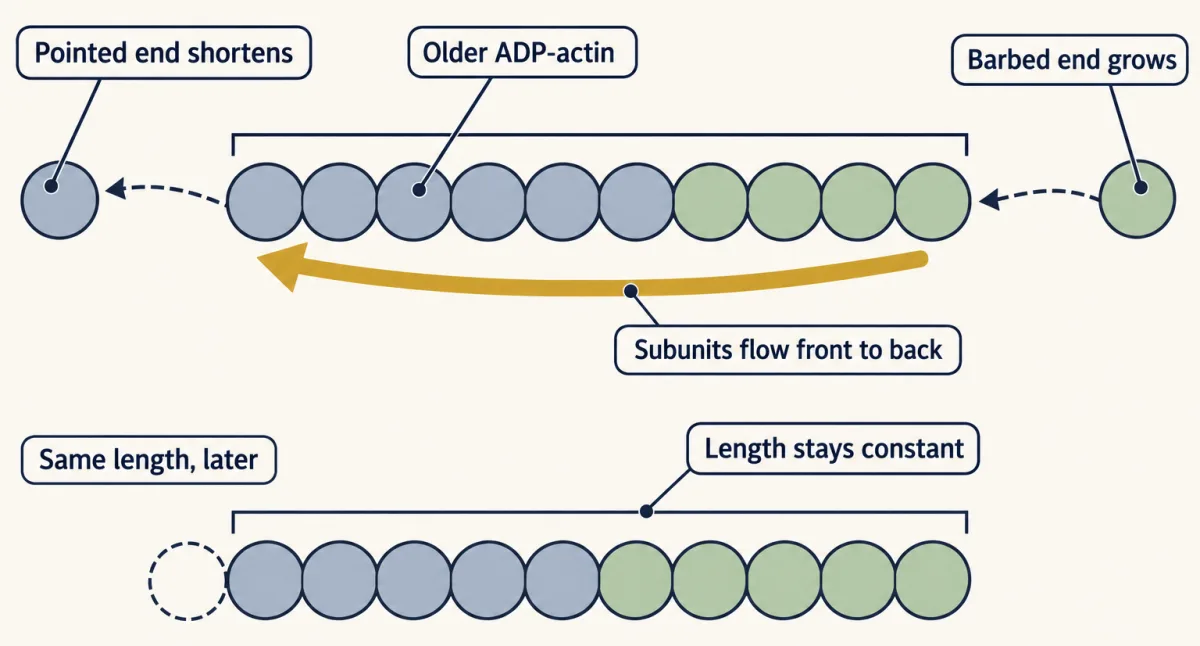
Binding drives assembly; hydrolysis afterwards turns the steady state into a flux. Pure treadmilling is slow, a small fraction of a micrometre per minute, so cells that rebuild actin quickly add proteins that start, speed, cap and sever filaments (see the design problem).
One end, two states: dynamic instability
A microtubule end switches between steady growth and rapid shortening. The switch to shortening is a catastrophe; the switch back is a rescue. This dynamic instability, a property of individual ends, is the rule’s time version: one end alternates between a tight (capped) and a loose (uncapped) state. (Microtubules can treadmill too, in vitro and in some cells, so treadmilling is not limited to actin.)
The standard explanation is the GTP cap that the first film animates. The GTP sits on β-tubulin and is hydrolysed soon after the dimer joins, inside the lattice, so the end stays intact only while a cap of not-yet-hydrolysed dimers remains.
- Capped, the end is tight. Its critical concentration is about 5 µM in one purified preparation (the worked example in the box above), so at typical test concentrations it grows.
- Uncapped, the end is loose. It sheds dimers so fast that its critical concentration is, by a rough estimate, far above any tubulin concentration tested (Going deeper, “Who first watched the switching”).
GDP-tubulin protofilaments are curved, which is why they peel outward once the cap is gone.
Because a cap can be lost by chance even at a steady growth rate, catastrophes are random events. Slowing hydrolysis lengthens the cap, as recombinant human tubulin mutants show (Roostalu and colleagues, 2020). Exactly what the cap looks like, and what final event commits an end to shortening, is still argued.
Mitchison and Kirschner inferred dynamic instability in 1984 from populations of microtubules; microscopy of single ends soon showed the switching directly, at both ends of purified microtubules (Going deeper). In cells, minus ends are often capped or anchored, for example at the centrosome.
Predict. A microtubule end has lost its cap and is shortening. Will doubling the free tubulin, say from 7 to 14 µM, slow the shortening?
Check your prediction
Barely. An uncapped end’s critical concentration is out of reach, so a bit more tubulin cannot turn loss into gain. A 1988 study of purified pig brain tubulin saw exactly this: rapid shortening did not change with tubulin concentration over 7 to 15.5 µM. The cap model also fits its second finding, that more tubulin made catastrophes rarer at both ends: a capped end grows faster with more tubulin and is thought to build a bigger cap that is harder to lose, though catastrophe also depends on the age of the end.
A worked problem: reading one trace
The data below are made up for teaching. They describe one plus end grown from a seed in purified tubulin; length is measured from the seed.
- 0 s, 5.0 µm; steady growth to 180 s, 9.5 µm; catastrophe
- 192 s, 3.5 µm; rescue; steady growth to 312 s, 6.5 µm; catastrophe
- 325 s, 0 µm (back to the seed)
Step 1, growth velocity. (9.5 − 5.0) µm over 180 s (3 min) = 1.5 µm per min. The second growth phase gives (6.5 − 3.5) µm over 120 s (2 min) = 1.5 µm per min, the same.
Step 2, shortening velocity. 6.0 µm in 12 s = 0.5 µm per s = 30 µm per min, twenty times faster than growth. The second shortening phase, 6.5 µm in 13 s, gives the same 0.5 µm per s.
Step 3, switching frequencies. Each frequency is the number of switches divided by the time spent in the state they leave. The end spent 180 + 120 = 300 s growing and had 2 catastrophes: 2/300 = 0.0067 per s, × 60 s = 0.4 per min. It spent 12 + 13 = 25 s shortening and had 1 rescue: 1/25 = 0.04 per s.
Step 4, dimers per second. Assume 13 protofilaments and 8 nm per dimer, so one micrometre holds 1,000 nm ÷ 8 nm = 125 dimers per protofilament, and 125 × 13 = 1,625 dimers in all. Growth of 1.5 µm per min is 0.025 µm per s, times 1,625 = about 41 dimers per s.
Check against real data. The box’s example, the 1988 study’s plus-end constants at 10 µM tubulin, gives +45 dimers per s, about 1.7 µm per min by the same conversion. The study’s rapid shortening, 733 dimers per s, is about 27 µm per min. The made-up trace is in the right range.
- Expected growth speed, kon × C − koff
- –
- Growth slope measured on the trace
- –
- Catastrophe frequency (in this sketch)
- –
- Catastrophes counted ÷ time growing (in this sketch)
- –
- Shortening speed (lesson: 733 dimers per s)
- –
- Shortening measured on the trace
- –
Move the slider or press Resample.
Rule to keep: growth is kon × C − koff, so it rises with tubulin, but rapid shortening does not depend on tubulin over 7 to 15.5 µM. Growth and shortening use the Walker et al. (1988) plus-end constants (pig brain tubulin free of associated proteins, 37 °C; in cells, associated proteins change them), applied across 7 to 15.5 µM by the rate law. Switching frequencies are invented for this sketch: catastrophe 0.4 per min at 10 µM scaled as 10/C, rescue fixed at 0.04 per s. The lesson supports only that more tubulin makes catastrophes rarer.
Drugs as tests
A drug tests a mechanism only if you know its target, its dose and the system. Each drug below acts on one piece of the rule: free subunits, ends, or the lattice. The second film asks when a perturbation becomes evidence about one of these moving parts.
Read the transcript of “When a Drug Becomes a Test”
Film transcript: When a Drug Becomes a Test
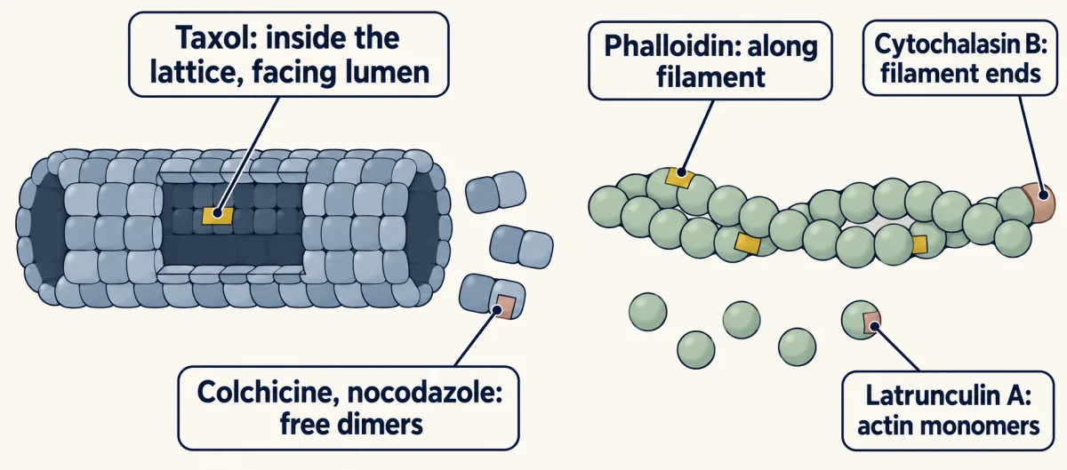
Chapter one linked free subunits, ends, and their lattice. How can a drug turn that rule into a clean test? A drug is informative only when three labels stay attached: its target, dose, and test system. Remove any label, and the same drug name can support the wrong conclusion. Keep the moving parts on screen. Free subunits feed an end; the end adds to or removes from a lattice. A perturbation can act at any of those three places. Latrunculin A binds actin monomers one to one, holding them so they cannot join. The available pool falls, so less actin enters polymer. Yet it can also speed loss at the ends of freshly built filaments. Sequestration is not the whole story.
Cytochalasin B acts elsewhere. Its binding follows filament-end number, not total actin, and it inhibits addition at barbed ends. Phalloidin instead binds along actin filaments. With purified muscle actin, it slows disassembly. Now predict. A drug lowers the amount of polymer. Does that observation alone reveal whether it trapped subunits, blocked ends, or changed the lattice?
No. The same amount change can begin at different sites. Add a second measurement that separates them. Holding total actin constant while changing end number can reveal binding that follows ends. That is the difference between an effect and a mechanism.
Polymer amount reports the outcome. A measurement tied to the proposed target tests how that outcome arose. Target is still not enough; dose changes the experiment. In tested cultured cells, nanomolar nocodazole can pause microtubules and slow growth and shortening. Micromolar doses commonly used on cultured mammalian cells remove most microtubules. The system can change a switching result too. With nocodazole, catastrophe became more frequent in cultured newt lung cells. In BSC-1 monkey kidney cells after an hour, catastrophe became less frequent. Phalloidin shows another system boundary. It slows disassembly with purified muscle actin, but enters living cells poorly.
Fluorescent phalloidin therefore mainly stains filaments after cells are fixed. Sometimes the drug is not selective enough. Withaferin A redistributes vimentin in human fibroblasts, but did not alter purified vimentin assembly. Removing vimentin genetically, then adding it back, is the cleaner test. Read every result as a complete sentence: this target, at this dose, in this system, changed this measurement. Then compare another perturbation aimed at a different step. Chapter one supplied the moving parts behind cytoskeletal turnover. This chapter adds the rule for testing them experimentally. If target, dose, or system changes, the conclusion may change too.
From the film
- A drug tests a mechanism only when its target, dose and test system stay attached.
- The same change in polymer amount can begin at free subunits, filament ends or the lattice.
- Latrunculin A sequesters actin monomers and can also speed loss at the ends of freshly built filaments.
- Nocodazole effects depend on dose and on the cultured cell system.
- A second measurement can separate mechanisms that produce the same outcome.
The table keeps the exact effects and caveats together for reference.
| Drug | Binds | Effect on the polymer | Evidence and caveats |
|---|---|---|---|
| Taxol (paclitaxel) | β-tubulin in the assembled lattice | stabilizes microtubules; drives extra assembly | low doses damp dynamics without adding polymer; higher doses add polymer (worked below) |
| Colchicine | free tubulin dimers | the drug-dimer complex adds to an end and blocks further growth there | small amounts suppress dynamics |
| Nocodazole | tubulin dimers | depends on dose and cell type | in the cultured cells tested, nanomolar doses pause microtubules and slow growth and shortening; the micromolar doses common on cultured mammalian cells remove most of them |
| Phalloidin | along actin filaments | slows disassembly (shown with purified muscle actin) | enters living cells poorly, so fluorescent phalloidin mainly stains filaments in fixed cells |
| Cytochalasin B | barbed ends of actin filaments | inhibits addition there | its binding follows the number of filament ends, not the amount of actin |
| Latrunculin A | actin monomers, 1:1 | sequesters them (holds them so they cannot join), lowering polymer | can also speed disassembly at filament ends, so sequestration is not the whole story |
The experiments behind most rows are in Going deeper.
Predict. Add taxol to growing microtubules at a dose too low to raise the total polymer. Sketch the new length-versus-time trace.
Check your prediction
Slower growth and slower, shorter shortening events, not more polymer: the sawtooth flattens. Taxol binds the assembled lattice, including the old GDP-tubulin behind the cap.
Intermediate filaments have no standard poison. Withaferin A redistributes vimentin in human fibroblasts, but it is not selective and did not alter the assembly of purified vimentin, so removing vimentin genetically and then adding it back is the cleaner test (experiments that distinguish). Neurons use both polar polymers in the growth cone and for neuronal polarity.
Design problem
In a cultured fibroblast, the total actin fluorescence in the lamellipodium, the thin sheet at the leading edge, looks constant over 5 minutes. Design an experiment to decide whether the filaments there are static or treadmilling. First ask: why can’t the total intensity decide?
Saved in this browser only.
One way to do it
A treadmilling network and a static one can hold the same polymer. Mark part of it and follow the mark (see microscopy and images and fluorescence recovery after photobleaching, or FRAP, in membrane organization).
- Express fluorescent actin and either bleach a narrow stripe across the lamellipodium, photoactivate a small patch of photoactivatable actin, or label sparsely so the network shows random speckles.
- Image every few seconds while the edge holds still. Treadmilling: the mark moves rearward and fades at the back while fresh fluorescence appears at the front. Static filaments: the mark stays put and does not recover. Exchange all along the filaments: the mark recovers in place without moving.
- Predict that, with the edge stationary, the mark’s rearward speed equals the speed at which new network is added at the front.
Controls: a fixed cell, where the mark should neither move nor recover; an unbleached region, to measure bleaching by imaging alone; and jasplakinolide, which stabilizes actin filaments and should slow the mark. With the edge still, a rearward-moving mark shows that the network flows back while new network is added in front; assembly pushing and myosin pulling both drive that flow. Fading of the mark, after correcting for bleaching, shows polymer being lost as it moves back. A lamellipodium is a branched network of short filaments rather than single long filaments, and in cells its fast turnover depends on accessory proteins such as ADF/cofilin, profilin and capping protein. Neither observation says which proteins drive either step.
Going deeper
Checking the rule against measured constants
Pollard (1986) measured elongation at each end of purified actin at 22 °C, using Limulus acrosomal seeds. Elongation at each end rose linearly with ATP-actin concentration up to 20 micromolar, and the slopes and intercepts gave separate rate constants for each end. For ATP-actin: barbed kon 11.6 per µM per s, koff 1.4 per s (Cc 0.12 µM); pointed 1.3 and 0.8 (Cc 0.62 µM). For ADP-actin: barbed 3.8 and 7.2 (Cc 1.9 µM); pointed 0.16 and 0.27 (Cc 1.7 µM). The ADP-actin pair checks the one-Cc argument. The steady-state concentration, (1.4 + 0.8)/(11.6 + 1.3) ≈ 0.17 µM, is an average of the two critical concentrations weighted by each end’s kon. The flux there is about 0.6 subunits per second, under a tenth of a micrometre per minute, likely an upper estimate, since real pointed ends carry older, ADP-containing subunits. (These constants are as tabulated in Pollard and Borisy, 2003.)
Who first watched the switching, and in what preparation
Mitchison and Kirschner proposed dynamic instability in 1984 from measurements on populations of microtubules in vitro: the way numbers and lengths changed over time fitted growing and shrinking microtubules that coexist and interconvert rather infrequently. Single microtubules were watched switching in real time soon after, by dark-field microscopy (Horio and Hotani, 1986) and by video light microscopy in Ted Salmon’s laboratory (Walker and colleagues, 1988). The Walker study used pig brain tubulin free of associated proteins, grown from axoneme fragments at 37 °C, and found both ends dynamic; rapid shortening at the plus end ran at 733 dimers per second.
If an uncapped end accepted dimers no faster than a capped one (kon no more than 8.9 per µM per s), its Cc would be at least 733 / 8.9 ≈ 82 µM plus the tubulin concentration itself, a rough bound far above the 7 to 15.5 µM tested.
How fast does the cap mature
In the 2020 mutant-tubulin study that lengthened the cap (Roostalu and colleagues), the binding of end-binding (EB) proteins, which recognize the cap region, suggested that the cap matures gradually, with a wild-type rate estimated near 0.2 per second. This is an estimate in one recombinant system.
Drug effects that depend on the system
Taxol. With purified microtubule protein, Schiff and Horwitz found in 1981 that taxol-treated microtubules resisted disassembly on dilution and that taxol drove extra assembly after an apparent steady state. In HeLa cells, taxol did not raise microtubule polymer mass below 10 nM but did at higher doses, and submicromolar taxol suppressed both growing and shortening of purified brain microtubules (Jordan and colleagues, 1993).
Cytochalasin B. Brown and Spudich showed in 1981 that its binding follows the number of filament ends, by changing the number of ends on actin-coated beads while holding total actin constant. That experiment shows end binding; it does not by itself show which end. That the bound end is the barbed end came from other experiments.
Nocodazole and latrunculin A. In cultured newt lung epithelial cells, 4 to 400 nM nocodazole introduced a paused state, slowed both growth and shortening and made catastrophes more frequent (Vasquez and colleagues, 1997); in BSC-1 monkey kidney cells after an hour the effects were similar, except that catastrophes became less frequent. So the direction of one switching parameter depends on the system. Latrunculin A binds actin monomers with an apparent Kd of about 0.2 µM in pure actin. Single-filament imaging in 2018 found it binds ATP-actin far more tightly than ADP-actin (0.1 versus 4.7 µM) and also speeds disassembly at the ends of freshly assembled ATP-actin filaments and slowly severs them.
Your notes
Select any text in the lesson to highlight it or add a note to it. Your highlights and notes are saved in this browser and collected in your notebook.
This browser is not letting the site save anything (a private window or strict privacy settings), so highlights and notes will be lost when you leave the page.
How we measure it
Seeded elongation with electron microscopy
Seeds whose ends can be told apart, such as Limulus acrosomal bundles for actin or axoneme fragments for tubulin, are mixed with purified subunits for a set time, then fixed and measured. Plotting growth at each end against subunit concentration gives kon from the slope and koff from the intercept.
Video-enhanced light microscopy of single microtubules
Differential interference contrast or dark-field optics with electronic contrast enhancement make single microtubules visible in a live preparation. Measuring the length of one end in every frame gives a length-versus-time trace, from which growth and shortening velocities and catastrophe and rescue frequencies are read.
TIRF microscopy of single actin filaments
Fluorescent actin filaments are tethered to a coverslip and lit only within about a hundred nanometres of the glass. Time-lapse images show each end growing or shrinking relative to fixed attachment points, which separates barbed-end and pointed-end behaviour on one filament.
Photobleaching, photoactivation and speckle marking
A laser bleaches or activates fluorescence in one region of a labelled network, or sparse labelling creates random fluorescent speckles. Following the mark shows whether the polymer moves, turns over in place or flows from one end to the other, which total intensity alone cannot show.
Practice questions on this lesson (3) → MCAT-style, original, each with an explanation
Where this lesson fits
Before thisReview first
- Transport Across MembranesThis course · Lesson 5
You need rate constants and the idea of a balance point, which this lesson reuses for the filament end.
- Experiments That Distinguish MechanismsThis course · Lesson 3
The drug section relies on tying a perturbation to its target, dose and test system.
After thisStudy next
- Molecular Motors and CargoThis course · Lesson 15
Motors walk along polar filaments, so the plus and minus ends and their dynamics set the tracks' direction.
- Cell Shape, Migration and CiliaThis course · Lesson 16
Actin treadmilling and network turnover drive protrusion and migration, and microtubules build cilia.
- Mitosis and Chromosome SegregationThis course · Lesson 19
Dynamic instability lets microtubules search for chromosomes and build the spindle.
Elsewhere on the siteFilms, simulations and method films in other courses
- Optical trap (optical tweezers)Method film
This lesson's polymer growth and shortening can be measured as single-filament forces and steps.
Suggested by an AI model from the lessons themselves, then checked. How we use AI
Finish line
Test yourself
8 questions
Practice and MCAT-style, with explained answers.
Next · Lesson 15
Molecular Motors and Cargo
A motor protein turns ATP into steps along a track, and every track has a direction. Which motors walk which way, and how do we know?
Continue →