Cell Signaling · Unit 2: The parts — switches and wiring
Ubiquitin and Regulated Proteolysis as Signals
Rewritten on : new text, flashcards and film. Your notes and highlights stay, but the text around them may have moved.
Why would a cell build a protein only to destroy it within minutes?
Pause when the reading reaches a film, and offer to play it. Saved on this device.
Watch the film now, or keep listening
Builds on:Phosphorylation: Kinases and PhosphatasesGTPase Switches: The TimerWhy these?
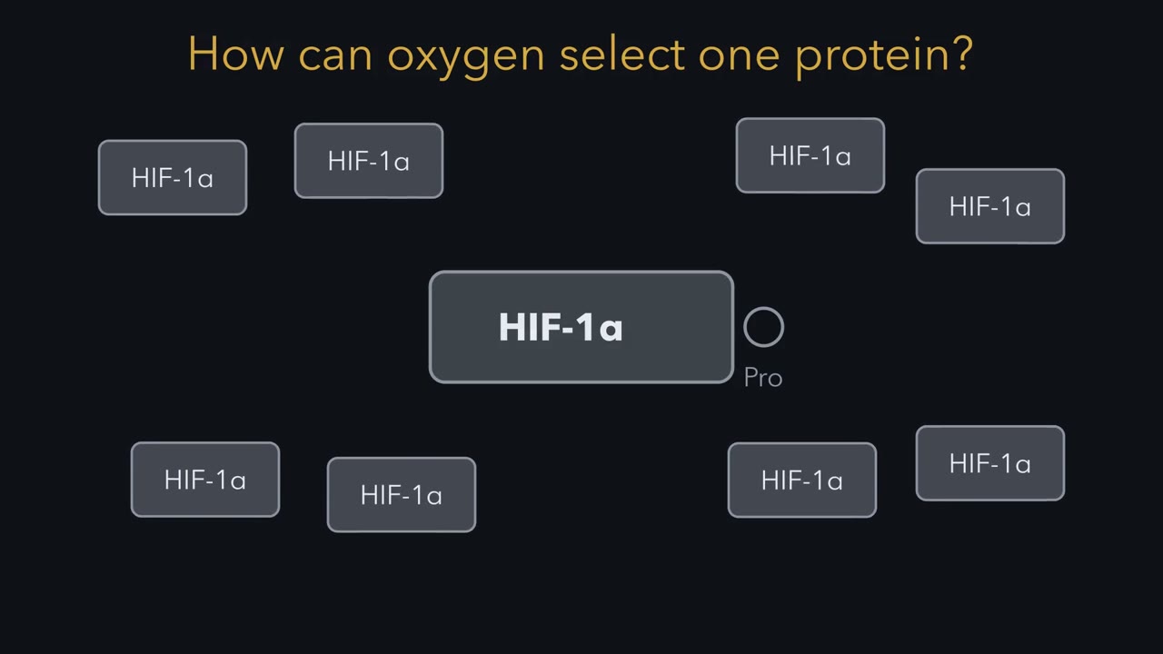 ▶ Watch How a Signal Marks One Protein · 3:29
Read the transcript of “How a Signal Marks One Protein”
Film transcript: How a Signal Marks One Protein
A cell holds many proteins. How can oxygen make it destroy HIF-1α and spare the rest? Watch the target, not the tag. Yellow ubiquitin is reusable. Grey proteins are possible targets. Green E3 is the selector. E1 spends ATP to activate ubiquitin, then holds it temporarily.
E2 receives and carries that activated tag. E3 brings loaded E2 beside a target it recognizes. Ubiquitin usually lands on a target lysine. The tag is common; recognition makes the transfer selective. The same relay can serve many targets because the selector can change. Different E3 receptors fit different target features. E1 and E2 keep ubiquitin moving. The E3 receptor decides which grey block enters the green grip. A degron is the target feature an E3 recognizes. It can be a short sequence, a chemical change, or both. Signals can create that feature or strengthen its contact. Signals change protein fate by changing what an E3 can recognize.
Now follow oxygen into this rule. HIF-1α is made continuously in the cells pictured here. When oxygen is available, a prolyl hydroxylase uses it as a reaction substrate. The enzyme adds a hydroxyl group to a proline on HIF-1α. That small badge completes a degron. Here are two HIF-1α targets. One carries the hydroxyl badge, and one does not. Which target will VHL grip?
VHL closes around the hydroxylated target. A peptide with hydroxyproline bound VHL tightly without cell extract. The same peptide with ordinary proline did not. The modification itself supplies the recognized feature. Once VHL holds HIF-1α, the ligase builds a ubiquitin chain. A red proteasome accepts the marked target and breaks it down. New HIF-1α keeps entering the system. With oxygen, removal keeps its level low. When oxygen falls, the hydroxylase completes its reaction less often. Fewer HIF-1α molecules gain the badge. VHL finds fewer targets, while synthesis continues.
HIF-1α accumulates, pairs with ARNT, and helps switch on low-oxygen genes. Notice what changed and what stayed shared. Oxygen changed the target feature VHL could read. It did not invent a new ubiquitin or proteasome. Signals change protein fate by changing what an E3 can recognize.
From the film
- Signals change protein fate by changing what an E3 can recognize.
- E1 activates ubiquitin, E2 carries it and an E3 brings it to a recognized target.
- In oxygen sensing, PHD hydroxylation lets VHL recognize HIF-1α.
- When oxygen falls, HIF-1α is hydroxylated less often and accumulates.
The first film showed oxygen marking one protein, HIF-1α, for destruction. HIF-1α is a transcription factor, a protein that switches genes on, and it turns on the genes a cell needs when oxygen runs short. This lesson is about that one-way switch and how a signal writes it.
One recognition step can change a protein’s fate
Ubiquitin is a small protein, 76 amino acids long, attached to other proteins as a tag. The proteasome is a barrel-shaped protease that destroys proteins carrying an appropriate ubiquitin signal. A degron is the feature the tagging machinery recognizes on a target: a short sequence, a chemical change or both.
The one idea. Signals change protein fate by changing what an E3 can recognize.
Scope: this rule covers the regulated ubiquitin pathways in this lesson. A signal may complete a degron or strengthen an E3-target contact. The mark built afterward also helps determine the outcome.
Proteolysis makes the choice costly to reverse. A phosphate can be removed, and a ubiquitin mark can be cut off before degradation. Once a protein is cut into peptides, only a newly made copy replaces it. Destroying an inhibitor can free a pathway until new inhibitor is made; destroying an activator can end a signal until it is replaced. A cell leaves mitosis by destroying cyclin B, which keeps it there.
The earlier lesson on protein turnover and amino acid catabolism covers proteasome mechanics and ubiquitin’s discovery.
The bonds and branches behind the relay
The film showed the handoff; here are the bonds. E1, the activating enzyme, spends ATP to join ubiquitin’s carboxyl end, glycine 76, to one of its own cysteines. That temporary thioester is an energy-rich sulfur-carbonyl bond. E1 passes ubiquitin to a cysteine on E2, the conjugating enzyme. E3, the ligase, completes the transfer, usually by an isopeptide bond: an amide between ubiquitin’s C-terminus and a target lysine side chain rather than the backbone.
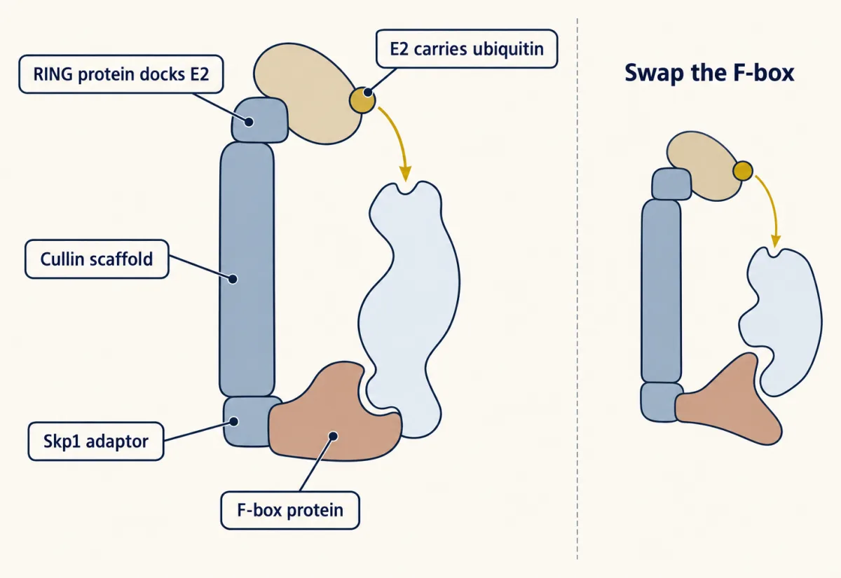
Human cells have two E1s, about forty E2s and roughly six hundred predicted E3s. One E1 supplies nearly the whole system, while each E3 recognizes a set of degrons. For the examples here, most target specificity sits in the E3 or its substrate receptor.
E3s have two main designs. A RING-type ligase holds loaded E2 beside the target, so ubiquitin passes directly across. A HECT-type ligase first takes ubiquitin onto its own cysteine. SCF ligases are RING-type: a cullin scaffold holds a small RING protein that docks E2, the adaptor Skp1 and an interchangeable F-box protein that binds the degron.
Predict. Mixed with ubiquitin-loaded E2, an E3 first carries ubiquitin as a short-lived thioester on one of its own cysteines before the target receives it. Is the E3 RING-type or HECT-type?
Check your prediction
HECT-type. A HECT ligase accepts ubiquitin from E2 onto its catalytic cysteine, then transfers it to the target. A RING ligase never holds ubiquitin itself; it promotes direct transfer from E2 to the target.
After the first ubiquitin, more can be added to a lysine on the preceding ubiquitin. Ubiquitin has seven lysines, and chains can also join through its first amino acid, methionine 1. Linkage, length and branching shape how a mark is read. A chain joined through lysine 48 and at least four ubiquitins long is a usual proteasome signal.
Signals write recognizable degrons
Some regulated targets carry an incomplete degron that a signal completes.
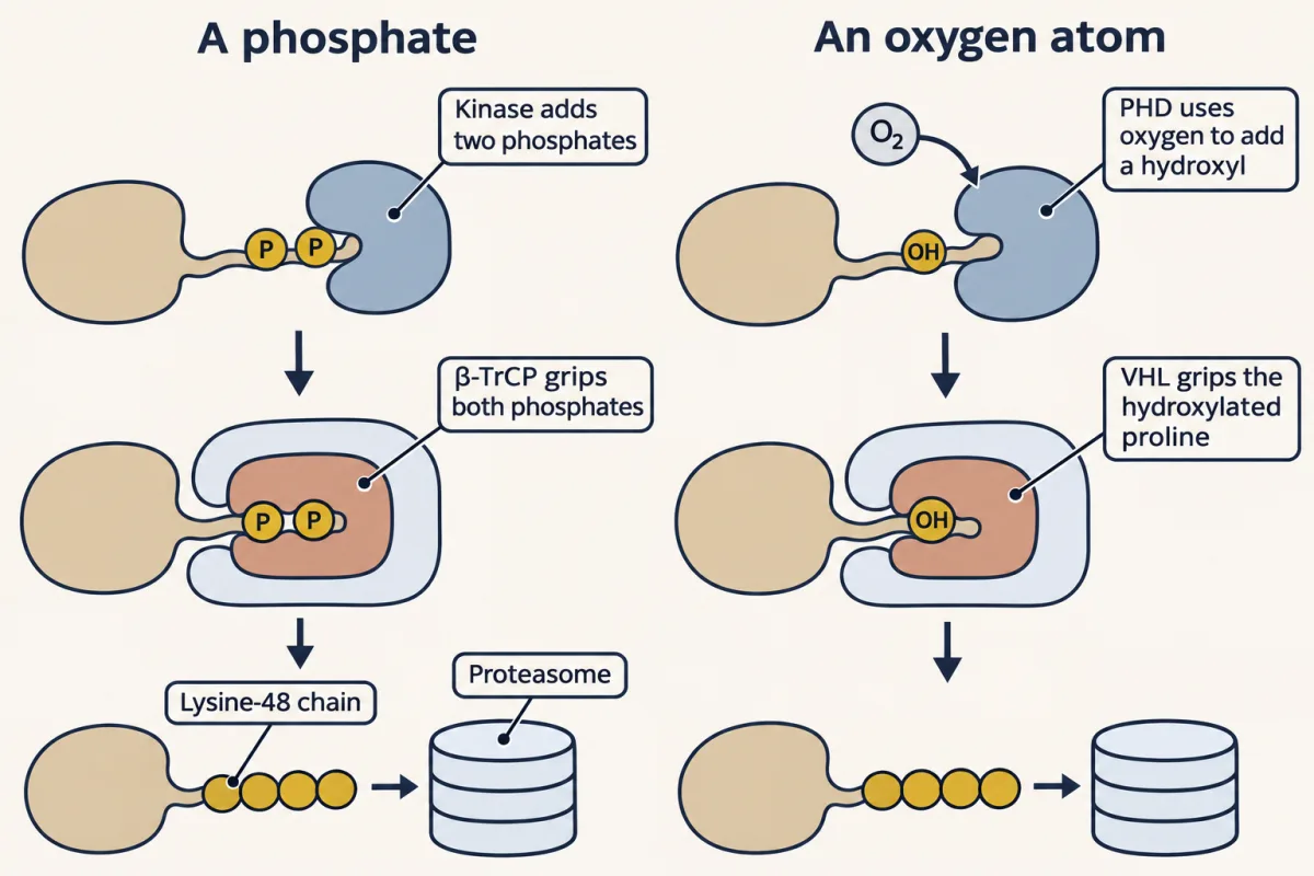
Phosphorylation can complete a docking site. Some inhibitors have a short sequence containing two serines. The F-box protein β-TrCP binds only when both serines are phosphorylated. A signal-activated kinase adds the phosphates, and β-TrCP brings the inhibitor to an SCF for a lysine-48 chain and proteasomal destruction. IκB, inhibitor of NF-κB, is the classic case, followed in the NF-κB lesson. The Wnt lesson uses the same logic for β-catenin, except its signal stops phosphorylation. The reversible phosphate reads the signal; destruction commits that target molecule.
Predict. Both degron serines are replaced by alanines, but the kinase and proteasome still work. What step fails first?
Check your prediction
Phosphorylation fails first: alanine has no hydroxyl, so the active kinase cannot add phosphates. Without them β-TrCP cannot recognize the degron, so the inhibitor never reaches the SCF.
Oxygen-dependent hydroxylation completes another docking site. Gregg Semenza found hypoxia-inducible factor 1 (HIF-1) while studying erythropoietin, the kidney hormone that tells bone marrow to make red blood cells. Its subunit HIF-1α was made continuously and destroyed rapidly when oxygen was available, so its standing amount changed with oxygen.
VHL is mutated in an inherited cancer syndrome, and cells lacking it made oxygen-regulated messages at high levels in ordinary air. VHL, the substrate receptor of a cullin-RING ligase, plays the role an F-box protein plays in an SCF, so it had to be reading some degron on HIF-1α. The film’s peptide test named it: a hydroxylated proline, recognized with no cell extract present.
Prolyl hydroxylases, or PHDs, add that hydroxyl. The reaction needs iron, 2-oxoglutarate and one molecule of oxygen. Oxygen is a substrate, used up like flour in bread; no separate oxygen receptor is required. Falling oxygen lowers how often PHDs finish the reaction, so VHL has less to grip and HIF-1α builds up while synthesis continues. It pairs with ARNT in the nucleus and switches on genes including VEGF, a growth factor that drives new blood vessels.
A close relative, HIF-2α, is destroyed through the same hydroxylation and VHL pathway. In the body it is the main factor switching on erythropoietin, and in kidney cancers lacking VHL it is the main tumour driver.
Keep the rule: HIF-1α is made all the time and destroyed all the time, so stopping the destruction, with low oxygen or with no VHL, raises it without any new transcription. In this sketch the level is relative to normal oxygen, the half-life of about five minutes applies only to normal oxygen, the destruction rate is taken as proportional to the oxygen level, and "on" means more than twice the normal level. The picture is a sketch of the logic, not a measurement. The lamp reads ON above 2 × normal, and destruction is set in proportion to oxygen: both are this sketch's choices. Where a slider changes nothing, the logic says nothing changes.
Production minus destruction
How fast can HIF-1α rise once destruction stops? Let a protein amount, P, be made at constant rate s and destroyed with first-order rate constant k:
dP/dt = s − k × P; at steady state P = s/k; half-life = 0.69/k.
A larger k gives a lower steady state and a shorter response time.
Predict. Synthesis stays fixed while k falls to half its original value. What happens to steady-state P and the half-life?
Check your prediction
Both double: the steady state becomes s/(k/2) = 2s/k, and the half-life becomes 0.69/(k/2), twice 0.69/k.
The second film asks what different ubiquitin marks make happen, then shows a hormone that changes target choice without modifying the target first.
Read the transcript of “One Tag, Different Destinations”
Film transcript: One Tag, Different Destinations
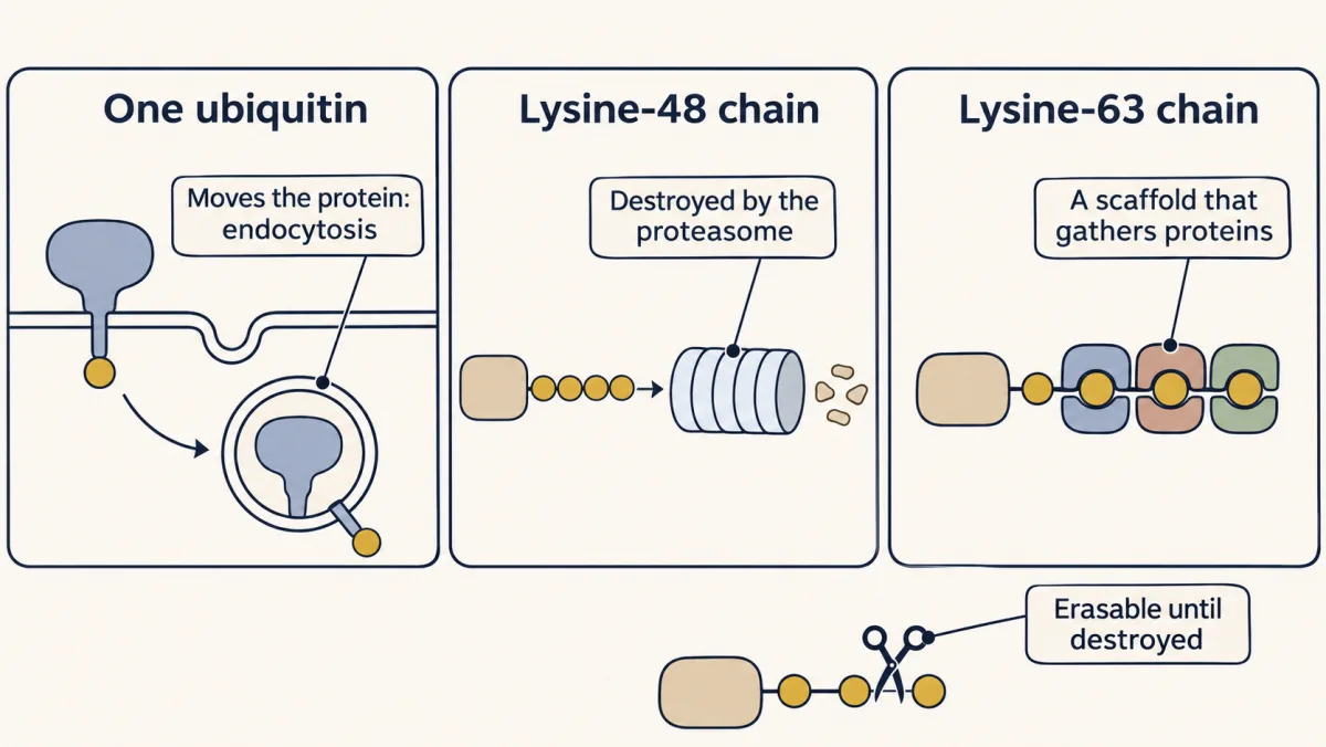
The first film showed oxygen changing what VHL could recognize. Why does one ubiquitin mark move a protein while another destroys it? Keep the same colors: yellow ubiquitin, grey targets, green E3 and red proteasome. Start with one ubiquitin attached to one lysine. This monoubiquitin mark seldom sends a protein to the proteasome. It more often changes location. On a growth-factor receptor, readers can help pull the receptor into the cell by endocytosis. Now reconnect the yellow blocks into the classic proteasome-directed linkage. A sufficiently long chain is a usual proteasome signal. The red barrel engages the marked protein and breaks it down.
This is the classic destruction route. Reconnect the same yellow blocks through another linkage, and the picture changes again. Ubiquitin-binding domains clamp onto this chain. Several proteins can gather around one scaffold. Signaling complexes and repair proteins at damaged DNA can assemble this way. Return to a receptor carrying only one yellow ubiquitin. Is that single mark enough to commit the receptor to the proteasome? Choose a destination before the picture moves.
Not usually. In this example, the single mark helps route the receptor into a vesicle. Movement and destruction can still cooperate later, because internalized receptors are often carried to lysosomes. The mark suggests an outcome; it does not guarantee one alone. So ubiquitin behaves as a context-dependent code. Linkage, chain length, branching, substrate and available readers all matter. A simple lookup table would show too much certainty. Keep the mark, its reader and the target together. A signal can also change recognition without modifying the target first. In plants, TIR1 is a green E3 receptor. An Aux/IAA repressor rests nearby, but their contact is weak without the hormone auxin.
Auxin fits into a pocket exactly where TIR1 and the repressor meet. The small gold signal fills their gap. Their grip tightens without building a separate cascade. Auxin acts as molecular glue at the target-receptor interface. The ligase now ubiquitinates the repressor, and the proteasome removes it. Auxin-responsive genes are released from repression. Here hormone detection and target recruitment happen in one binding step. The signal triggers destruction by tightening an E3 grip. These two ideas work together. The E3 chooses a recognized target, while the ubiquitin mark recruits particular readers. Ubiquitin marks route proteins; molecular glues change what an E3 grips.
Follow both questions to predict a target's fate.
From the film
- Ubiquitin marks route proteins; molecular glues change what an E3 grips.
- A single ubiquitin more often changes a protein's location than destroys it.
- A lysine-48 chain is a classic proteasome signal; a lysine-63 chain can assemble a complex.
- Auxin tightens TIR1's grip on a repressor, coupling detection to destruction.
Ubiquitin marks remain editable
Deubiquitinating enzymes cut ubiquitin off and can trim lysine-48 chains.
Predict. A deubiquitinating enzyme removes a target’s lysine-48 chain before the proteasome engages it. Can the same protein molecule still be rescued? What if proteolysis has begun?
Check your prediction
Before engagement, yes: chain removal rescues that molecule from this destruction signal. Once proteolysis has cut the protein, removing ubiquitin cannot rebuild it; a replacement must be made.
SUMO, a small ubiquitin-like modifier of about 100 amino acids, is folded like ubiquitin and attached to lysines by its own E1, E2 and E3. It usually changes binding or location, though some SUMO chains are read by ubiquitin ligases that send the protein to the proteasome.
Evidence that TIR1 is receptor and selector
The film called auxin a molecular glue, a small molecule that strengthens or creates a protein-protein contact. Auxin tells shoots which way to bend and roots where to grow. Botanists long sought its receptor at the cell surface, but the receptor behind its gene response is part of an SCF destruction machine.
Arabidopsis plants with a broken TIR1 gene respond poorly to auxin. TIR1 is an F-box protein, then thought to grab targets rather than sense hormones. In 2005 two laboratories independently showed that TIR1 binds auxin itself (see Going deeper). The relevant targets are Aux/IAA repressors, which hold auxin-responsive genes off.
A 2007 crystal structure from Mark Estelle’s group with Ning Zheng confirmed the film’s picture: auxin sits where TIR1 and an Aux/IAA repressor meet.
Predict. Auxin and TIR1 bind normally, but the proteasome is blocked. Will the repressor disappear and the genes switch on by this route?
Check your prediction
No. Recognition and ubiquitination can occur, but the repressor remains if the proteasome cannot destroy it, so the genes stay off.
Drug makers use the same principle. Lenalidomide, a thalidomide relative used in multiple myeloma, binds cereblon, a cullin-RING substrate receptor. The drug reshapes cereblon’s surface so it grips the transcription factors Ikaros and Aiolos, which it would otherwise ignore, and they are then ubiquitinated and destroyed. The drug was used clinically before this mechanism was known. PROTACs are two-headed molecules: one end binds a target, while the other binds a ligase such as VHL, using a small molecule built around the hydroxyproline VHL recognizes in HIF-1α.
Design problem
A transcription factor must be nearly absent when oxygen is plentiful and enter the nucleus within minutes when oxygen falls. Design the control. Why destroy it continuously rather than make it on demand?
Saved in this browser only.
One way to do it
Make the factor continuously and destroy it through an oxygen-dependent tag. Give it a five-minute half-life in air. Then k is about 0.14 per minute and 1/k about seven minutes, so the steady pool equals about seven minutes of synthesis.
In this simple model, when oxygen falls, hydroxylation and destruction stop while synthesis continues. Each minute adds s to the existing s/k pool, about 14 percent of baseline. After 30 minutes the pool is about five times baseline, without waiting for new transcription.
Making it on demand would wait tens of minutes to hours for transcription, RNA processing and translation. Continuous destruction also makes the off-switch as fast as the on-switch: when oxygen returns, the excess above baseline halves every five minutes, and after 25 minutes only about 3 percent of the excess is left. The cost is ATP and amino acids spent on discarded protein.
Going deeper
How VHL was tied to oxygen
Peter Ratcliffe’s group showed in 1999 that HIF-1α destruction depends on VHL and occurs efficiently when oxygen is available. William Kaelin reached VHL from the other direction, through the inherited cancer syndrome above.
The degron and oxygen bookkeeping
In 2001 the two competing laboratories independently identified the hydroxylated degron. Proline 564 in human HIF-1α is one site; proline 402 is modified too. One oxygen atom enters the new hydroxyl. The other enters succinate when 2-oxoglutarate loses carbon dioxide.
Where target choice really sits
E2-E3 pairing and the rest of the machinery can also influence ubiquitin transfer and chain architecture, so the E3 is the main selector in these pathways, not the only one.
The auxin receptor papers
On 26 May 2005, Estelle’s laboratory at Indiana University and, independently, Ottoline Leyser and Stefan Kepinski at the University of York reported in the same issue of Nature that TIR1 binds auxin itself.
Your notes
Select any text in the lesson to highlight it or add a note to it. Your highlights and notes are saved in this browser and collected in your notebook.
This browser is not letting the site save anything (a private window or strict privacy settings), so highlights and notes will be lost when you leave the page.
How we measure it
Forward genetics with a hormone-resistant mutant
Treat plants so that their genes mutate, then look for individuals that respond abnormally to the hormone or to a drug that disturbs it. TIR1 came from a screen for plants resistant to a drug that blocks auxin transport, and the mutants turned out to respond poorly to auxin. The method finds the parts of a pathway without needing to guess in advance what they are.
Northern blot
RNA from cells is separated by size on a gel, transferred to a membrane and probed with labeled DNA for one gene. Band intensity reports how much messenger RNA of that gene the cells contain. Comparing cells with and without VHL at 21 and 1 percent oxygen showed that VHL is needed for the oxygen dependence.
Binding assay with a synthetic peptide
Chemists make a short peptide that carries exactly the modification they want, such as a hydroxyproline, and test whether a purified partner such as VHL binds it. With a defined peptide and no cell extract, the modification itself is shown to be the signal.
X-ray crystallography
A crystal of a protein complex scatters X-rays, and the pattern gives the positions of the atoms. Crystals of TIR1 grown with auxin and a repressor peptide showed the hormone sitting between the two proteins.
Practice questions on this lesson (3) → MCAT-style, original, each with an explanation
Where this lesson fits
Before thisReview first
- Phosphorylation: Kinases and PhosphatasesThis course · Lesson 6
You need to know that a phosphate can be added and removed to see why destruction is a different, one-way switch.
- GTPase Switches: The TimerThis course · Lesson 7
It shows how a switch gets its timing, which helps you see why a pathway might use a timer or a one-way step.
After thisStudy next
- Wnt: β-Catenin and Planar PolarityThis course · Lesson 24
It applies the phosphate-then-destruction mark to β-catenin, where the signal works by stopping the phosphorylation.
- Innate Immunity: Toll-like Receptors and NF-κBThis course · Lesson 28
It uses the phosphorylated-inhibitor degron on IκB, which β-TrCP and an SCF ligase send to the proteasome.
- Stress Signals: p38 and JNK, p53, Unfolded Proteins and OxidantsThis course · Lesson 33
It takes up p53, which the cell builds and destroys rapidly until a signal protects it.
Elsewhere on the siteFilms, simulations and method films in other courses
- Choose, Recycle, SplitFilm · Biochemistry
It shows how E3 ligases give ubiquitin tagging its target specificity and how the proteasome handles tagged proteins.
- One Rise, One FallFilm · Cell Biology
Cyclin is destroyed at every division, a clear case of regulated destruction used as a one-way step.
- A Brake Wired to a SensorFilm · Cell Biology
Destruction of securin is a one-way trigger for the next cell-cycle step.
Suggested by an AI model from the lessons themselves, then checked. How we use AI
Finish line
Test yourself
8 questions
Practice and MCAT-style, with explained answers.
Next · Lesson 9
Protein Modules: SH2, SH3, PTB, PDZ and PH
A receptor has just phosphorylated its own tyrosines. What reads the marks, and how does each reader pick its mark?
Continue →