Organismal Biology · Unit 1: Organisms and their diversity
New to this? Review it at high school level: Living or Not? Viruses and Bacteria, Domains and Kingdoms
Microbes, Protists and Fungi
Rewritten on : new text, flashcards and film. Your notes and highlights stay, but the text around them may have moved.
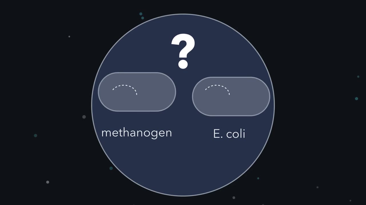
A methane-making microbe and E. coli look alike under a microscope. How can we tell whether they are close relatives?
Pause when the reading reaches a film, and offer to play it. Saved on this device.
Watch the film now, or keep listening
Builds on:What an Organism Has to DoSize, Scale and SurfaceDomains and KingdomsWhy these?
 ▶ Watch Look Alike, Far Apart · 3:18
Read the transcript of “Look Alike, Far Apart”
Film transcript: Look Alike, Far Apart
A methanogen and E. coli look almost the same under a microscope. Are they close relatives? Both are small cells with no nucleus. A methanogen makes methane, and this one lives in a cow's rumen. For a long time, biologists lumped all cells without a nucleus together as prokaryotes. But that grouping rests on something missing. Two cells can both lack a nucleus and still be distant relatives. Carl Woese looked for a part that every cell carries, doing the same job in each. He chose the ribosome, the particle that makes proteins.
Inside its small subunit lies a strand of RNA. His laboratory pulled this small-subunit ribosomal RNA from many microbes. They cut each strand into pieces and compared the pieces, organism by organism. Each pair got a similarity score. Here is the logic. Small changes pile up in the RNA as lineages separate. So fewer differences usually mean a more recent common ancestor. Now the scores fill a grid, one cell for every pair. A stated grouping method can then propose a tree from the grid. Different methods make different assumptions, so the grid alone does not fix one tree. Methanogens look like bacteria. So where do you think they landed on the tree?
Close to E. coli, or somewhere else? Not among the bacteria. Woese and George Fox reported that methanogens formed their own group. It sat about as far from bacteria as from eukaryotes, the cells with a nucleus. They first called the new group archaebacteria.
Later, Woese and colleagues proposed three domains: Bacteria, Archaea and Eukarya. Those names are still the standard labels. What do the scores tell us, and what don't they? They rank closeness. They do not date a split, and they do not reveal an ancestor. Across the deepest splits, repeated changes at one site can blur the signal in a single molecule. Many genes and better models of change sharpen the branching pattern. But which branch came off first also needs an outside reference. Most recent trees of this kind place eukaryotes inside the archaea, closest to a group called the Asgard archaea. Exactly where is still debated. On that view, life has two primary branches, Bacteria and Archaea.
So, back to the methanogen and E. coli. Their looks hid how far apart they are. A molecule every cell shares revealed it.
From the film
- Cells that look alike can be distant relatives; looks do not reveal descent.
- Woese compared small-subunit ribosomal RNA, a molecule every cell carries, and scored each pair of organisms.
- Methanogens formed their own group, about as far from bacteria as from eukaryotes: the archaea.
- Most recent trees built from many genes place eukaryotes inside the archaea, closest to the Asgard archaea; the exact position is debated.
- Fewer differences usually mean a more recent common ancestor; the scores rank closeness but do not date splits or identify an ancestor.
The first film answered the opening question: a methane-producing microbe (a methanogen) and the gut bacterium Escherichia coli belong to different domains. This text adds what the film left out, then asks how bacteria divide and swap genes, why protist is only a convenience word, how fungi feed and partner, and why viruses stand apart. It builds on what an organism has to do and on the surface and size limits in size and scaling.
The one idea. Relatedness is read from differences in a molecule every cell shares, not from how a cell looks: small changes pile up in small-subunit ribosomal RNA as lineages separate, so fewer differences usually mean a more recent common ancestor.
Scope: cells only; the last section shows why. “Usually,” because changes pile up faster in some lineages than others, and over the deepest splits repeated changes at one site blur the count. So the scores rank closeness roughly; they do not date splits or identify an ancestor, and the deepest splits need many genes and models of change.
Three domains from one molecule
Every cell has a plasma membrane (the lipid boundary of the cell), cytoplasm (the contents inside the membrane, outside any nucleus), DNA as its genetic material, and ribosomes, the particles that make proteins. A eukaryote is a cell, or an organism made of cells, with a nucleus: a membrane-enclosed compartment holding the DNA. Animals, plants, fungi and protists are eukaryotes. Bacteria and archaea have no nucleus, and for a long time they were lumped together as prokaryotes, the “before the nucleus” cells. Eukaryotic organelles are in the measured eukaryotic cell.
Three things the film left out:
- The data were pieces, not whole sequences. In 1977 Woese and George Fox compared catalogues of short RNA pieces, scored pair by pair (see Going deeper).
- Eukaryotes were compared by their cytoplasmic RNA. Mitochondria and chloroplasts carry their own bacteria-like ribosomal RNA, so the comparison used the ribosomal RNA of the cytoplasm.
- A second, independent line of evidence agrees. Archaea also differ from bacteria in their membranes and walls (below).
The three domains proposed in 1990 by Woese, Otto Kandler and Mark Wheelis, Bacteria, Archaea and Eukarya, are still the standard labels, even though many-gene trees nest eukaryotes inside the archaea, closest to the Asgard archaea, a group first known from DNA in marine sediments. Whether eukaryotes branch within the Asgard group or beside it, and which Asgard lineage is closest, is still debated. None of these comparisons observed an ancestor or showed how eukaryotic cells arose. See reading phylogenetic trees and history of life.
The second line of evidence is chemistry. Archaeal membrane lipids are branched isoprenoid chains joined to glycerol by ether bonds, where bacteria and eukaryotes mostly use fatty acids joined by ester bonds (see membrane structure and function). Ether bonds alone do not mark archaea, since some bacteria and some eukaryotic lipids carry them too. The archaeal mark is the combination: isoprenoid chains, ether bonds, and a glycerol that is the mirror image of the bacterial one, like a left hand to a right. Archaeal walls also lack peptidoglycan, the mesh of sugars and short peptides that forms most bacterial walls. Some methanogens have a different but similar polymer, pseudomurein; many archaea are covered only by a surface layer of protein. Nor are archaea simply “extreme bacteria”: many live in soils, oceans and sediments, and methanogens live in the guts of cattle and humans.
Bacteria: shape, envelope, fission and gene swapping
Bacteria come in three common shapes: cocci (spheres), bacilli (rods) and spirilli (spirals). A cell of E. coli is a rod roughly 1 micrometre (um) wide and 2 um long.
The cell envelope is everything that surrounds the cytoplasm. In 1884 Hans Christian Gram published a stain that sorts bacteria by their envelopes. In the method used today, a smear is stained with crystal violet, treated with iodine, washed with alcohol and counterstained with red safranin. Gram-positive cells, such as Staphylococcus aureus and Bacillus subtilis, have a thick peptidoglycan wall that keeps the crystal violet-iodine complex through the wash, so they stay purple. Gram-negative cells, such as E. coli, have a thin peptidoglycan wall beneath an outer membrane; the wash removes the violet and the safranin turns them pink. Not every bacterium has a wall at all: Mycoplasma species lack peptidoglycan and give no useful Gram reaction.
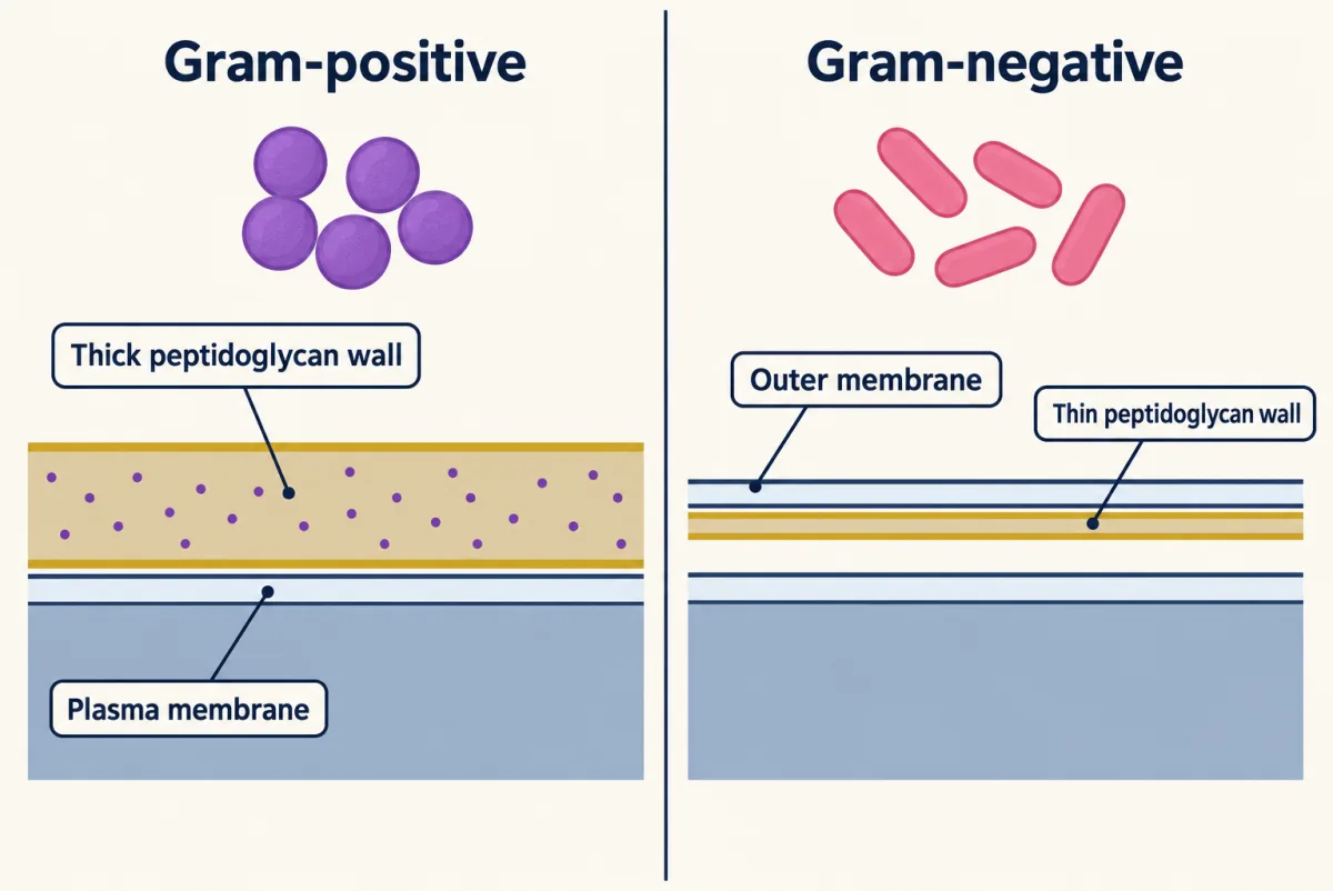
Predict. Archaea have no peptidoglycan, yet some of them stain purple. Could a Gram stain tell you which domain a new cell belongs to?
Check your prediction
No. Gram results for archaea are not fully consistent. In some methanogens an intact pseudomurein wall keeps the dye, so the cell stains purple, but in archaea the colour does not by itself report how thick the wall is or what it is made of. The stain reads features of the envelope, not the domain, and a purple or pink cell can belong to either.
Bacteria and archaea usually reproduce by binary fission: the cell copies its DNA, grows, and divides into two. The doubling time, or generation time, is the time one round takes. Bacteria also gain genes from other cells: horizontal gene transfer. In transformation a cell takes up DNA from its surroundings. In conjugation DNA passes between two cells in contact. In transduction a bacteriophage, a virus that infects bacteria, carries bacterial DNA from one cell to another. Their genetics are in bacterial and phage genetics, and the experiments showing DNA is the genetic material in DNA, the genetic material.
Worked problem: how fast does fission add cells? In rich laboratory medium at 37 degrees C, E. coli typically doubles about every 20 minutes. Start with 100 cells. How many are there after 3 hours?
Three hours is 180 minutes, or 180 / 20 = 9 doublings, and each doubling multiplies the count by 2: 100 x 29 = 100 x 512 = 51,200 cells. In general, if every cell divides once per doubling time, none die, and food and space never run short (exponential growth),
N = N0 x 2g, where g = elapsed time / doubling time
g counts generations; 2g is how many-fold the population has grown.
Predict. The Asgard archaeon MK-D1 doubles roughly every 20 days. Starting from the same 100 cells, how long would it need to reach 51,200?
Check your prediction
The same nine doublings, on a slower clock: 9 x 20 days = 180 days. Growth is set by the number of generations, not by the hours. In E. coli’s 3 hours, MK-D1 grows by a factor of only about 1.004. And if a culture shows the reported lag phase, 30 to 60 days before it begins to grow at all, the dish would need about 210 to 240 days.
Twenty days is 20 x 1,440 = 28,800 minutes, so an MK-D1 generation lasts 28,800 / 20 = 1,440 times as long as an E. coli one.
- Generations
- Doublings
- Cells
- The same number of doublings on each clock
In this sketch: exponential growth, every cell divides once per doubling time, none die, food and space never run short, constant doubling time, with the curve drawn smoothly so a count between whole doublings is an average; counting for MK-D1 starts after its lag.
Rule to keep: g = elapsed time / doubling time, and N = N0 × 2g, so equal numbers of doublings give equal cell numbers; only the clock differs. This is a sketch of the logic, not a prediction for a real culture.
The model fails once food runs short or cells die. Beyond the lab, each generation is another chance for a mutation or a transferred gene to be copied, so a short generation gives a population more generations in which to change.
Protists: a word for “everything else”
Protist is an informal name for any eukaryote that is not an animal, a plant or a fungus, not a natural group: protists belong to many separate branches of the eukaryote tree, and their classification keeps changing. Nor are protists “early” eukaryotes; every living lineage has had as long to evolve as animals have.
Compare four ways of life:
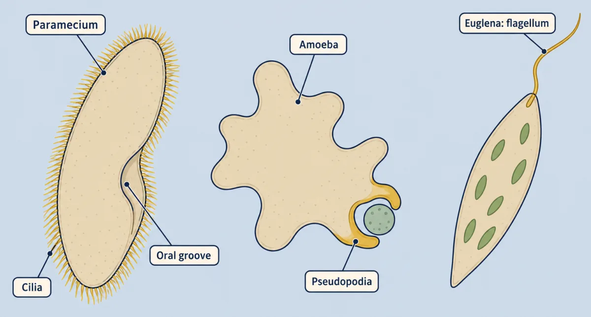
- Paramecium swims and sweeps food into an oral groove with cilia, many short hair-like projections that beat together.
- Amoeba crawls and engulfs prey by extending pseudopodia, temporary bulges of cytoplasm.
- Euglena swims with one long flagellum, a whip-like projection (a second stays short, inside the cell), and is mixotrophic: it can photosynthesize in light and absorb organic food when light is poor.
- Plasmodium, the malaria parasite, passes between mosquitoes and vertebrates including humans, and in humans multiplies inside liver and red blood cells.
None of these four is closer to the others because it lacks something, and even mitochondria are not universal: the gut protist Monocercomonoides has no detectable mitochondrion, and its genome suggests the organelle was lost in that lineage, not that it never had one.
Fungi make the same point the other way. They were long grouped with plants, but their sequences put them closer to animals: the shared molecule, not the look, places an organism. Yet a fungus eats like neither a plant nor an animal.
Read the transcript of “Digest Outside, Fuse in Two Steps”
Film transcript: Digest Outside, Fuse in Two Steps
Sequences put fungi closer to animals than to plants. Yet a fungus eats like neither. So how does a mould on bread eat, with no mouth and no gut? Watch a spore land on the bread. It grows a single thread, called a hypha. The hypha branches again and again into a mass called a mycelium. That fuzz is the mould. Each thread is wrapped in a wall that contains chitin, a tough polymer also found in insect exoskeletons. Fungi do not photosynthesize. So this mould's food must come from the bread. At the growing tip, the hypha releases enzymes into the bread. The enzymes cut starch, a large molecule, into small sugars outside the fungus. Then the hypha takes the sugars in.
So a fungus digests first and takes food in second. Most animals do the reverse. They take food into a gut, and digest it there. A thin thread suits this way of eating. Every part of its cytoplasm stays close to the surface where the food comes in. Some fungi skip the threads. A yeast is a single cell. Baker's yeast reproduces by budding, pinching a small daughter cell off a larger one. Now, how many fungi mate. Two hyphae of different mating types grow toward each other and meet. Their cytoplasm joins. This step is called plasmogamy. Each partner brought a haploid nucleus, carrying one set of chromosomes.
Now the joined cell holds both nuclei, one from each partner, side by side. Two sets of chromosomes, in one cell. So is this cell diploid? Think about where the two sets are. Not yet. A diploid nucleus holds both sets in one nucleus. Here the two sets sit in two separate nuclei. A cell like this is called a dikaryon. In mushroom-forming fungi, this stage can last a long time. Only a later step fuses the two nuclei into one diploid nucleus. That step is karyogamy. Plasmogamy joins cells. Karyogamy joins nuclei. Then meiosis follows, and the cycle comes back to where it began. It makes haploid spores, each carrying one set of chromosomes again.
So a fungus eats by digesting outside itself and absorbing what it frees. And it mates in two steps. Cells fuse first, and nuclei later.
From the film
- A fungus grows as threads called hyphae, or as single yeast cells, with walls containing chitin.
- Fungi release enzymes outside themselves and absorb the small molecules those enzymes free.
- Plasmogamy joins two partners' cytoplasm; karyogamy fuses their nuclei later.
- In between, a cell with two separate haploid nuclei is a dikaryon, not a diploid cell; in mushroom-forming fungi this stage can last a long time.
- Meiosis then makes haploid spores.
Fungi digest outside the body and partner with roots and algae
Beyond the second film’s bread mould: fungal walls hold glucans as well as chitin, a mycelium spreads by growing at its hyphal tips, and baker’s yeast is Saccharomyces cerevisiae.
The film’s mating cycle, from plasmogamy through the dikaryon and karyogamy to meiosis, is a schematic found in many fungi, not the bread mould’s own (see chromosomes and meiosis). How long the dikaryon lasts varies: in many fungi, including mushroom-forming species, each cell keeps its two nuclei for long periods before karyogamy. Some fungi have no known sexual cycle at all.
Fungi also live in partnerships. Mycorrhizae are associations between plant roots and fungi. In arbuscular mycorrhizae the fungus grows into cells of the root cortex (the layer of cells between the root’s surface and its central core), inside the cell wall but outside the cell membrane, and forms branched exchange structures called arbuscules; the plant membrane folds around each one, so the two never share cytoplasm. The mycelium extends far into the soil, and plant and fungus trade carbon from the plant for mineral nutrients, especially phosphorus, from the fungus.
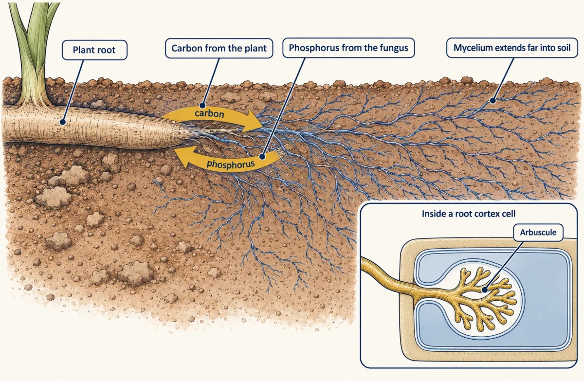
Predict. A crop is grown in soil heavily fertilized with phosphate. Does it gain more or less from its mycorrhizal fungus?
Check your prediction
Usually less, if the fertilizer raises the phosphorus the plant can actually take up (soil chemistry can lock phosphate away). Then the fungus’s phosphorus benefit falls, colonization often declines, and the carbon the plant pays can exceed that benefit. Total benefit need not always fall, because the fungus may still supply other nutrients or protection against stress.
Lichens pair a fungus with a photosynthetic alga or cyanobacterium. For the ecology, see mutualism, parasitism and coevolution, nutrient cycling and soil, nutrients and partners.
Viruses as the contrast case
A virus is a genome, either DNA or RNA, usually enclosed in a protein coat called a capsid. Bacteriophage T2, which infects E. coli, has a DNA genome; influenza viruses have RNA genomes. A virus has no ribosomes and no cell structure, and it is copied only inside a host cell, using that cell’s machinery.
Predict. Where would you place a virus on a tree built from small-subunit ribosomal RNA?
Check your prediction
Nowhere. A virus has no ribosomes, so it has no small-subunit ribosomal RNA to compare, which is why the one idea is scoped to cells.
This course defines an organism as a living individual made of one or more cells. On that definition a virus is not an organism. That is a choice of definition, not a final verdict on what counts as life. How animals detect and fight microbes and viruses is in innate immunity.
Design problem
A student cultures a microbe from a cow’s rumen. Under the microscope it is small and has no nucleus. How would you decide whether it is a bacterium or an archaeon?
Saved in this browser only.
One way to do it
Start with the molecule Woese used: score its small-subunit ribosomal RNA against known bacteria, archaea and eukaryotes. A methanogen groups with the archaea, scoring about as low against E. coli and Bacillus as against the cytoplasmic RNA of yeast.
Then check the second line of evidence: lipids for the archaeal combination, and the wall for the absence of peptidoglycan. The RNA comparison is the main test, and lipids and wall should agree with it. Looking or staining like a bacterium tells you nothing about the answer.
Going deeper
How Woese compared the RNA
Woese’s laboratory at the University of Illinois and its collaborators cut the RNA of methanogens and typical bacteria with an enzyme, ribonuclease T1, into short pieces and catalogued the sequence of each; a pair of organisms scored higher when their catalogues shared more pieces. Beyond the film’s point that a grid of scores does not fix one tree, placing the root needs an outside reference or a model of change. The 1990 paper spelled the third domain Eucarya.
Plant and fungus rewards: a contested mechanism
Kiers and colleagues reported in 2011 that, in experiments with the barrel medic Medicago truncatula and arbuscular mycorrhizal fungi, plants allocated more carbohydrate to fungal partners that cooperated better, and fungi passed more nutrient to roots that supplied more carbohydrate. Their full methods were not part of this lesson’s sources. Later work questions how general the result is: a 2024 review concluded that current evidence does not show plant carbon transfer to be directly regulated by the nutrients a fungus delivers. How would you test whether a root system rewards the better fungal partner, and whether a fungus rewards the better root?
One way to do it
Give one root system two fungal partners that differ in how much phosphorus they deliver, one generous and one poor.
Label each resource. Feed the leaves carbon dioxide carrying a carbon isotope and measure how much label reaches each fungus. Give each fungus phosphorus carrying a separate label, placed in a compartment its hyphae can reach but roots cannot, and measure how much reaches the plant.
Add controls. A no-choice control colonizes plants with only one fungus each, which shows whether a fungus draws more carbon wherever it grows or only when the plant can compare partners. In a second run, swap which fungal species is the generous one, separating the partner’s behaviour from its species identity.
Predictions: if plants reward the better partner, the generous fungus receives more carbon in the mixed plants than the poor one does, beyond any difference in the no-choice plants. If fungi reward the better root, more phosphorus flows to roots that supply more carbon.
Limit the conclusion to the species tested: a result in Medicago and two fungi does not cover all mycorrhizal partnerships.
Exceptions and updates
A 2019 revision of eukaryote classification by Sina Adl and colleagues dropped Excavata as a formal supergroup.
A few viruses of fungi have no capsid at all.
The slowest clock: MK-D1
The Asgard archaeon MK-D1, grown by Hiroyuki Imachi and colleagues in 2020, had a doubling time estimated at about 14 to 25 days. The worked problem’s 20 days sits inside that range. Its 30 to 60 day lag came before any growth, and growth was measured as qPCR gene copies, not cell counts.
Your notes
Select any text in the lesson to highlight it or add a note to it. Your highlights and notes are saved in this browser and collected in your notebook.
This browser is not letting the site save anything (a private window or strict privacy settings), so highlights and notes will be lost when you leave the page.
How we measure it
Gram staining
Stain a smear of bacteria with crystal violet, fix the dye with iodine, wash with alcohol, then counterstain with safranin. Bacterial cells that stay purple have a thick peptidoglycan wall (Gram-positive); cells that turn pink have a thin wall and an outer membrane (Gram-negative). Cells with no wall, such as Mycoplasma, give no useful reaction.
Comparative ribosomal RNA analysis
Extract the RNA of the small ribosomal subunit from many organisms, read part or all of its sequence, and score each pair by how similar the sequences are. A grouping method applied to these scores proposes a tree of relationships that does not depend on how the cells look.
qPCR growth monitoring
Measure the number of copies of one organism's ribosomal RNA gene in a culture over time by quantitative PCR. A straight line on a semilogarithmic plot gives a doubling time even for a slow microbe in a mixed culture, though gene copies are not the same as a direct cell count.
Fluorescence in situ hybridization with microscopy
Add a fluorescent probe that binds the ribosomal RNA of one lineage, then look at intact tissue under a fluorescence microscope. It shows where cells of that lineage sit, as in the yeast cells found in the outer layer of some lichens.
Method films · the methods library
Practice questions on this lesson (3) → MCAT-style, original, each with an explanation
Where this lesson fits
Before thisReview first
- What an Organism Has to DoThis course · Lesson 1
This lesson builds on what every organism must do, such as exchanging materials and growing, before comparing microbes.
- Size, Scale and SurfaceThis course · Lesson 2
The small size of bacterial cells and their surface-to-volume limits are used here without being re-taught.
- Domains and KingdomsBiology · High school
You should already know the three domains and how to sort a cell into one before this deeper comparison.
After thisStudy next
- Plants on LandThis course · Lesson 5
The fungal cycle of fusion and meiosis prepares you to read haploid and diploid stages in plant life cycles.
- Soil, Nutrients and PartnersThis course · Lesson 24
It takes the mycorrhizal partnership between roots and fungi further, with phosphorus uptake from soil.
- Innate Immunity: Recognize and RespondThis course · Lesson 17
It shows how the body detects and fights the bacteria and viruses introduced here.
Elsewhere on the siteFilms, simulations and method films in other courses
- The Molecule Behind the TreeFilm · Biology
It shows how ribosomal RNA comparison redrew the tree of life when looks failed to show ancestry.
- Bacterial and phage geneticsMethod film
It covers how bacteria swap genes, the gene exchange this lesson names beside fission.
Suggested by an AI model from the lessons themselves, then checked. How we use AI
Finish line
Test yourself
8 questions
Practice and MCAT-style, with explained answers.
Next · Lesson 4
Animal Body Plans: Similar Bodies, Different Branches
A nematode and an earthworm are both soft, tube-shaped worms with a gut from mouth to anus. Genes put the nematode with insects and other moulting animals, and the earthworm with snails and flatworms. What does a body plan tell you, and what does it not?
Continue →