Organismal Biology · Unit 5: Defense, movement and reproduction
New to this? Review it at high school level: Germs, Vaccines and Antibiotics
Innate Immunity: Recognize and Respond
Rewritten on : new text, flashcards and film. Your notes and highlights stay, but the text around them may have moved.
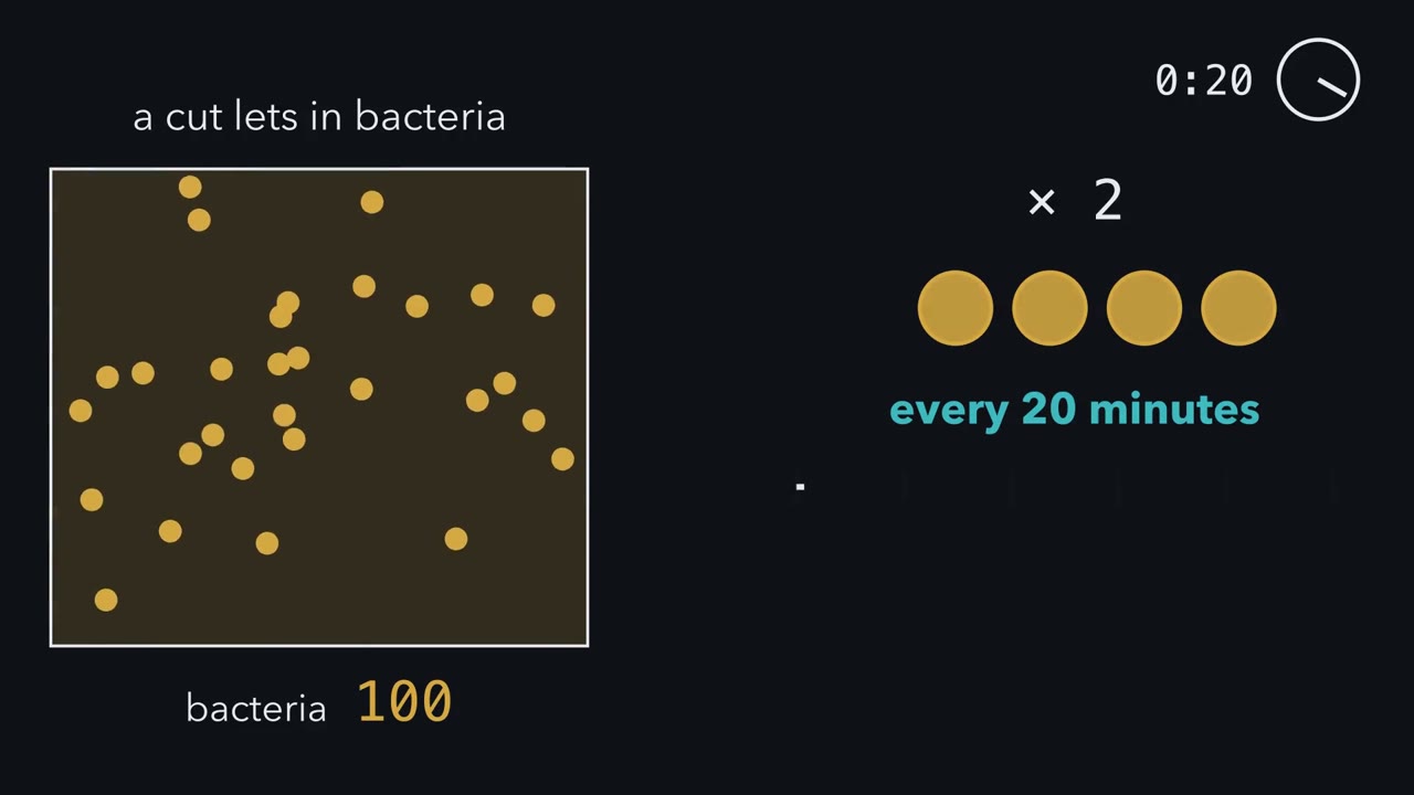
A cut lets in 100 bacteria that double every 20 minutes, and new antibodies take days. What does your body have to do in the first few hours to stay ahead?
Pause when the reading reaches a film, and offer to play it. Saved on this device.
Watch the film now, or keep listening
Builds on:Germs, Vaccines and AntibioticsHow Microbes MultiplyTissues, Organs and HomeostasisWhy these?
 ▶ Watch Winning the First Hours · 3:17
Read the transcript of “Winning the First Hours”
Film transcript: Winning the First Hours
A cut lets in 100 bacteria, and each one doubles every 20 minutes. New antibodies take days, so what has to happen in the first few hours? Start with no defense at all. Every 20 minutes, the count doubles. The clock runs for four hours, and the counter climbs past 400,000. Now add a defense. In each doubling time, it removes a fraction of the bacteria. Call that fraction k. The survivors then double, just as before. So each doubling time multiplies the count by 2 × (1 − k). That factor is the whole race. Above 1, the count grows. Below 1, it shrinks. Suppose the defenses remove three quarters of the bacteria every 20 minutes. Does the count rise or fall?
Decide before the counter moves. It falls. The factor is 1/2, so every 20 minutes the pile halves. After four hours, the expected count is far below one bacterium. The infection is very likely cleared. Now slide the dial down. At exactly one half removed the factor is 1, and the counter freezes. So in this model, the rule is simple: remove more than half every doubling time. But when does the removal start? Keep the same strong defense and make it wait one hour. The pile reaches 800 before it turns. Wait two hours, and it reaches 6,400.
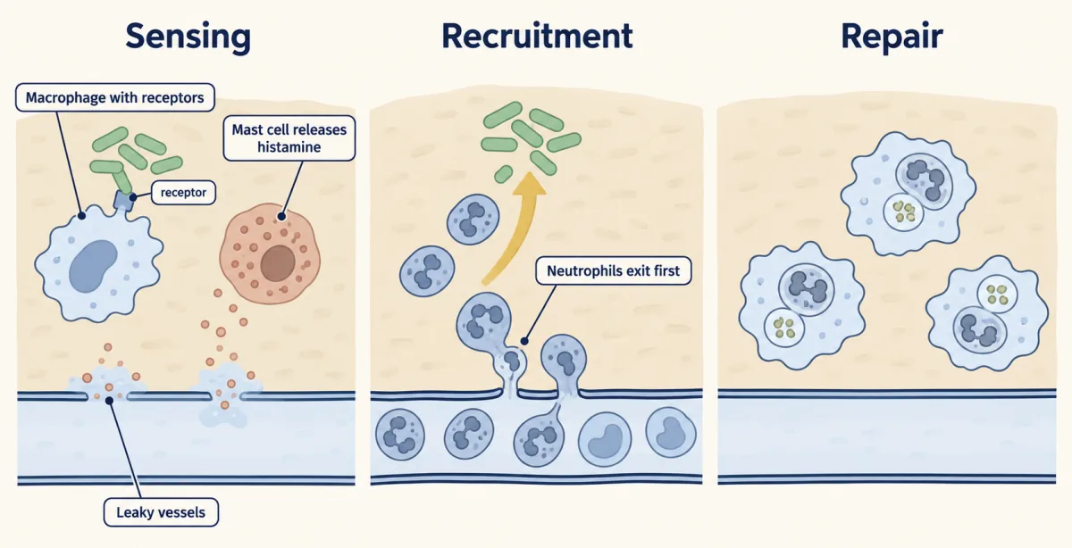
Each hour of waiting multiplies the pile eightfold. That is why the receptors must be ready before infection. In this model of a bacterial wound, the defenses we follow do three jobs. First, fewer get in. Skin and other barriers lower the starting number. Second, the clock starts early. A macrophage, a long-lived eating cell in the tissue, carries ready-made receptors. One, Toll-like receptor 4 senses a molecule from the outer membrane of many bacteria. Third, more are removed per doubling.
Sensing sets off inflammation. A nearby vessel widens and leaks. Neutrophils pour out first, then monocytes, which become macrophages. Inflammation runs in four steps: sense, vessels change, cells arrive, then clear and repair. As the eating cells gather, the fraction removed in each doubling time climbs. Keep them out, see them at once, and remove more than half every doubling time. This is an illustrative model of one wound, not a measured infection.
From the film
- In the first hours of an infection, defenses race bacteria that can double every 20 minutes or so, long before new antibodies can be made.
- If defenses remove a fraction k of the bacteria each doubling time, the count changes by 2 × (1 − k) per doubling, so it shrinks only when more than half are removed. This is an illustrative model, not a measured wound.
- Start early: each hour of delay lets bacteria that double every 20 minutes grow eightfold before killing begins, leaving a bigger pile to bring down, which is why innate receptors are ready before infection.
- In this model of a bacterial wound, the defenses the film follows do three jobs: barriers lower the number that get in, ready-made receptors start the response at once, and inflammation brings neutrophils and then monocytes that remove more per doubling.
Innate defenses are inherited and ready before an organism meets a particular microbe. Adaptive immunity builds new antibodies and T cells against each microbe, which takes days the first time. Animals and plants meet microbes constantly, and those days belong to innate defenses. They are not “nonspecific”: their receptors recognize particular classes of microbial molecules.
The one idea. Early in an infection, innate defense is a race against doubling. If defenses remove a fraction k of the bacteria in each doubling time (the time one bacterium takes to become two), each doubling time multiplies the count by 2 × (1 − k): 2 for the doubling, (1 − k) for the survivors. Once removal starts, the count shrinks only when that factor is below 1, so defenses must remove more than half every doubling time. Starting early keeps the peak smaller and the time to clear it shorter. In this model of a bacterial wound, the defenses this lesson follows do three jobs: fewer get in (barriers), the clock starts early (ready-made receptors sense at once), or more are removed per doubling (inflammation brings eating cells, which complement speeds).
Scope: an illustrative model of expected counts in one well-mixed wound with constant rates, not a measured infection; real infections add tissue damage it omits. The three jobs are not a complete list: some defenses slow growth or spread instead of removing microbes, as interferons do against viruses.
Near the threshold
The first film ran the extremes, no defense and k = 0.75. Here is the middle, with the same 100 bacteria, a 20-minute doubling time, four hours (12 doubling times).
| k, fraction removed | Factor, 2 × (1 − k) | Expected bacteria after 4 hours |
|---|---|---|
| 0.40 | 1.2 | about 892 |
| 0.45 | 1.1 | about 314 |
| 0.55 | 0.9 | about 28 |
Moving k by 0.1, from 0.45 to 0.55, turns about 314 into about 28. Near the threshold a small loss, such as one missing complement protein, can decide the outcome. Each row comes from one line (N0 is the starting number, n the number of doubling times):
N after n doubling times = N0 × [2 × (1 − k)]^n
- Intervals elapsed, n = time ÷ 20 min
- 12
- Multiplier per interval, 2 × (1 − k)
- 2 × 0.25 = 0.5
- Expected bacteria, N = N0 × [2 × (1 − k)]n
- —
- With no defense, N = N0 × 2n
- —
—
In this sketch: illustrative numbers, not a measured infection; constant rates, a well-mixed wound, and expected values with no chance.
The population shrinks only if 2 × (1 − k) is below 1, so the fraction killed must exceed 50% per doubling time.
Predict. Defenses manage only k = 0.4, but a barrier lets in 100 times fewer bacteria. Does the barrier clear the infection or only buy time, and how much time?
Check your prediction
In the model it buys time: the factor is still 1.2, so the expected count climbs from 1 back to 100 in about 25 doubling times (1.225 is about 100), or 8.4 hours, perhaps long enough for recruitment to raise k above one-half. With no killing that takes 6.6 doubling times (26.6 is about 100), 2.2 hours, so below the threshold a barrier buys more time the closer killing is to break-even: at k = 0.45 (factor 1.1), about 16 hours. The model tracks averages, though: one bacterium can also die out by chance, so at low numbers a barrier also raises the odds of clearance.
Fewer get in: barriers
The cheapest defense lowers the starting number. In humans, skin and the linings of the airways and gut do this (epithelia). The outer skin is dead, tightly packed cells that few microbes can cross unbroken.
Where a lining must stay thin and moist, it relies on moving fluids. In mammalian airways, a sheet of mucus, a sticky gel, traps inhaled particles and microbes, and beating cilia, hair-like projections on the lining cells, carry it up and out. In the human stomach, strong acid kills many swallowed microbes (see digestive systems). Lining cells, such as the airways’, release antimicrobial peptides, short proteins, many positively charged, that damage microbial membranes, some all the time and more when they sense microbes. The mostly harmless resident microbes of skin and gut take up space and nutrients that a newcomer would need.
Other organisms build walls too: an insect’s tough cuticle, a leaf’s waxy cuticle over cell walls. Many microbes enter through a wound or a natural opening, though some fungi can push straight through an intact cuticle.
Starting the clock: sensing and inflammation
Resident cells such as macrophages (long-lived eating cells in tissues that also clear debris) carry pattern-recognition receptors, which bind molecules typical of microbes and molecules released by damaged host cells. The first film’s sensor, TLR4, is a Toll-like receptor (named after the fly protein Toll, met below) for bacterial lipopolysaccharide. The bound receptor activates NF-κB, a protein that turns on inflammatory genes in the nucleus (TLRs and NF-κB).
In mammals those genes start inflammation, which gathers the other defenses. The signals sit in the middle two of its four steps:
- Vessels widen, leak and turn sticky. Mast cells, tissue cells packed with granules of signals, release mediators such as histamine, which widen small vessels and briefly loosen their lining’s junctions. Fluid and plasma proteins, complement among them (met below), leak into the tissue (see blood and vessels). TNF-α, a signal protein made by activated macrophages, makes the lining display adhesion molecules that passing white cells can grab.
- Cells arrive. Neutrophils, short-lived white blood cells, roll along the vessel wall, stick, crawl and squeeze through; the macrophages the monocytes become also clear spent neutrophils as the tissue rebuilds.
In human medicine the visible signs are redness, heat, swelling and pain. Fever, a raised set point for body temperature, is treated in thermoregulation.
Eating the invader: phagocytes
A phagocyte engulfs particles; in mammals the main ones are neutrophils and macrophages, the cells inflammation gathers. The engulfed microbe sits in a membrane bubble, the phagosome, which fuses with lysosomes, sacs of digestive enzymes, to form a phagolysosome, where the microbe is usually killed, though some pathogens survive there.
Do such cells defend the body at all? In the 1880s and 1890s that was open, and proteins dissolved in the blood were the rival answer. The second film follows one experiment on each side; each supplied part of the answer.
Read the transcript of “Cells or Serum?”
Film transcript: Cells or Serum?
In the first film's model, defenses must remove more than half the bacteria every doubling time. But who does the removing: cells, or something dissolved in the blood? In the 1880s and 1890s, immunologists argued over exactly this. Elie Metchnikoff, a zoologist, championed the cells. He chose an animal he could see through.
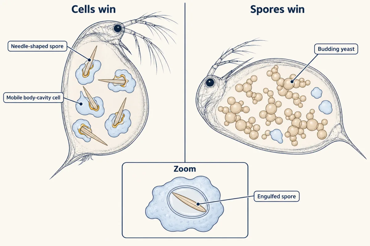
The water flea Daphnia is small and transparent. Some fleas swallow the needle-shaped spores of a budding yeast with their food. The spores pierce the gut wall into the body cavity. Suppose the flea's blood cells were passive, or even carried the infection. What would you expect them to do with the spores?
Passive cells should ignore the spores, or spread them. Instead, Metchnikoff saw mobile cells crowd onto the spores, surround them, and engulf them. The outcome tracked the flea's fate. Where the cells destroyed the spores, the infection stopped. Where spores escaped and grew into budding yeast, the flea died. His 1884 paper made the eating cell, the phagocyte, an active defender. But it did not show that the cells work alone. In Paris in the 1890s, Jules Bordet studied serum, the fluid that remains once blood clots. Serum from an animal immunized against a cholera bacterium killed those bacteria. Heated to about 55 °C, the immune serum lost its power.
Fresh serum from an animal never immunized could not kill the bacteria on its own either. Then Bordet mixed the heated immune serum with the fresh normal serum. The mixture killed again. So the killing needs two separate parts. One part survives heat and is raised by immunization. We now call it antibody. The other is destroyed by heat and is present in all fresh serum. We call it complement. So which side was right? Watch a bacterium meet complement. Small green tags, a piece called C3b, coat its surface. A neutrophil grabs the coated bacterium quickly. Coated microbes are eaten far more readily. In the first film's race, that raises k. The cells eat, and the proteins make the eating faster.
In 1908, Metchnikoff shared the Nobel Prize with Paul Ehrlich, who stressed the defenses in serum. Each side had described part of one system.
From the film
- Metchnikoff watched mobile cells in transparent water fleas engulf swallowed yeast spores: the outcome tracked the flea's fate, as fleas whose cells destroyed the spores lived and those where spores grew into budding yeast died.
- That showed eating cells, phagocytes, are an active defense; it did not show that they work alone.
- Bordet found that immune serum's killing needs two parts: a heat-stable one raised by immunization, antibody, and a heat-sensitive one present in all fresh serum, complement.
- Cells and serum proteins are one system: complement coats microbes so phagocytes eat them faster, which raises the fraction removed per doubling.
Metchnikoff’s 1884 paper named the yeast Monospora bicuspidata. Showing that the flea needs those cells takes disabling them and measuring survival, the loss-of-function logic of the design problem below.
Complement: mark, alarm, puncture
Complement, Bordet’s heat-sensitive factor, is a set of proteins, most made in the liver, that circulate inactive in blood plasma. The second film showed it coating a bacterium; it has three jobs.
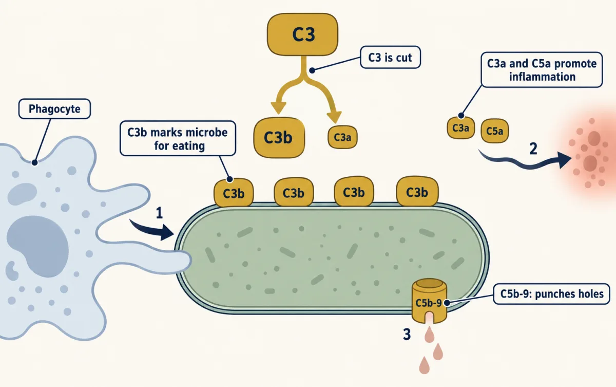
Three routes switch it on (see Going deeper), and all converge on one protein, C3, by cutting it in two. For C3’s two pieces, remember: C3b binds, C3a alarms.
- Mark (C3b). The large piece, C3b, and its trimmed form iC3b stick to the microbe, and phagocytes carry receptors for both. Coating a microbe for eating is opsonization, the coat the film showed raising k.
- Alarm (C3a and C5a). The small piece, C3a, drifts away, as does C5a, cut a step later from the next protein, C5. Both promote inflammation, and C5a also draws neutrophils to the site.
- Puncture (C5b-9). C5’s larger piece, C5b, starts the membrane attack complex, C5b to C9, a ring that can kill some microbes by punching a hole in their membrane. Bacteria with an outer membrane over a thin wall are its targets, Neisseria especially; bacteria with a thick wall and no outer membrane mostly resist it.
Predict. A person makes normal C3 and C5 but lacks C6, C7 or C8, late proteins of the C5b-9 ring. Which jobs still work, and which infections should trouble this person most?
Check your prediction
Marking and alarm still work, because C3b, C3a and C5a are made before the late proteins act. Puncture fails, so the gap shows with the microbes the ring kills best: people lacking a late protein are prone to repeated Neisseria infections. Losing C9, the last protein, is milder: C5b-8 alone still makes small holes and kills Neisseria, only more slowly.
Complement also helps clear dying cells quietly (see cell death) and prunes synapses in the brain (microglia and complement).
Viruses: interferons and natural killer cells
A virus multiplies inside a cell, out of easy reach of phagocytes and complement.
Interferons are signals released by virus-infected cells, and by some immune cells, that put nearby cells into an antiviral state, so each infected cell infects fewer new ones. Alick Isaacs and Jean Lindenmann named them in 1957 after finding that chick membrane exposed to heat-killed influenza virus released a factor that protected fresh membrane. Their receptors signal through Janus kinase (JAK) and signal transducer and activator of transcription (STAT) proteins (JAK-STAT).
Natural killer (NK) cells are lymphocytes, the white-cell family of B and T cells, but they kill infected or abnormal body cells without B and T cells’ rearranged antigen receptors. They weigh activating against inhibitory signals; major histocompatibility complex (MHC) class I, a surface protein most body cells display, is an inhibitory one, so a cell that loses it releases a brake. NK cells then release granules of perforin and granzymes, which can trigger the target’s self-destruction.
Three organisms, one problem: fly, plant, mouse
Every lineage has ready-made recognition; the receptors and responses differ.
The fruit fly Drosophila melanogaster makes no antibodies and has no lymphocytes. Its blood cells, hemocytes, can engulf bacteria, and infection switches on genes for antimicrobial peptides such as drosomycin (mainly through the Toll pathway) and diptericin (mainly through the Imd pathway). Fly Toll is activated by Spätzle, a fly protein cut during infection, not by fungal molecules directly (TLRs and NF-κB tells how Toll’s role in defense was found).
The plant Arabidopsis thaliana has no mobile defender cells; every cell defends itself. Surface pattern receptors start pattern-triggered immunity (PTI). Mostly inside the cell, NLR receptors detect effectors, molecules a pathogen delivers into plant cells, many of which suppress defense, directly or by the changes they make to plant proteins, and start effector-triggered immunity (ETI). In Arabidopsis infected with Pseudomonas syringae, the two cooperate. A strong response often includes the hypersensitive response, death of plant cells at the infection site, which can limit the pathogen’s spread. Signals that spread resistance through the plant are in plant signals.
The mouse, like humans, has barriers, complement, neutrophils, macrophages, natural killer (NK) cells, interferons and fever, and on top of them T and B lymphocytes. None of these three is more primitive than the others; each lineage has met microbes for the same length of time, and the arms race is in mutualism, parasitism and coevolution.
What innate immunity lacks, and how it can change
Innate immunity lacks receptors built anew in each lymphocyte, so it does not make the receptor-based memory of B and T cells. What it has is a head start: remove more than half every doubling time, and start before the pile grows.
Its responses can still change: in a 2020 study, human blood cells responded more strongly to an unrelated fungus 90 days after BCG vaccination. This trained immunity is not specific to one microbe, and a stronger response in a dish is not protection from infection. How it works is not fully understood; Going deeper looks at the evidence.
Design problem
Lipopolysaccharide provokes strong inflammation in most mice. Design a test of the claim that mice need the receptor TLR4 to sense lipopolysaccharide and start inflammation.
Saved in this browser only.
One way to do it
Start with a loss-of-function comparison: give mice lacking a working Tlr4 gene and normal mice the same dose of purified lipopolysaccharide, with a salt-solution control injection.
Measure steps of the sequence, not only survival: inflammatory signals from resident cells, and neutrophils arriving at the injection site. If TLR4 is needed, mutants should show little of either after lipopolysaccharide, and match normal mice after the control.
Rule out a general defect: if mutants respond normally to a microbial molecule sensed by another receptor, the defect is specific to sensing lipopolysaccharide.
Use more than one mutation: the strains C3H/HeJ and C57BL/10ScCr barely respond to lipopolysaccharide and carry different Tlr4 defects, and two independent mutations in one gene beat one strain, which could differ in other genes too.
State the limits: a loss-of-function test shows that TLR4 is needed, not how it recognizes the molecule; showing that it binds lipopolysaccharide takes a different experiment.
Going deeper
What the race model leaves out
Real tissues break the model’s constant rates and mixing: recruitment ramps up, and some microbes hide. The threshold is not an artefact of whole doubling times: with smooth growth against smooth killing, break-even is still exactly one-half per doubling time.
In the model, delay does not change whether removal above one-half wins. It costs more when killing is weak: at k = 0.75 each doubling lost before killing starts takes one more doubling time to undo; at k = 0.6, a factor of 0.8, it takes about three.
Receptor and complement details
Most Toll-like receptors signal to NF-κB through the adaptor MyD88, though TLR3 uses TRIF (TLRs and NF-κB). The three complement routes are the classical (usually started by antibody, Bordet’s route), lectin (mannose-binding lectin, which binds microbial sugars) and alternative (constant low activation, switched off on host cells by host regulators) pathways. Many pathogens have evolved ways to block complement on their surfaces. C3 alone is around 1 mg per mL of plasma.
Memory-like innate responses
Some mouse natural killer (NK) cells show a memory-like response to cytomegalovirus: after one infection, a subset that recognizes a viral protein expands and responds more strongly next time. Unlike trained immunity, this special case is specific to one virus.
Exercise. Cirovic and colleagues gave the tuberculosis vaccine BCG to 15 healthy adults and a placebo to 5. Ninety days later, blood cells from the vaccinated group responded more strongly in a dish to an unrelated fungus, monocytes showed changes in which parts of their DNA were accessible, and marrow progenitor cells showed changes in which genes were active. Design a test of the claim that innate cells “remember”.
One way to do it
Compare each volunteer with their own blood before vaccination, and keep a placebo group handled identically, so seasons or sampling stress are less likely to explain a change.
Use a heterologous (unrelated) stimulus such as the fungus Candida albicans, since a stronger response to BCG itself could be ordinary adaptive memory. Mixed blood cells contain T cells, so rule out cross-reactive ones, for example by testing purified monocytes.
Separate numbers from behaviour: count each cell population in the same samples, since more monocytes would release more signal with no cell changing. Cirovic and colleagues found no significant change in the measured myeloid population counts. The study was small, so this makes a change per cell likely rather than proven.
Ask how long it lasts by sampling at several times beyond 90 days, which that study did not do.
State the limits: no antigen receptor was rebuilt, the response is not specific, and showing protection needs infections measured in vaccinated and placebo groups.
Your notes
Select any text in the lesson to highlight it or add a note to it. Your highlights and notes are saved in this browser and collected in your notebook.
This browser is not letting the site save anything (a private window or strict privacy settings), so highlights and notes will be lost when you leave the page.
How we measure it
Watching infection in a transparent living animal
Choose an animal you can see through, such as Daphnia or a starfish larva, and follow its cells and the invading microbe under a microscope over time. It shows which cells act and when, though it cannot by itself show that those cells are necessary for survival.
Heat inactivation and serum mixing
Heat serum to destroy its heat-labile parts, then add back fresh serum from an untreated animal that cannot kill on its own. If the mixture kills, the activity needs at least two components with different heat sensitivity, as Bordet found for complement and antibody.
Loss-of-function mutants with a survival readout
Compare wild-type and mutant organisms (flies lacking Toll pathway parts, mice lacking a working Tlr4, Arabidopsis lacking several surface receptors) for a molecular output and for survival or resistance after infection. It tests whether a part is needed, not how it recognizes the microbe.
Ex vivo restimulation of blood cells
Take blood cells from a person or animal before and after a treatment, challenge them in a dish with an unrelated microbe, and measure what they release, such as the signal IL-1β. Counting the cells in the same samples helps tell a change in cell numbers from a change in each cell's response.
Practice questions on this lesson (8) → MCAT-style, original, each with an explanation
Where this lesson fits
Before thisReview first
- Germs, Vaccines and AntibioticsBiology · High school
It introduces how germs spread and how the body's defenses and antibiotics fight them, which this lesson's race with bacteria assumes.
- How Microbes MultiplyBiology · High school
You need bacterial doubling from there to see why the count grows by 2 each doubling time.
- Tissues, Organs and HomeostasisThis course · Lesson 6
It introduces feedback and tissue organization, which the inflammation response here builds on.
After thisStudy next
- Adaptive ImmunityThis course · Lesson 18
It takes up the antibodies and T cells that arrive days later, once the innate race has bought time.
- Cell Death and ClearanceOne level up · Cell Biology
It shows how dying cells are cleared, a job complement and phagocytes share with those met here.
Suggested by an AI model from the lessons themselves, then checked. How we use AI
Finish line
Test yourself
13 questions
Practice and MCAT-style, with explained answers.
Next · Lesson 18
Adaptive Immunity
How can your body already hold cells that fit a virus you have never met?
Continue →