Organismal Biology · Skim version
Microbes, Protists and Fungi
Read the full lesson → Skim the whole course →
The guiding idea
A methane-making microbe and E. coli look alike under a microscope. How can we tell whether they are close relatives?
Relatedness is read from differences in a molecule every cell shares, not from how a cell looks, and cells that look alike can be only distantly related.
From the film: Look Alike, Far Apart
- Cells that look alike can be distant relatives; looks do not reveal descent.
- Woese compared small-subunit ribosomal RNA, a molecule every cell carries, and scored each pair of organisms.
- Methanogens formed their own group, about as far from bacteria as from eukaryotes: the archaea.
- Most recent trees built from many genes place eukaryotes inside the archaea, closest to the Asgard archaea; the exact position is debated.
- Fewer differences usually mean a more recent common ancestor; the scores rank closeness but do not date splits or identify an ancestor.
From the film: Digest Outside, Fuse in Two Steps
- A fungus grows as threads called hyphae, or as single yeast cells, with walls containing chitin.
- Fungi release enzymes outside themselves and absorb the small molecules those enzymes free.
- Plasmogamy joins two partners' cytoplasm; karyogamy fuses their nuclei later.
- In between, a cell with two separate haploid nuclei is a dikaryon, not a diploid cell; in mushroom-forming fungi this stage can last a long time.
- Meiosis then makes haploid spores.
-
Three domains from one molecule
-
Bacteria: shape, envelope, fission and gene swapping
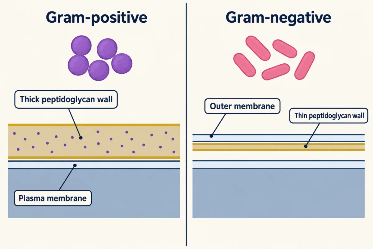
Gram-positive cells have a thick peptidoglycan wall that keeps the crystal violet-iodine complex through the alcohol wash, so they stay purple. Gram-negative cells have a thin wall beneath an outer membrane, so the violet washes out and the safranin turns them pink. AI-generated illustration -
Protists: a word for “everything else”
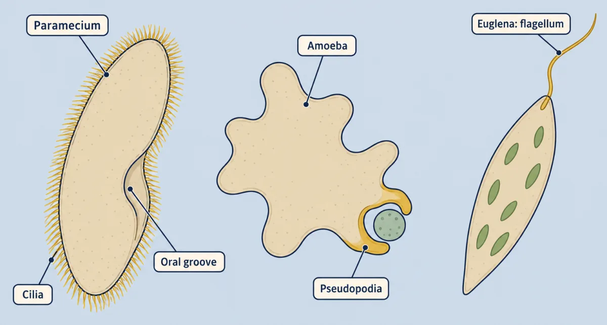
Protists share no single way of moving or feeding: Paramecium sweeps food in with cilia, Amoeba engulfs prey with pseudopodia, and Euglena swims with one long flagellum and can photosynthesize. AI-generated illustration -
Fungi digest outside the body and partner with roots and algae
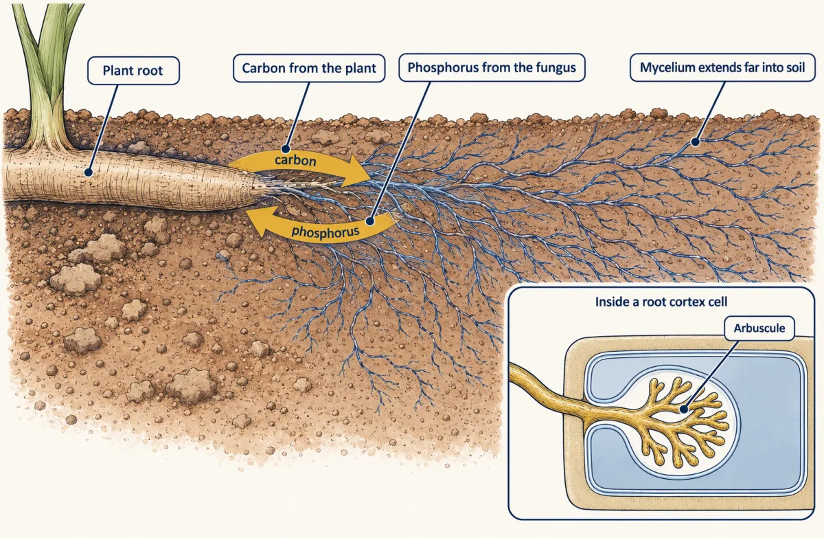
An arbuscular mycorrhizal fungus grows inside root cortex cells' walls but outside their membranes, and reaches far into the soil: the plant pays carbon for mineral nutrients, especially phosphorus. AI-generated illustration -
Viruses as the contrast case
-
Going deeper
Where this lesson fits
Before thisReview first
- What an Organism Has to DoThis course · Lesson 1
This lesson builds on what every organism must do, such as exchanging materials and growing, before comparing microbes.
- Size, Scale and SurfaceThis course · Lesson 2
The small size of bacterial cells and their surface-to-volume limits are used here without being re-taught.
- Domains and KingdomsBiology · High school
You should already know the three domains and how to sort a cell into one before this deeper comparison.
After thisStudy next
- Plants on LandThis course · Lesson 5
The fungal cycle of fusion and meiosis prepares you to read haploid and diploid stages in plant life cycles.
- Soil, Nutrients and PartnersThis course · Lesson 24
It takes the mycorrhizal partnership between roots and fungi further, with phosphorus uptake from soil.
- Innate Immunity: Recognize and RespondThis course · Lesson 17
It shows how the body detects and fights the bacteria and viruses introduced here.
Elsewhere on the siteFilms, simulations and method films in other courses
- The Molecule Behind the TreeFilm · Biology
It shows how ribosomal RNA comparison redrew the tree of life when looks failed to show ancestry.
- Bacterial and phage geneticsMethod film
It covers how bacteria swap genes, the gene exchange this lesson names beside fission.
Suggested by an AI model from the lessons themselves, then checked. How we use AI