Biology · Unit 3: Bacteria and viruses
Living or Not? Viruses and Bacteria
Rewritten on : new text, flashcards and film. Your notes and highlights stay, but the text around them may have moved.
A cold virus on a light switch does nothing on its own for hours, yet inside your nose it gets cells to build new copies of it. Is it alive?
Pause when the reading reaches a film, and offer to play it. Saved on this device.
Watch the film now, or keep listening
Builds on:One Cell or ManyThe Cell Theory and Its EvidenceRespiration and ATPWhy these?
Where this lesson sits
- Virginia SOL BIO.4a
- Virginia SOL BIO.4c
- Practices BIO.1c, BIO.1d, BIO.1f
Essential question: What makes something alive, and do bacteria and viruses pass the test?
-
Life Science (grade 7)
Review it at grade 7 level
Virginia's grade 7 Life Science course does not study viruses in depth. It does teach that living things are made of cells. It sorts organisms into groups by whether their cells have a nucleus, how many cells they have, what cell parts they have, and how they get energy. Bacteria are in that course as living things made of one cell with no nucleus. Biology adds viruses, which are not cells at all, and asks whether they belong on the list of living things.
-
Biology BIO.4a, BIO.4c You are here
This lesson
-
Biology II Biology II: Advanced Survey of Biology Topics, BIOII.7 c (human health option); Genetics, GEN.2 a
Preview where it goes next
Virginia's Biology II courses take this lesson in two directions. In the human health option of Advanced Survey of Biology Topics (BIOII.7 c), you study infectious disease: how viruses and bacteria spread between people and how the immune system responds to each. In Genetics (GEN.2 a), you compare how DNA is packed. Prokaryotes such as bacteria typically have one circular chromosome, while eukaryotes have linear chromosomes. Both can carry plasmids, small extra rings of DNA. Biology II also explains why the three energy groups in this lesson are a simplification: energy source and carbon source are two separate questions, and some bacteria use light for energy but take their carbon from other living things.
-
College
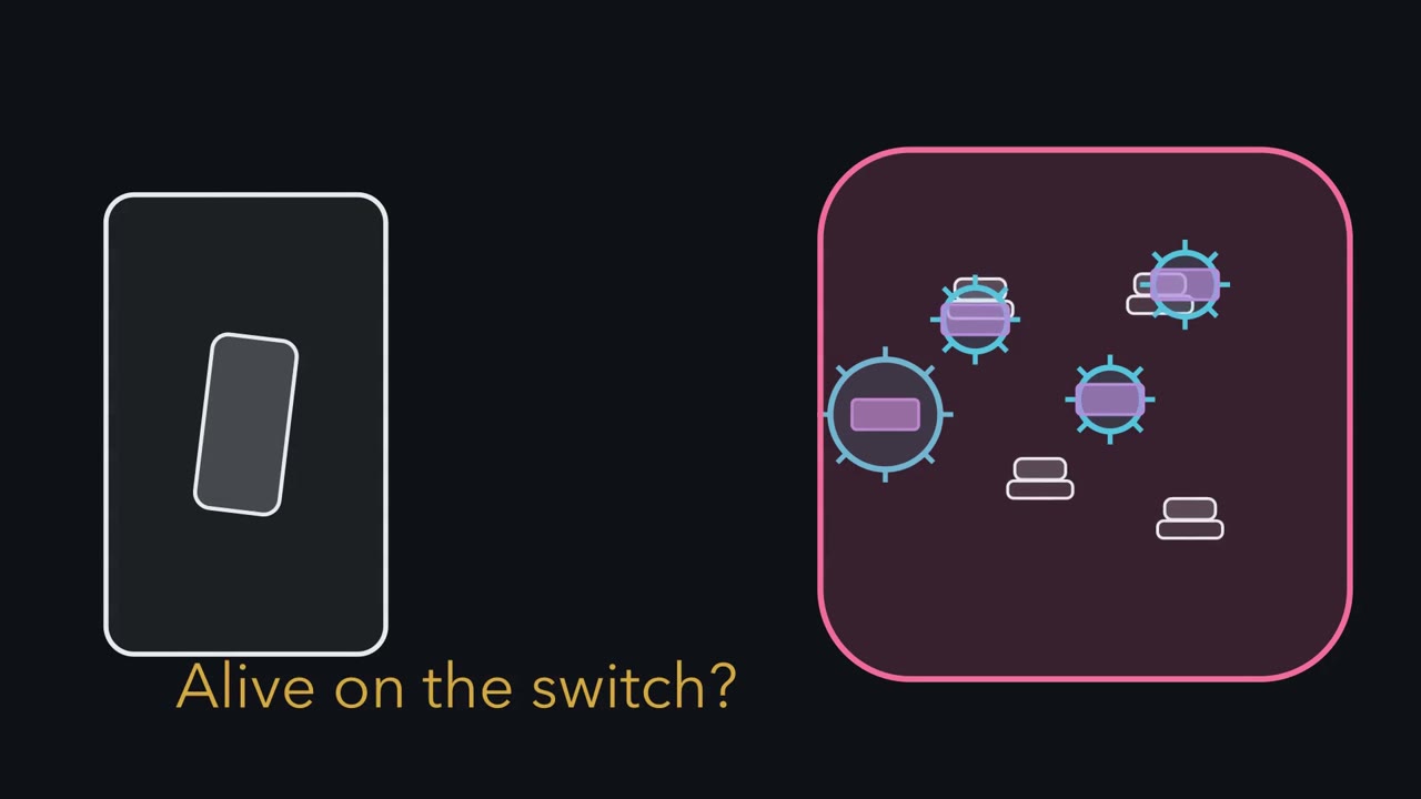 ▶ Watch Who Can Act Alone? · 3:19
Read the transcript of “Who Can Act Alone?”
Film transcript: Who Can Act Alone?
A virus waits on a light switch, then reaches a cell that makes new copies. Was the virus alive on the switch? Watch what each object can do, and who supplies the work. Biologists organize evidence with characteristics of life. Living things have cells, use energy, grow, respond, keep conditions steady, reproduce, and evolve. The list guides a decision. It is not a score where an individual tick wins. Bring in a bacterium. It is a single cell with a membrane, cytoplasm, genes, and ribosomes. This prokaryote has no nucleus and, typically, no membrane-covered organelles. In suitable conditions, materials cross its membrane. Its ribosomes build proteins.
It uses energy, grows, responds, and divides. Now bring back the virus. It carries genes inside a protein capsid, but it is not a cell. The capsid protects the genes. Some viruses add a fatty envelope outside the capsid. It has no cytoplasm or ribosomes. Outside a host, it does not take in food, use energy, or grow. Predict what changes when the virus enters the host cell. Does the virus gain cell machinery, or does the host supply what it lacks? Keep your eye on the ribosomes.
The host supplies what the virus lacks. It provides ribosomes, energy, and building blocks. Viral genes use them to make virus parts. Host enzymes or viral enzymes copy the viral genes, depending on the virus. The parts assemble around the copies. The virus never gains its own metabolism. Edge cases show why a box cannot decide alone. A mature red blood cell cannot divide, yet it remains a cell. A crystal gets bigger, but it has no cells and no metabolism.
Growth alone is not enough. Viruses carry genes and evolve, so scientists still discuss their place near life's edge. Virginia biology classes viruses as nonliving because they are not cells and have no metabolism of their own. They depend on a host cell to be copied. Return to the switch. The virus can wait there and later be copied, but waiting is not living. The host cell supplies the ribosomes and energy. The bacterium carries its own cell machinery and can act when conditions suit it. So ask these questions. Is it a cell, and does it have its own ribosomes and metabolism? That leads to the course rule. Bacteria are living cells; viruses are not cells and are classed nonliving.
From the film
- Bacteria are living cells; viruses are not cells and are classed nonliving.
- The characteristics of life organize evidence, but no single checked box decides the case.
- A bacterium is a cell that uses energy, grows, responds and divides when conditions suit it.
- A virus is not a cell and has no metabolism of its own.
- Inside a host cell, viral genes use the cell's machinery to make new virus parts.
The first film ended on a rule. Test it against three common ideas, then the simulation.
What do you think? Decide before you read.
-
Scientists disagree: Most biology courses, including Virginia’s, class viruses as nonliving infectious agents. A virus is not a cell, has no metabolism of its own, and is copied only inside a host cell. Scientists still discuss the question, because viruses carry genes and change over generations.
What scientists think
Scientists disagree: Most biology courses, including Virginia’s, class viruses as nonliving infectious agents. A virus is not a cell, has no metabolism of its own, and is copied only inside a host cell. Scientists still discuss the question, because viruses carry genes and change over generations.
-
Scientists disagree: Electron microscope images show a virus as a protein coat, often with spikes, that holds its genetic material. There is no cell membrane around cytoplasm, and there are no ribosomes. Some viruses also have a fatty outer layer, but that layer does not make them cells.
What scientists think
Scientists disagree: Electron microscope images show a virus as a protein coat, often with spikes, that holds its genetic material. There is no cell membrane around cytoplasm, and there are no ribosomes. Some viruses also have a fatty outer layer, but that layer does not make them cells.
-
Scientists disagree: Some bacteria and some viruses cause disease, so people call both germs. But a bacterium is a whole living cell that can grow and divide by itself in the right conditions. A virus can only be copied inside a host cell. Most bacteria are also many times larger than most viruses.
What scientists think
Scientists disagree: Some bacteria and some viruses cause disease, so people call both germs. But a bacterium is a whole living cell that can grow and divide by itself in the right conditions. A virus can only be copied inside a host cell. Most bacteria are also many times larger than most viruses.
Explore: run the living-or-not checklist
In the simulation, pick a bacterium, a virus outside or inside a cell, a human red blood cell or a salt crystal. Switch on the characteristics it shows, then check your boxes.
Rule to keep: no single box decides it. In this sketch, each answer follows the lesson for that thing; the shapes are labelled schematics, not to scale.
Predict. Tick the boxes for a virus outside a cell, then for a virus inside one. Which box changes, and does that change make the virus alive?
Check your prediction
The reproduce box changes to “only with a host cell,” because new viruses are built only inside a host cell. The cell and metabolism boxes stay unmet, so the classification does not change.
Explain: what counts as alive
- virus
- host cell
- capsid
- envelope
- prokaryote
- nucleoid
- ribosome
- cell wall
- flagellum
- heterotroph
- photoautotroph
- chemoautotroph
The first film’s characteristics of life make a checklist. Living things:
- are made of one or more cells
- take in and use energy (all the chemical reactions that do this are called metabolism)
- grow and develop
- respond to their surroundings
- keep their inside conditions steady
- reproduce
- change over many generations (evolve)
As the first film’s red blood cell and crystal showed, no single box decides it. The key evidence here is different. A bacterium is a cell with its own ribosomes and metabolism. A virus is not a cell and must use a host cell’s.
A virus is an infectious agent made of genetic material inside a protein coat. A host cell is the living cell a virus infects; the first film showed it supplying ribosomes, energy and building blocks. Picture a factory running someone else’s blueprints. The picture has one limit. Depending on the virus, either the host’s enzymes or enzymes made from the virus’s own genes copy its genetic material.
The one idea. Bacteria are living cells; viruses are not cells and are classed nonliving. A virus has no metabolism of its own, and new copies are built only inside a host cell.
Scope: this is the classification in Virginia’s Biology standards (the SOL). Scientists still discuss definitions of life, and some living bacteria also depend on host cells, so needing a host does not decide the case by itself.
Many scientists agree, though viruses carry genes and evolve, and some giant viruses carry more than a thousand genes. Under a definition based on cells and metabolism, they are nonliving.
If a virus can carry that many genes, genes cannot be what separates it from a bacterium. The second film opens both to find what does.
Read the transcript of “Parts Reveal the Difference”
Film transcript: Parts Reveal the Difference
The first film classified bacteria and viruses by what they can do. If both carry genes, why can a bacterium grow while a virus waits? Open them and follow the work. Start with the bacterium, a prokaryotic cell. Its membrane controls what enters and leaves.
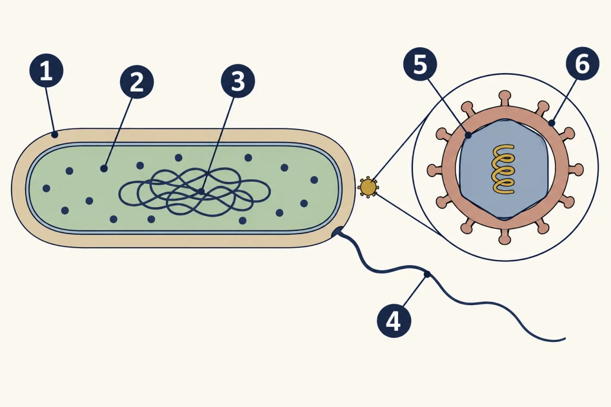
Cytoplasm fills the inside. Ribosomes build proteins from instructions in DNA. That DNA sits in a nucleoid, a region without a membrane. Many bacteria also have a cell wall that helps prevent bursting when water enters. Some have a capsule outside. Some have flagella that spin to push them through water. These parts vary, but the working cell remains. Now open the virus. Its DNA or RNA sits inside a protein capsid. Some viruses add a lipid envelope outside. There is no cytoplasm and no ribosome workbench. A host cell must build every viral protein. Genes are the shared part, not proof that both are cells. The bacterium reads its genes with its own ribosomes.
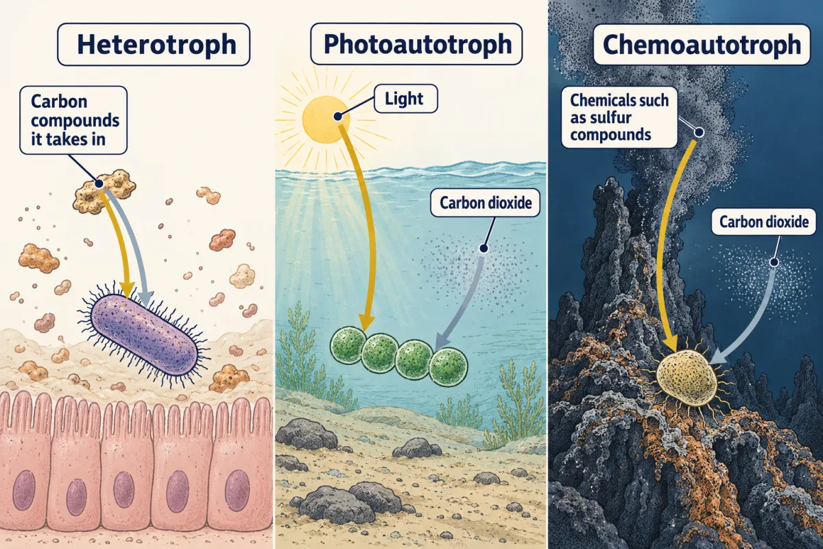
Viral genes can be read only after they reach a suitable host cell. A working cell also needs energy and carbon. Energy powers the work. Carbon supplies material for new parts. For a useful first model, sort bacteria into common patterns by where those inputs come from. Heterotrophs take in carbon compounds for both energy and carbon.
Photoautotrophs use light for energy and carbon dioxide for carbon. Chemoautotrophs use chemicals for energy and carbon dioxide for carbon. Predict the pattern in a dark spring. The bacterium uses sulfur compounds for energy and carbon dioxide for carbon. Which name fits, and which input tells you?
Chemoautotroph fits. Chemical energy selects chemo, and carbon dioxide supplies the carbon. The model is a simplification. Energy source and carbon source are separate questions, and a few bacteria mix the patterns. Dependence alone does not make a cell nonliving. Some bacteria grow only inside other cells, yet keep ribosomes and metabolism.
Viruses remain different because they are not cells and have no ribosomes of their own. Size does not decide the case either. Most viruses are smaller than typical bacteria. But some giant viruses are longer than small bacteria, so that shortcut breaks. Parts and work matter more than length. The comparison now has a reason. Bacterial parts let the cell obtain inputs and build proteins.
Viral parts carry genes between hosts. Bacteria are living cells; viruses are not cells and are classed nonliving.
From the film
- Bacteria are living cells; viruses are not cells and are classed nonliving.
- A bacterium has a membrane, cytoplasm, ribosomes and DNA in a nucleoid.
- A virus has DNA or RNA in a capsid, sometimes inside an envelope.
- Bacteria can get energy from carbon compounds, light or chemicals.
- Size does not decide whether something is living.
Explain: parts reveal the difference
The second film opened both; this table lines up each part with its job. Bacteria are prokaryotes: one-celled living things with no nucleus and, typically, no membrane-covered organelles. Your cells are eukaryotes, which usually have a nucleus; mature red blood cells lose theirs. One Cell or Many compares them.
| Part | Typical bacterium | Virus | What the part does |
|---|---|---|---|
| Genetic material | DNA in the nucleoid, a region with no membrane around it | DNA or RNA, inside the capsid | Carries the instructions |
| Coat | Cell membrane around cytoplasm | Capsid, a protein coat | Membrane controls what enters and leaves; capsid protects the genes |
| Ribosomes | Yes | No | Build proteins; a virus uses the host cell’s |
| Cell wall | Often | No | Helps keep the cell from bursting when water rushes in |
| Outer layer | Some have a capsule | Some have an envelope, a fatty layer outside the capsid | Envelope helps some viruses enter cells |
| Flagellum (plural flagella) | Sometimes | No | A corkscrew-shaped tail that spins to push the cell through water |
Viruses infect all the main kinds of living thing: bacteria, archaea, plants, animals, fungi and protists. A virus that infects bacteria is called a phage. Each virus can enter only certain cells, because proteins on its surface must fit receptors on the host cell.
- cell wall
- ribosomes
- nucleoid
- flagellum
- capsid
- envelope
- cell wall
- blank
- blank
- flagellum
- blank
- blank
Word bank: capsid, envelope, nucleoid, ribosomes
- blank
- blank
- blank
- blank
- blank
- blank
Check your labels
- cell wall
- ribosomes
- nucleoid
- flagellum
- capsid
- envelope
Explain: three bacterial energy patterns
Every living cell needs energy to do work and carbon to build its parts. The table adds an example of each pattern from the second film.
| Group | Energy comes from | Carbon comes from | Example |
|---|---|---|---|
| Heterotroph | Carbon compounds it takes in | The same compounds | E. coli in your gut; bacteria that decompose dead matter |
| Photoautotroph | Light | Carbon dioxide | Cyanobacteria in ponds and oceans |
| Chemoautotroph | Chemicals such as sulfur, iron or nitrogen compounds | Carbon dioxide | Bacteria near deep-sea vents and in soil |
Predict. Some bacteria use light for energy but take their carbon from other living things. Which row of the table do they fit?
Check your prediction
Neither row fits cleanly: the energy column matches a photoautotroph, the carbon column a heterotroph. Energy source and carbon source are separate questions, so the three groups are a useful simplification.
The second film’s bacteria that grow only inside other cells include Rickettsia, which cause diseases like Rocky Mountain spotted fever. They are still living cells with their own ribosomes and metabolism, though they take some energy molecules and nutrients from the host. Most bacteria carry out every life process themselves when conditions suit them; fully dried bacteria do not grow. How Microbes Multiply shows how bacteria divide.
Elaborate: compare real sizes
A micrometer (μm) is a thousandth of a millimeter, and a nanometer (nm) is a thousandth of a micrometer.
| Object and what was measured | Reported size | Size in nanometers (nm) | Kind of sample |
|---|---|---|---|
| SARS-CoV-2 (COVID-19 virus), spike on the surface | about 9 to 12 nm | 9 to 12 | Virus grown from patient samples, electron microscope |
| SARS-CoV-2, particle diameter | about 60 to 140 nm | 60 to 140 | Virus grown from patient samples, electron microscope |
| Influenza virus, diameter of round particles | 80 to 120 nm | 80 to 120 | Range from a review of studies |
| T4 phage head, after drying | about 75 × 60 nm | 75 × 60 | Atomic force microscope |
| T4 phage head, in liquid | about 100 × 85 nm | 100 × 85 | Atomic force microscope |
| T4 phage, whole particle length | about 200 nm | 200 | Earlier electron microscope work |
| Tupanvirus (a giant virus), average length | about 1.2 μm | 1,200 | Electron microscope |
| E. coli bacterium, length × width | about 2 × 0.5 μm | 2,000 × 500 | Reference size for normal cells |
| Human red blood cell, mean diameter | about 7.5 μm | 7,500 | Medical reference for normal cells |
These values come from different studies, not one experiment. The SARS-CoV-2 sample was virus grown from patients admitted to a Wuhan hospital in December 2019. The sources are Zhu and colleagues (2020), a 2006 influenza review by Machała and Brydak, Kuznetsov, Chang and McPherson (2011), Abrahão and colleagues (2018), Maki and colleagues (2000), and a 1990 medical textbook chapter by Lynch. The last column uses 1 μm = 1,000 nm.
Most viruses are about 20 to 300 nm across. Typical bacteria are about 1 to 5 μm (1,000 to 5,000 nm) long, so they are larger. The two groups overlap only at the extremes. A giant virus such as Tupanvirus is longer than the smallest typical bacteria, and a few bacteria are big enough to see without a microscope.
Predict. About how many of the largest round influenza particles (120 nm) fit along an E. coli cell? Convert before dividing.
Check your prediction
Convert 2 μm to 2,000 nm, then divide by 120 nm. The result is about 17. A typical 100 nm particle gives 20.
- The T4 head measured about 100 × 85 nm in liquid and 75 × 60 nm after drying. Why must a scientist report sample preparation?
- Tupanvirus is longer than an E. coli cell is wide. Does that make it alive? What evidence matters instead?
Claim, evidence, reasoning
Make a claim that answers the essential question: are bacteria and viruses living? Use the characteristics of life and this data.
Start with: Bacteria are blank, and viruses are classed as blank.
Start with: A bacterium has blank and can blank by itself. A virus has only blank, and the table shows it is usually blank than a bacterium.
Start with: This matters because living things blank, and a virus can only be copied blank.
Compare with a model answer
Bacteria are living, and viruses are classed as nonliving. An E. coli cell is a cell with a membrane, cytoplasm and ribosomes, and it can grow and divide by itself in suitable conditions. A virus is genetic material in a protein coat with no ribosomes, and new viruses are built only inside a host cell. The table also shows most viruses are much smaller: an influenza particle is 80 to 120 nm, while E. coli is about 2,000 nm long. Size is not the test, though; the giant Tupanvirus is 1,200 nm and still is not a cell. One limit: these sizes come from different studies and preparations, and the T4 head shrank when dried.
Saved in this browser only.
Design problem
A jar of pond mud with a loose lid slowly grows dark specks of a mineral. Your partner says living bacteria are making the specks. Plan a test of that claim. Name your groups, what you keep the same, what you measure, how many jars you need, and how you stay safe.
Saved in this browser only.
One way to do it
- Groups: jars of fresh mud (living microbes present) and jars of the same mud heated in a pressure cooker by your teacher to kill the microbes.
- Independent variable: whether living microbes are present.
- Dependent variable: the area covered by dark specks, measured from photos each week.
- Constants: same amount of mud and water, same temperature, same light, same jar size and same number of weeks.
- Repeated trials: at least three jars in each group, so one odd jar does not decide the result.
- Safety: keep jars closed with a loose lid so gas can escape, wear gloves, never smell or taste the contents, and let your teacher dispose of them. Never grow microbes taken from people.
- What would mean what: specks only in jars with living microbes support the claim. Specks in the heated jars too would point to a chemical reaction that needs no life. Even then, the specks do not prove the bacteria use that chemical for energy. A 2020 study of manganese-forming bacteria answered that by tracing whether carbon from carbon dioxide ended up in new cells.
Evaluate: check your thinking
Return to the switch. The virus there is not a cell and has no metabolism of its own. Only living host cells, such as the cells lining your nose, can build new copies of it. The course answer: bacteria are living cells; viruses are not cells and are classed nonliving.
Science practices you used: testing predictions in a model, converting units and comparing sizes in a data table, making a claim from evidence and naming its limits, and planning a fair test with a control group and repeated trials.
Your notes
Select any text in the lesson to highlight it or add a note to it. Your highlights and notes are saved in this browser and collected in your notebook.
This browser is not letting the site save anything (a private window or strict privacy settings), so highlights and notes will be lost when you leave the page.
How we measure it
Electron microscopy
A beam of electrons instead of light makes images of things far smaller than a light microscope can show, such as single virus particles. Researchers measure particle size and shape from these images, as in a 2020 report on the COVID-19 virus grown from patient samples.
Atomic force microscopy
A tiny tip moves across a sample and traces its bumps, like a finger reading a surface in the dark. It can measure a virus particle in liquid and again after drying, which shows how preparation changes a measured size.
Tracking how cells swim
A microscope records bacteria swimming before and after a chemical is added. A change in how they swim is evidence that living cells respond to a stimulus.
Where this lesson fits
Before thisReview first
- One Cell or ManyThis course · Lesson 11
It compares prokaryotes and eukaryotes, which you need to sort bacteria from your own cells.
- The Cell Theory and Its EvidenceThis course · Lesson 6
The rule that living things are made of cells is what separates bacteria from viruses here.
- Respiration and ATPThis course · Lesson 5
You need to know how cells get energy to sort bacteria into energy groups and to understand metabolism.
After thisStudy next
- How Microbes MultiplyThis course · Lesson 13
It shows how bacteria divide and how a virus makes copies inside a host cell.
- Microbes in Bodies and EcosystemsThis course · Lesson 14
It puts the bacterial energy groups to work in soil and water ecosystems.
- Germs, Vaccines and AntibioticsThis course · Lesson 15
It uses the difference between bacteria and viruses to explain why antibiotics work on one and not the other.
Elsewhere on the siteFilms, simulations and method films in other courses
- Set Up: Alive or Not?Film · Biology Together
It shows the basic checklist of what living things do, which this lesson applies to viruses and bacteria.
Suggested by an AI model from the lessons themselves, then checked. How we use AI
Finish line
Test yourself
8 questions
Practice and MCAT-style, with explained answers.
Next · Lesson 13
How Microbes Multiply
One bacterium can become millions of cells in an afternoon, yet a virus never divides at all. How, then, can a cold sore heal and come back in about the same spot?
Continue →