Biochemistry · Membranes & Transport
New to this? Review it at high school level: Crossing the Membrane
Membranes: The Impermeable Sheet That Builds Itself
Rewritten on : new text, figures and flashcards. Your notes and highlights stay, but the text around them may have moved.
Lipids assemble on their own into a sealed, nearly impermeable sheet, and proteins in that sheet decide what crosses.
Pause when the reading reaches a film, and offer to play it. Saved on this device.
Watch the film now, or keep listening
Builds on:Water, Weak Bonds, and the Generation of OrderLipids: The Building Blocks That Refuse to PolymerizeProtein Three-Dimensional Structure: The Sequence Sets the ShapeWhy these?
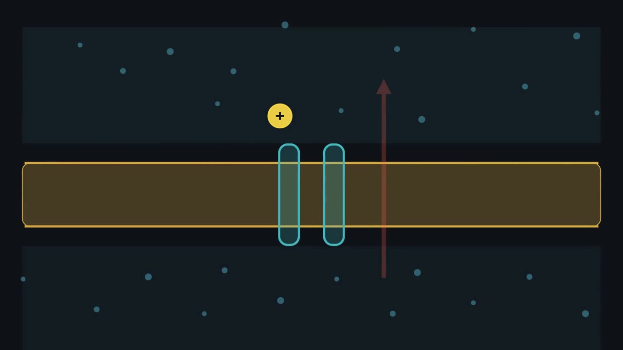 ▶ Watch A Path Is Not a Pump · 3:24
Read the transcript of “A Path Is Not a Pump”
Film transcript: A Path Is Not a Pump
If a membrane protein opens a path, can it make a solute travel uphill? The answer separates a doorway from a pump. Follow one charged solute across this membrane. In water, a charged solute wears a water shell. To enter the oily bilayer core, it must lose that help. The core offers almost nothing in return. This energy cost is the toll for leaving water. A channel offers a polar pathway through the oil. Its polar groups replace some help from water, lowering the toll.
Solute now crosses much faster. Yet the open channel supplies no directional energy. Direction comes from the solute's electrochemical gradient. Concentration pushes from the crowded side. Voltage pulls opposite charges and pushes like charges. Together, those pushes define downhill. Here, Na⁺ is more concentrated outside, while the cell interior is negative. If a Na⁺ channel opens, which way will Na⁺ move? Follow both pushes before you answer.
Concentration pushes Na⁺ inward, away from the crowded outside. The negative interior also pulls positive Na⁺ inward. Both pushes agree, so opening the channel speeds Na⁺ downhill into the cell. Now reverse the combined electrochemical gradient. The same open channel lets Na⁺ run the opposite way. The protein changes speed, not the downhill direction. A protein can change speed, but uphill transport requires energy coupling. Channels are pores, open to both sides when their gates open. Carriers instead bind cargo, change shape, and release it across the membrane. Either mechanism can move a solute downhill without supplying energy. A pump is a carrier coupled directly to an energy source.
This view shows only Na⁺ from the Na⁺-K⁺ pump. ATP drives its shape change and Na⁺ uphill, charging the gradient battery. In one animal-cell example, a coupled carrier spends that stored gradient. Na⁺ runs downhill through the carrier. Glucose travels uphill in the same direction. No ATP is spent there, but the Na⁺ gradient ultimately depends on the pump. Classify any crossing with three questions. Is the route a pore or alternating access? Is the solute moving downhill or uphill? If uphill, what supplies energy: ATP or another downhill flow? Opening a channel or cycling a passive carrier cannot push a solute against its gradient. Look for energy coupling before calling transport uphill.
Only coupled energy can drive transport uphill.
From the film
- Charged solutes pay an energy toll to leave water for a bare bilayer.
- An electrochemical gradient combines concentration and voltage to set a solute's downhill direction.
- Channels and passive carriers speed downhill transport but cannot drive it uphill.
- In this lesson, uphill transport couples to ATP or another solute moving downhill.
A sodium ion is smaller than a water molecule, yet water crosses a bare lipid bilayer about ten billion times faster.
A sodium ion is about 1.0 Å in radius. A water molecule is about 1.4 Å. If a membrane worked like a sieve, sodium would win. It loses by ten orders of magnitude, and the reason explains most of what a membrane does. In most cystic fibrosis, one chloride channel, cystic fibrosis transmembrane conductance regulator (CFTR), mostly never reaches the cell surface, and salt and water stop moving as they should.
The membrane and its puzzles
A cell membrane is a lipid bilayer: two sheets of lipid molecules, back to back. Each lipid has a polar head that sits comfortably in water and two greasy hydrocarbon tails that do not. The two sheets are called leaflets. Their tails meet in the middle of the bilayer, and their heads face the water on both sides. Proteins sit in this sheet and do most of the membrane’s work.
That simple sheet raises five puzzles.
- Self-assembly. Why does the sheet build itself, with no ATP and no covalent bonds holding it together? And why does it reseal when you puncture it?
- The blocked ion. Why does the small sodium ion barely cross, while the larger water molecule crosses easily?
- Moving ions anyway. How do cells move ions across at all, and what decides which way they go?
- The picky channel. How does a potassium channel reject sodium, the smaller ion?
- The missing channel. What goes wrong in cystic fibrosis, and how do drugs repair part of it?
One idea answers puzzles 2 to 4 and frames puzzle 5. It needs one term. A solute’s electrochemical gradient is the combined push it feels from two differences across the membrane: a difference in its concentration and, if it is charged, a difference in voltage.
Every crossing answers two separate questions: how fast, set by the toll for leaving water, and which way, set for a solute moving on its own only by its electrochemical gradient. The film above separates them: a route around the toll changes speed, while only coupled energy drives an uphill move. The text below supplies the measurements, mechanisms and cases the film skips.
A sheet that builds itself
Phospholipids form bilayers on their own. Drop lipids in water and the tails cluster to escape it, the heads face out, and a bilayer forms. Closed vesicles form on their own as well.
The driving force is the hydrophobic effect, and it is entropic. Water molecules next to an oily tail are held in an ordered cage around it. When the tails huddle together, that caged water is released into disorder, and the entropy of the system rises. The energy comes from water: once the lipids are made, the cell spends no ATP to assemble them, though making the lipids and inserting proteins does cost energy.
The same force sets the sheet’s shape. A bilayer always forms closed compartments with no free edges, because an edge would leave tails exposed to water. Noncovalent forces hold it together, not covalent bonds. The lipid bilayer itself is about 5 nanometers (50 Å) thick overall (about 4 nm between the phosphate groups of the two leaflets), with an oily core near 3 nm. A plasma membrane looks thicker in electron micrographs, around 7 to 8 nm, partly because the stain and its proteins and attached sugars add to its apparent width. For scale, a ribosome is about 25 nm across, so a membrane as it appears in micrographs is roughly a quarter of that.
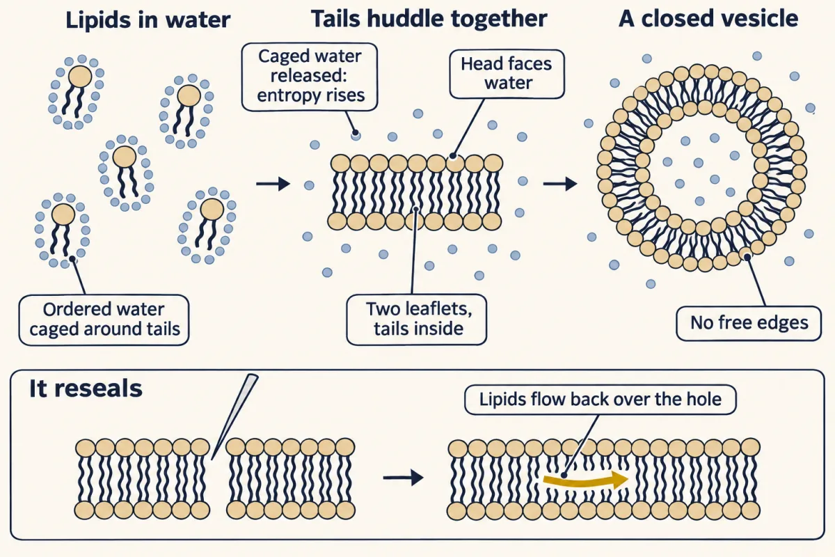
Predict. Push a fine glass needle through a cell membrane, then pull it out. What happens to the hole, and why? Would a plastic bag do the same?
Check your prediction
The hole seals. No bonds broke when the needle went in, because none held the lipids together. The lipids at the edge of the hole now have tails facing water, and the hydrophobic effect drives them to flow back and close the gap. A living cell speeds the repair by fusing vesicles at the wound. A plastic bag is covalently bonded plastic. It tears and stays torn.
The toll for leaving water
Now the second puzzle. If size does not decide, something else must.
The film above shows a charged solute’s water shell meeting the bilayer’s oily core. Water molecules are polar: each has a slightly negative oxygen end and slightly positive hydrogen ends. An ion’s charge orients and holds those ends in a tight shell around it. That attraction makes salts dissolve, and pulling the shell off costs energy. Because hydrocarbon tails offer no partial charges in return, this trade carries the toll for leaving water, also called the desolvation penalty.
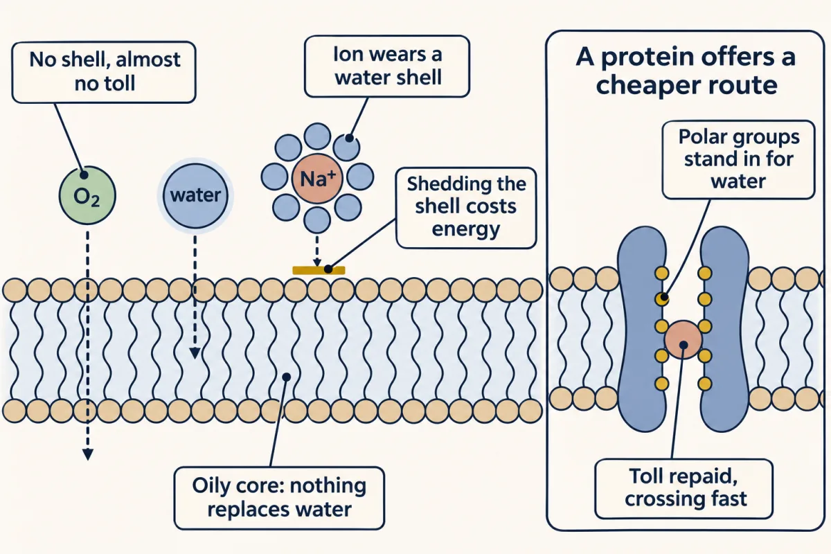
Predict. Rank these by how easily they cross a bare bilayer: O₂, water, glucose, Na⁺. What one feature are you using to rank them?
Check your prediction
O₂ crosses fastest, then water, then glucose, then Na⁺, far behind. The feature is how strongly each one holds water. O₂ is nonpolar and holds almost none, so its toll is close to zero. Water and glucose are polar and pay a moderate toll; glucose has more polar groups and is larger. Na⁺ has a full charge and pays the largest toll of all. Size matters a little. Charge matters most.
How large is the toll for an ion? The energy released when an ion gains its water shell, its hydration energy, is a few hundred kilojoules per mole: roughly 300 to 400 kJ/mol for K⁺ and Na⁺, with about 70 kJ/mol more for the smaller Na⁺, whose charge is packed more tightly. The exact values depend on the convention used (Going deeper has a table), but the roughly 70 kJ/mol difference in hydration free energy does not, and only that difference matters later, at the potassium channel. For comparison, a hydrogen bond is about 20 kJ/mol. A toll that large means an ion essentially never dissolves in the oil and crosses directly. The few ions that do leak through probably take a cheaper route: they drag some water into the bilayer with them, or slip through rare, short-lived water-filled defects. Even that cheapest route is extremely slow.
To feel the gap between water and sodium, scale it in time. If a water molecule took one millisecond to cross, a sodium ion would take about four months. Chemists measure how readily a solute crosses with a permeability coefficient, in centimeters per second. Across the solutes a cell cares about, it spans about twelve orders of magnitude. Small gases such as O₂ and CO₂ sit at the top, at about 1 cm/s and above. Water comes next, then small polar molecules such as urea and glycerol, then glucose. Ions sit at the bottom, near 10⁻¹² cm/s for Na⁺.
A natural pair isolates the variable. Indole is the aromatic ring of tryptophan on its own. Tryptophan carries the same ring plus an amino group and a carboxyl group, which at pH 7 are both charged. Indole crosses many orders of magnitude faster. The ring is the same and the weight differs by less than twofold, so neither size nor weight can explain the gap. Each charged group on tryptophan pays the toll. The ring crosses fine; the charge is what costs. Test the ranking yourself below. Send molecules at the bilayer and watch each one land on a log-scale plot. The gap that opens between small neutral molecules and ions is the toll.
Click a molecule to test it. It will try to cross the bilayer, and its permeability lands on the plot.
Pick a molecule above and watch it try to cross.
crossing rate: —
Each molecule you test lands in its own column, fastest on the left. Test them all and the cliff opens up: every gridline down is 100× slower.
The full toll for stripping an ion is 300–400 kJ/mol; real ions leak by a cheaper route (dragging water along, or through brief defects), so the barrier that sets the leak is far smaller.
This plot answers only how fast: the toll for leaving water sets the rate across bare lipid. Which way an ion moves is set by its electrochemical gradient (the Nernst calculator below). The same toll returns at the K⁺ channel filter, which repays nearly all of K⁺'s toll and falls short on Na⁺'s.
A separate question: keeping the sheet fluid
This is the one topic outside the guiding idea: it is about how tightly lipid tails pack, not about crossing.
A membrane has to stay fluid, and the cell tunes that fluidity with chemistry. Below a transition temperature, Tm, the tails pack into an ordered, nearly frozen gel. Above it they are disordered and the sheet is fluid. Tm is the midpoint of that change. Two features of the tails set it. Longer fatty-acid tails pack more snugly and raise Tm. A cis double bond puts a roughly 30-degree kink in the tail that prevents tight packing and lowers Tm. Trans fats straighten the tail back out, so they pack like a saturated fat despite being technically unsaturated.
Cholesterol is the membrane’s thermostat, and it works in both directions. When the cell gets cold, its rigid four-ring body jams between lipids and stops them from packing into a gel, so the membrane does not freeze solid. When the cell warms up, the same flat rings press against the upper stretch of neighboring tails and restrain their motion, so the membrane does not get too runny. The thermostat is a teaching image: cholesterol does not hold the membrane at a set temperature, but it broadens and flattens the gel-to-fluid change, so fluidity changes less as temperature changes.
What moves, and how fast
Membrane proteins and lipids move, and FRAP lets us watch them do it. In fluorescence recovery after photobleaching, tag the membrane with a fluorescent dye, bleach a small spot dark with a laser, and watch unbleached molecules diffuse back in. How fast the spot recovers gives the diffusion coefficient. How completely it recovers gives the mobile fraction. If recovery stalls at 50%, half the labeled molecules cannot move, often because they are anchored to the cytoskeleton.
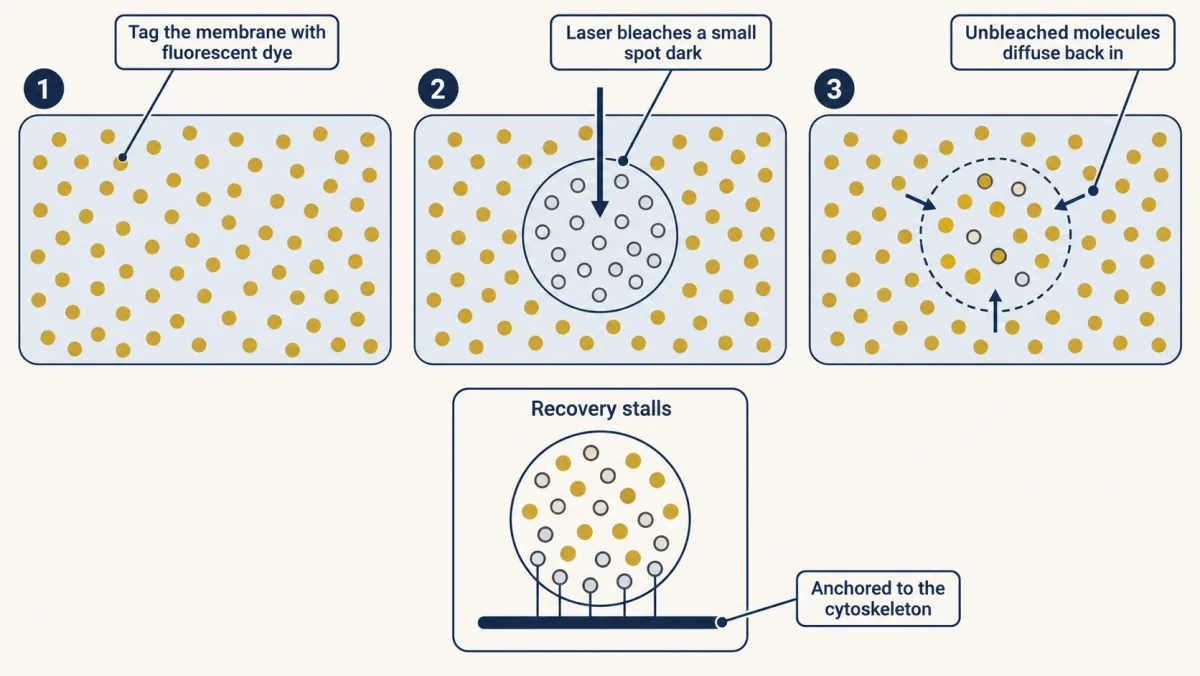
The two kinds of motion are not equal. Lateral diffusion, within one leaflet, is fast: a lipid can travel the length of a bacterium in about a second. Transverse motion, a lipid flipping from one leaflet to the other, is glacially slow. Here the toll returns. To flip, the polar head would have to cross the oily core. Flip-flop happens at a useful rate only with dedicated enzymes: flippases move lipids to the inner leaflet, floppases to the outer, and scramblases either way.
That difference in rates is what keeps the two leaflets different. The membrane is asymmetric: the outer leaflet differs from the inner one. Phosphatidylserine, for instance, stays on the inside of a resting cell, because flip-flop is slow on its own and ATP-driven flippases keep returning it to the inner leaflet. When a cell dies, phosphatidylserine flips outward, and that becomes an “eat me” signal for the immune system.
Proteins in the sheet
Most of what a membrane does, it does through protein. Most membrane proteins are integral, embedded in the bilayer; the rest are peripheral, perched on the surface. The test for telling them apart is operational. Peripheral proteins come off with high salt or a pH change. Integral proteins release only when detergent dissolves the membrane around them.
Proteins that cross the bilayer face a toll of the same kind as ions, though far smaller per group. Every peptide bond in a protein’s backbone carries a polar N–H and a polar C=O. Burying those groups in oil without a partner would be costly. An alpha-helix solves this: its backbone groups hydrogen-bond to each other along the helix, so the backbone pays its own toll, and its hydrophobic side chains face the oil. A helix rises 1.5 Å per residue, so about 20 hydrophobic residues span the 3 nm oily core once, and multi-pass proteins string several together. A beta-barrel rolls a beta sheet into a tube, in which each strand hydrogen-bonds to its neighbors. Beta-barrels are found in the outer membranes of bacteria, mitochondria and chloroplasts.
Some proteins skip spanning entirely and attach by a lipid anchor: a GPI anchor on the outside, a prenyl or fatty-acyl group on the inside. The signaling protein Ras, for example, is prenylated so it can dock at the inner membrane. Other enzymes, such as aspirin’s target COX-1, sit in only one leaflet.
Which way: the electrochemical gradient
So far the toll has told us how fast things cross. Now the second question, which way. The electrochemical gradient has two parts.
The first part is concentration. A solute tends to move from where it is more concentrated to where it is less. The second part is voltage, and it applies only to charged solutes. The membrane potential, Vm, is the voltage inside the cell relative to outside. In a typical neuron the inside sits around 60 to 70 millivolts negative. Most cells fall between about −35 and −80 mV, and some sit far shallower: red blood cells are about −10 mV. A negative inside pulls positive ions in and pushes negative ions out.
Predict. Sodium is 145 mM outside a cell and 12 mM inside, and the membrane potential is −70 mV. Which way does the concentration difference push sodium? Which way does the voltage push it? Do they agree?
Check your prediction
Both push sodium in. The concentration is about twelve times higher outside, so sodium tends to move in. The inside is negative, and sodium is positive, so the voltage pulls it in too. The two pushes agree.
The two parts add up in one equation.
In words The free-energy change for moving a solute in equals a concentration term, RT times the log of inside over outside concentration, plus a voltage term, the charge times Faraday's constant times the membrane potential.
- ΔGin“delta G sub in”
- the free-energy change for moving one mole of the solute from outside to insidekJ/mol
- RT“R T”
- the gas constant times the absolute temperature; about 2.58 kJ/mol at 37 °CkJ/mol
- cin“c sub in”
- the solute's concentration inside the cellmM
- cout“c sub out”
- the solute's concentration outside the cellmM
- z“z”
- the solute's charge: +1 for Na⁺ and K⁺, −1 for Cl⁻, 0 for glucose
- F“F”
- Faraday's constant, the charge on one mole of +1 ions: about 96.5 kJ per mole per voltkJ/(mol·V)
- Vm“V m”
- the membrane potential, inside relative to outside, in volts (−70 mV is −0.070 V)V
Sanity check If the concentrations are equal and Vm is zero, ΔG is zero and there is no net push. For an uncharged solute such as glucose, z = 0 and only the concentration term counts. Moving the solute out instead gives the same size with the opposite sign.
Try one number
Try potassium, 4 mM outside and 140 mM inside, at Vm = −70 mV and 37 °C.
Concentration term: 2.58 × ln(140 / 4) = 2.58 × 3.56 = +9.2 kJ/mol. Potassium is much more concentrated inside, so this term opposes moving it in.
Voltage term: (+1) × 96.5 × (−0.070) = −6.8 kJ/mol. The negative inside favors moving it in.
Total: 9.2 − 6.8 = +2.4 kJ/mol. Moving potassium in is uphill, so on its own potassium tends to leave the cell.
Need a refresher? e and ln · units and prefixes
Now the sodium from the prediction, 145 mM outside and 12 mM inside. Concentration term: 2.58 × ln(12 / 145) = −6.4 kJ/mol. Voltage term: (+1) × 96.5 × (−0.070) = −6.8 kJ/mol. Total: about −13.2 kJ/mol for moving one mole of sodium in. Both parts are negative and about equal, so the move is well downhill.
For each ion there is one voltage where the two parts cancel exactly, so the ion has no net push in or out. That voltage is the ion’s equilibrium potential. Set ΔG to zero and solve for the voltage, and you get the Nernst equation.
In words An ion's equilibrium potential equals RT over its charge times Faraday's constant, times the log of outside over inside concentration.
- Eion“E sub ion”
- the ion's equilibrium potential, the membrane potential at which its ΔG for crossing is zeromV
- RT“R T”
- the gas constant times the absolute temperaturekJ/mol
- z“z”
- the ion's charge
- F“F”
- Faraday's constantkJ/(mol·V)
- cout“c sub out”
- the ion's concentration outsidemM
- cin“c sub in”
- the ion's concentration insidemM
Sanity check If the concentrations are equal, ln(1) = 0, so E is zero: nothing needs balancing. At 37 °C, for an ion with charge +1, each tenfold excess outside adds about 61 mV.
Try one number
Potassium again: 4 mM outside, 140 mM inside, 37 °C. RT/F is 2.58 ÷ 96.5 = 0.0267 V, about 26.7 mV.
ln(4 / 140) = −3.56.
EK = 26.7 × (−3.56) = about −95 mV.
The same arithmetic gives ENa of about +67 mV (145 outside, 12 inside) and ECl of about −64 mV (110 outside, 10 inside, with z = −1).
Where it comes from optional
Step 1, set the free-energy change to zero at Vm = E: RT × ln(cin / cout) + zF × E = 0.
Step 2, move the concentration term across: zF × E = −RT × ln(cin / cout) = RT × ln(cout / cin).
Step 3, divide both sides by zF.
Need a refresher? e and ln · log scales
At rest the membrane is most permeable to K⁺, so the resting potential sits near K⁺‘s equilibrium potential. Pick an ion in the calculator below, change its inside and outside concentrations, and see where its equilibrium potential lands relative to rest.
- Equilibrium potential EK
- —
- Concentration ratio out/in
- —
- Driving force (Vm − E)
- —
- ΔG to move one mole in
- —
Potassium is high inside and low outside, so it tends to leak out, leaving the inside negative, which is why the resting cell sits near EK.
E = (61.5 / z) · log₁₀([out]/[in]) mV at body temperature. Each ion pulls the membrane toward its own E; the resting potential is the weighted compromise, dominated by whichever ion the membrane lets through most.
Animal cells keep sodium high outside and potassium high inside. Those gradients store energy, like a charged battery.
Moving things across: channels, carriers and paying for uphill
Charged or large solutes use transport proteins, whose mechanism and energy source are separate. The film above animates that split. A channel is a pore open to both sides when its gate opens. Channels pass typically millions of ions per second, up to about 10⁸ per second for the fastest potassium channels; voltage (voltage-gated channels) or a bound molecule (ligand-gated channels) gates many of them. A carrier uses alternating access: it binds cargo on one side, changes shape and releases it on the other. It is never open to both sides at once, and it is slower than a channel. Separately, the solute moves either downhill with its electrochemical gradient or uphill against it.
| Downhill: speeds the move toward equilibrium | Uphill: against the gradient | |
|---|---|---|
| Channel (a pore, open to both sides when its gate is open) | Potassium channels, aquaporins, CFTR | None. A pore open to both sides only lets solutes run downhill. |
| Carrier (alternating access, never open to both sides at once) | Uniporters, which carry one solute down its gradient | Pumps spend ATP (the Na⁺-K⁺ ATPase). Coupled transporters spend another solute’s downhill run (SGLT1, the Na⁺/Ca²⁺ exchanger). |
The pump that charges the battery
Pumps are slow, around a hundred cycles per second, but they push solutes uphill. The flagship pump is the Na⁺-K⁺ ATPase, a P-type ATPase that ejects 3 sodium ions and imports 2 potassium ions per ATP. The “P” marks its trick: it phosphorylates one of its own aspartate residues, and that phosphoaspartate intermediate drives the shape change from inward-facing to outward-facing.
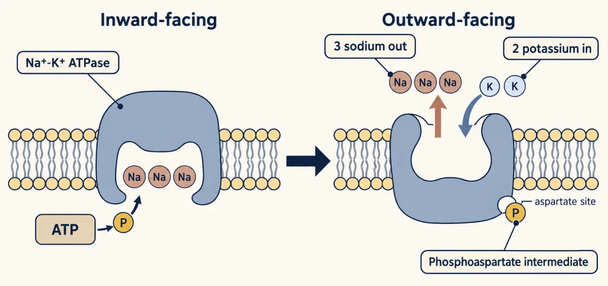
Can one ATP pay for that? Use the numbers from the last section. Pushing one mole of sodium out against its gradient costs about +13.2 kJ, the reverse of the −13.2 kJ/mol for moving it in, so three cost about 39.6 kJ. Bringing one mole of potassium in costs about 2.4 kJ, so two cost about 4.8 kJ. One cycle costs about 44 kJ/mol. ATP hydrolysis under cell conditions releases roughly 50 to 57 kJ/mol. The pump fits inside one ATP, with a modest margin.
It runs constantly and burns a large fraction of your resting ATP, on the order of a quarter, and up to half in neurons, to maintain the gradients that almost everything else depends on.
Spending the battery: coupled transport
The film above shows a sodium gradient acting as a battery. Spending one solute’s downhill run to drag another uphill, with no ATP spent at that step, is secondary active transport. Symporters move both solutes in the same direction: Sodium-glucose cotransporter 1 (SGLT1) hauls glucose into intestinal cells on the back of sodium. Antiporters move them in opposite directions: the Na⁺/Ca²⁺ exchanger uses sodium flowing in to push calcium out.
The coupling ratio matters. SGLT1 carries two sodium ions with each glucose. Two sodiums running in release about 2 × 13.2 = 26.4 kJ/mol. Glucose is uncharged, so only its concentration term counts, and 26.4 kJ/mol is enough in principle to hold glucose about 28,000-fold higher inside than outside. (26.4 / 2.58 is about 10.2, and e raised to 10.2 is about 28,000.) One sodium per glucose would allow only about 170-fold. Carrying two sodiums is what lets the transporter pull glucose so far uphill.
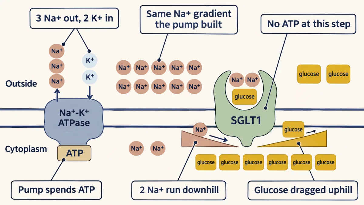
In animal cells, these sodium-coupled transporters depend on the gradient the Na⁺-K⁺ ATPase builds. Other secondary transporters, such as those in plant cells and lysosomes, spend proton gradients made by proton pumps.
Two drugs test this logic. The diabetes drugs called gliflozins block sodium-glucose cotransporter 2 (SGLT2), the kidney’s sodium-glucose symporter that normally reclaims filtered glucose, so glucose spills into the urine and blood sugar falls. Digitalis, from the foxglove plant, inhibits the Na⁺-K⁺ ATPase. Sodium then backs up inside the heart cell, the Na⁺/Ca²⁺ exchanger slows because the sodium gradient it spends has shrunk, calcium accumulates, and each heartbeat hits harder. It is a treatment for failing hearts with a narrow therapeutic window, since too much triggers calcium-overload arrhythmias.
Lowering the toll exactly: the potassium channel filter
Now the fourth puzzle. A potassium channel passes K⁺ more than a thousand times more readily than Na⁺, and Na⁺ is the smaller ion.
The part of the channel that chooses is the selectivity filter, the narrowest stretch of the pore. It is a short tube lined mostly with backbone carbonyl oxygens, turned inward by the signature sequence Thr-Val-Gly-Tyr-Gly (TVGYG). Along the tube sit four binding sites in a row, called S1 to S4. Each site is a cage of eight oxygens; in the innermost site, S4, four of them come from threonine side chains.
Predict. Na⁺ is smaller than K⁺. If the filter were a sieve, which ion would pass more easily? Since K⁺ wins instead, what must the filter be doing? Use the toll.
Check your prediction
A sieve would pass Na⁺, so the filter is not a sieve. It works on the toll. A K⁺ ion entering the filter sheds its water, and the eight oxygens of a site take the water’s place closely, so K⁺ pays little net energy to enter. In the classic snug-fit picture, Na⁺ is too small to touch all eight oxygens at once. It cannot be paid back the energy it spent shedding its own water, so it stalls. The filter repays nearly all of K⁺‘s toll and falls short on Na⁺‘s.
How exact must the fit be? Less exact than it sounds. A preference of 1000 to 1 corresponds to a free-energy difference of RT × ln(1000), only about 17.8 kJ/mol at 37 °C. The hydration energies of Na⁺ and K⁺ differ by about 70 kJ/mol. So the filter does not have to be a perfect stand-in for water. It can even give Na⁺ more energy back than K⁺, as much as about 52 kJ/mol more, and still pass K⁺ a thousand times better, because that falls about 18 kJ/mol short of Na⁺‘s extra 70. Small differences in a huge toll are enough. Simulations refine the snug-fit picture: they suggest the oxygens do give Na⁺ more energy than K⁺, just not enough more. The filter’s carbonyls are flexible, and their number and electrical properties matter as much as the fit, a point still being worked out.
That raises one more puzzle. If the filter binds K⁺ well enough to be that selective, why does K⁺ ever leave? The answer is probably crowding. Several K⁺ ions sit in the filter at once, and like charges repel. When a second ion enters, the repulsion between the ions lowers the barrier for the leading ion to leave. This is the knock-on mechanism. A Newton’s cradle is a fair image of it, but only an image: the energy is not passed along like a collision. Whether water molecules separate the ions in the filter is still argued, though recent evidence favors direct ion-to-ion contact. The point to keep: repulsion among the crowded ions lifts each ion out of the deep well it would sit in if it were alone, so K⁺ leaves fast. K⁺ moves at near the diffusion limit, as fast as ions can diffuse to the pore, around 10⁸ ions per second, selective and fast at the same time. Roderick MacKinnon solved this structure and won the 2003 Nobel Prize in Chemistry for it.
Aquaporins give water its own cheap path in the same spirit, single file, while excluding protons. How voltage-gated Na⁺ and K⁺ channels use the gradients from this lesson to fire a nerve impulse is the subject of the Action Potential lesson.
CFTR: a channel built from a pump family
Now the last puzzle. Cystic fibrosis transmembrane conductance regulator (CFTR) belongs to the ABC transporter family, and its family is mostly pumps. ABC transporters share an architecture: two transmembrane domains that form the path across, and two nucleotide-binding domains that act as the ATP-powered engine. When ATP binds, the two engine domains clamp together and flip the protein from inward-facing to outward-facing. Most family members use that flip to pump solutes; P-glycoprotein, which throws drugs out of cells, is one.
CFTR is the family’s odd member. It has the same core domains plus a regulatory domain, and it works as a chloride channel. Once protein kinase A phosphorylates the regulatory domain, ATP binding gates its pore open.
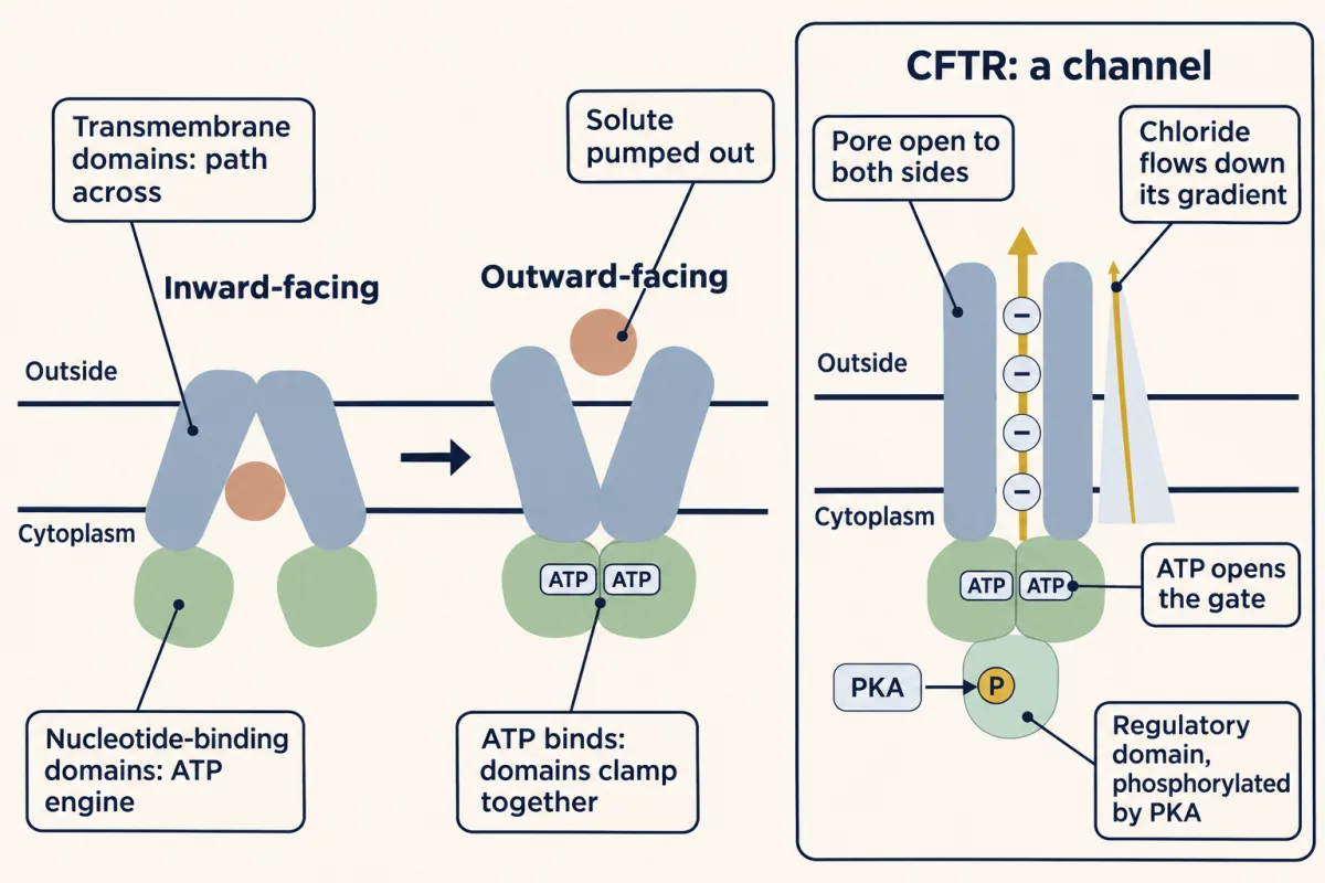
CFTR uses ATP, so why is it a channel and not a pump? Apply the “which way” question. Once the pore is open, chloride flows down its own electrochemical gradient, the way it would through any channel. ATP opens the gate. It does not pay for the move.
The disease comes from a missing protein. In ΔF508, the most common mutation, one phenylalanine is deleted. The protein misfolds, and most of it is destroyed in the endoplasmic reticulum, so in most cystic fibrosis CFTR mostly fails to reach the membranes of cells lining the lungs and gut. The main defect is not a broken channel at the membrane. Most of it never arrives, and the little that does opens poorly. Everything downstream, the thick mucus, the chronic infections, the shortened life, follows from that one missing protein.
Predict. Two kinds of drug could help. A corrector helps the protein fold and reach the surface. A potentiator helps a channel that is already at the surface open. Which does a patient with ΔF508 need?
Check your prediction
Both. The corrector is the one that cannot be skipped: a potentiator alone has little to work on, because most of the channel never arrives. Treatment follows the split in the defect, which is precision medicine aimed at the protein-level consequence of a mutation.
Putting the two questions together
Every case in this lesson came down to the two questions: how cheap is the route around the toll, and which way does the gradient point. Use them on one new case.
Design problem
A drug candidate crosses cell membranes poorly. It carries a carboxylate group, with a pKa of 4. Its target is inside the cell.
- Predict the effect of masking the carboxylate as an ester.
- Suppose a transporter in the membrane happens to carry the drug. Why can the drug now act on its target, and what sets the direction in which the transporter moves it?
Saved in this browser only.
Check your answer
- At pH 7, three units above the pKa, only about one molecule in a thousand is uncharged. The charged carboxylate pays the toll for leaving water, like the charged groups on tryptophan. An ester carries no charge, so the masked drug should cross far faster. Once inside, cellular esterases can cut the ester back to the active acid. This is the speed question.
- The transporter offers a cheap route around the toll, so speed is no longer the problem. Direction is a separate question. If the transporter is a uniporter or a channel, it can only move the drug down its electrochemical gradient. For a negatively charged drug the voltage counts too: the negative inside pushes an anion out, so a uniporter alone would leave the drug less concentrated inside than outside. A carrier coupled to sodium could pull it in uphill, the way SGLT1 pulls in glucose.
Going deeper
Hydration energies depend on the convention
The toll for an ion is often quoted as a single number, but tables differ, because absolute hydration energies depend on a reference convention. All values below are magnitudes, in kJ/mol.
| Quantity | K⁺ | Na⁺ | Na⁺ minus K⁺ |
|---|---|---|---|
| Hydration free energy, Marcus scale | 295 | 365 | 70 |
| Hydration free energy, absolute scale (Tissandier) | 352 | 424 | 72 |
| Values often quoted in teaching (about 330 and 400), between the free-energy and enthalpy figures | 330 | 400 | 70 |
The difference on the free-energy scales is the number the filter section uses. Tabulated hydration enthalpies are a different quantity; they differ by somewhat more, about 80 kJ/mol.
How ions actually leak through a bare bilayer
If an ion had to pay its whole toll to cross, it would never cross. The chance that a molecule has enough energy to cross a barrier is about e−ΔG/RT, the Boltzmann factor. For a toll of 330 to 400 kJ/mol that is 10⁻⁵⁶ to 10⁻⁶⁷. Even if the oil’s slight polarizability is counted (the Born model with a dielectric constant of 2, and effective cavity radii of 1.68 Å for Na⁺ and 2.17 Å for K⁺, larger than the ionic radii in the opening), the cost of moving the ion into the core is about 201 and 156 kJ/mol, giving factors of 10⁻³⁴ and 10⁻²⁶. The observed water-to-sodium ratio of 10¹⁰ corresponds to an extra barrier for Na⁺, over and above water’s own, of only about 59 kJ/mol (RT × ln 10¹⁰). So real ions must take the much cheaper water-dragging or defect route described in the main text, and which picture fits best depends on how thick the bilayer is. That route sets the measured leak.
Liposomes as drug carriers
Liposomes, closed lipid vesicles, carry drugs. Load a water-soluble drug in the aqueous core, a fat-soluble one in the bilayer, and you have a delivery vehicle. Doxil packages the chemotherapy drug doxorubicin in liposomes and spares the heart much of its toxicity. The COVID-19 mRNA vaccines used lipid nanoparticles to protect fragile mRNA long enough to deliver it. Closed vesicles form on their own, but making small, uniform liposomes usually needs extra steps such as sonication or extrusion.
Lipid rafts
Cholesterol and sphingolipids are thought to cluster into lipid rafts, small, short-lived patches roughly 10 to 200 nm across. Their size and roles in living cells are still debated. They may act as gathering points where signaling proteins find their partners, and HIV and influenza appear to use them to assemble and bud.
Statins and muscle pain
The pathway that makes cholesterol may also explain why statins can cause muscle pain in some people. Statins block the mevalonate pathway, which makes not only cholesterol but also isoprenoids and coenzyme Q10 that muscle cells depend on. The mechanism is still debated.
COX-1, aspirin and ibuprofen
The enzyme COX-1 is a monotopic membrane protein: it sits in one leaflet of the endoplasmic reticulum and nuclear membranes, without spanning the bilayer. It pulls arachidonic acid in through a hydrophobic channel to make prostaglandins. Aspirin works by irreversibly acetylating a serine (Ser530) in that substrate channel, which blocks arachidonic acid from reaching the catalytic site. Because the block is permanent, it lasts until the cell makes fresh enzyme. Platelets have no nucleus and cannot make new COX-1, so a blocked platelet stays blocked for its whole life, which is the basis of low-dose aspirin’s lasting antiplatelet effect. Ibuprofen competes for the same site reversibly, so taking ibuprofen before aspirin can shield the site and blunt aspirin’s antiplatelet effect; how much heart protection is lost is still unclear.
Toxins and sensor channels
Two famous toxins, tetrodotoxin from pufferfish and saxitoxin from the dinoflagellates behind paralytic shellfish poisoning, both plug the outer mouth of voltage-gated sodium channels, so nerves cannot depolarize and the victim is paralyzed. TRP channels sense the environment: Transient receptor potential vanilloid 1 (TRPV1) opens to heat and to capsaicin, and transient receptor potential melastatin 8 (TRPM8) opens to cold and to menthol. Capsaicin activates the heat-sensing channel directly, which is why chili registers as heat and mint as cold even though nothing has changed temperature.
From gradients to a nerve impulse
The action potential is where all of this comes together: voltage-gated sodium and potassium channels firing in sequence, the Na⁺-K⁺ pump maintaining the gradients, an inactivation gate that closes the sodium channel shortly after it opens, and a refractory period that keeps the signal traveling in one direction. The region the impulse just passed cannot fire again yet, so the signal does not run backward. The membrane, the impermeable sheet that builds itself, turns out to be the substrate on which a nerve fires.
P-glycoprotein and multidrug resistance
P-glycoprotein is the ABC family’s promiscuous pump, accepting structurally diverse drugs. Tumor cells that overexpress it can become multidrug-resistant, pumping chemotherapy out before it can work.
How an aquaporin passes water but not protons
Aquaporins are channels that pass about 3 billion (3 × 10⁹) water molecules per second through a pore just 2.8 Angstroms wide, single file. Protons normally race through water by hopping along hydrogen bonds, but at the pore’s center two asparagines, helped by the electric field of two short pore helices, reorient the water and break that chain, and a positively charged arginine at the narrowest point repels protons. The channel passes water in a torrent and still excludes the proton. In the kidney, the hormone vasopressin inserts aquaporin-2 into the collecting-duct membrane; when that regulation fails, the kidneys cannot concentrate urine and a person can pass up to about 20 liters of dilute urine a day, the condition called diabetes insipidus.
Trikafta and the clinical picture for CFTR
The drug Trikafta, approved in 2019, combines two correctors (elexacaftor and tezacaftor) with a potentiator (ivacaftor). It is approved for people with at least one copy of ΔF508, and for some other mutations. The projection of decades of added expected survival is for people who carry two copies of ΔF508.
Your notes
Select any text in the lesson to highlight it or add a note to it. Your highlights and notes are saved in this browser and collected in your notebook.
This browser is not letting the site save anything (a private window or strict privacy settings), so highlights and notes will be lost when you leave the page.
How we measure it
FRAP (fluorescence recovery after photobleaching)
Bleach a spot on a fluorescently labeled membrane, then watch it refill. How fast it recovers gives the lateral diffusion coefficient; how much never recovers gives the immobile, anchored fraction. A 50% recovery means roughly half the labeled molecules are immobile, often tethered to the cytoskeleton.
Detergent vs. salt extraction
The operational test for integral versus peripheral membrane proteins. Peripheral proteins release with high salt or a pH change; integral proteins, which sit in the hydrophobic core, come out only when detergent dissolves the bilayer around them.
Planar lipid bilayer recording
Fuse a single purified channel into an artificial bilayer and record its current, resolving one molecule opening and closing in real time. This single-channel approach underpinned decades of channel biophysics.
Membrane-protein X-ray crystallography
Crystallizing a membrane protein (notoriously hard) and solving its atomic structure by X-ray diffraction. The KcsA structure transformed a long argument about how a channel selects ions by showing the carbonyl-oxygen geometry of the selectivity filter directly.
Method films · the methods library
Practice questions on this lesson (8) → MCAT-style, original, each with an explanation
Where this lesson fits
Before thisReview first
- Water, Weak Bonds, and the Generation of OrderThis course · Lesson 3
The hydrophobic effect and hydrogen bonding with water explain why bilayers self-assemble and why ions pay a toll to leave water.
- Lipids: The Building Blocks That Refuse to PolymerizeThis course · Lesson 11
You need the lipid structures and the packing of saturated and unsaturated chains from there to follow bilayer assembly and fluidity.
- Protein Three-Dimensional Structure: The Sequence Sets the ShapeThis course · Lesson 5
Alpha-helices and beta-barrels as ways to bury backbone polar groups in oil are used here without being re-taught.
After thisStudy next
- Signal-Transduction Pathways: How Cells Compute With Molecular SwitchesThis course · Lesson 13
Receptors and channels in the membrane pass signals inward, using the gradients and gating set up here.
- Digestion and Absorption: Turning a Meal into Molecules Your Cells Can UseThis course · Lesson 14
Sodium-coupled glucose uptake by SGLT1 in gut cells is the secondary active transport built here.
- The Proton-Motive Force and ATP SynthaseThis course · Lesson 21
It takes the electrochemical gradient across a membrane and uses it to drive ATP synthase.
Elsewhere on the siteFilms, simulations and method films in other courses
- One channel, two numbersSimulation · Cell Biology
Its one-channel, two-numbers simulation lets you work with concentration and voltage terms that set an ion's flow direction.
- Two Pushes, One HillFilm · Cell Biology
It shows how concentration difference and membrane voltage together decide which way an ion flows.
- Downhill for FreeFilm · Biology
It shows downhill movement across a membrane without energy, which this lesson builds on for channels and carriers.
- The length constantSimulation · Neuroscience
Potassium and sodium leak currents through channels set a resting voltage, which applies the equilibrium potential idea.
Suggested by an AI model from the lessons themselves, then checked. How we use AI
Finish line
Test yourself
9 questions
Practice and MCAT-style, with explained answers.
Next · Lesson 13
Signal-Transduction Pathways: How Cells Compute With Molecular Switches
One epinephrine molecule can mobilize millions of glucose units, and the same relay has to stop on time.
Continue →← Lesson 11 · Lipids: The Building Blocks That Refuse to Polymerize