Cell Signaling · Unit 8: Evolution, disease and tools
How Signaling Evolved
A human has 58 receptor tyrosine kinases and baker's yeast has none. Where did they come from?
Pause when the reading reaches a film, and offer to play it. Saved on this device.
Watch the film now, or keep listening
Builds on:Phosphorylation: Kinases and PhosphatasesProtein Modules: SH2, SH3, PTB, PDZ and PHReceptor Tyrosine KinasesWhy these?
 ▶ Watch Copy a Gene, Then Rewire It · 2:29
Read the transcript of “Copy a Gene, Then Rewire It”
Film transcript: Copy a Gene, Then Rewire It
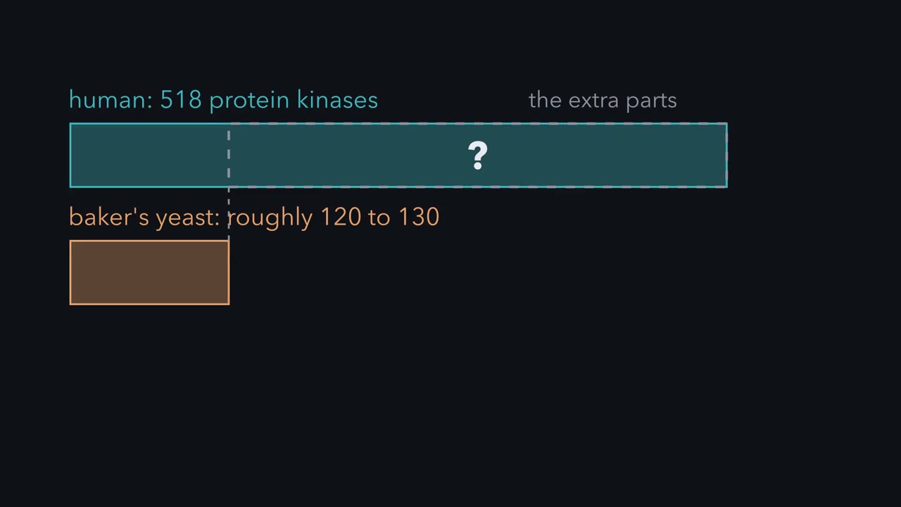
A human has 518 protein kinases. Baker's yeast has roughly 120 to 130. Animals share a short list of signaling pathways, so where did the extra parts come from? The record does not show animals inventing one new pathway after another. Kinase totals roughly track genome size, but the receptors multiplied more than the core. Mistakes in copying DNA sometimes duplicate a single gene. Say a receptor gene is copied. The original keeps doing the job, so the spare copy is free to change. A fly has one EGF receptor and a human has four. Jawed vertebrates carry traces of two whole genome doublings, the likely route from one to four. A spare copy has three main fates.
The common one is loss. A mutation disables the copy, nothing selects against that, and it decays into a pseudogene. In the second fate, the copies divide the old job. Suppose the old receptor worked in gut and in nerve. If one copy loses the regulatory DNA for gut and the other loses it for nerve, the animal needs both. In the third fate, one copy takes a new job. Its tail may change, so a different set of tyrosines recruits different SH2 proteins. A docking site is only a few amino acids. The SH2 domains of PI 3-kinase read a phosphotyrosine followed three positions later by methionine. One or two point mutations in the tail can make such a site, and with it a line to PI 3-kinase.
Now carry the duplication through a whole pathway. In one outcome, everything downstream is copied too, and the four cascades stay apart like four wires. In the other, the receptors go on sharing downstream parts and gain cross-connections. Many inputs then funnel into a few hubs, and Ras is one. The counts fit shared wiring, but they cannot show it, and the core was copied too. So the extra kinases are not a list of new pathways. The record shows old pathways copied, mostly at the receptor end, and the copies changed. New wiring can cost only a few amino acids, because signaling proteins are built from parts that work on their own. Keep this rule: old pathways get copied, and the copies get rewired.
Your genome holds genes for 518 protein kinases, and 58 of them are receptor tyrosine kinases (RTKs). Baker’s yeast runs on roughly 120 to 130 kinases and has no receptor tyrosine kinase at all. So ask: “How did we get such rapid expansion of RTKs and signaling pathways??” The short answer is that animals did not invent one new pathway after another. They kept a small set of old pathways, copied the genes, mostly at the receptor end, and changed the copies. The tyrosine-kinase parts that the receptor pathway uses were already present in their single-celled relatives. A second question holds the rest of the answer: “How are new pathways coupled to receptors??” Movable domains and short docking sites make new connections cheap.
The idea: a few old pathways, copied many times
You have now met the core animal pathways in this course, and the list is short. Comparisons of animal genomes keep returning the same core set. A widely cited review of animal development (Pires-daSilva and Sommer, 2003) names seven, and a 2007 review of receptor tyrosine kinase networks (Amit, Wides and Yarden) adds G-protein-coupled receptors to make eight. This lesson uses the eight:
- G-protein-coupled receptors (lesson)
- nuclear receptors (lesson)
- TGF-beta (lesson)
- Notch (lesson)
- Janus kinase (JAK) and signal transducer and activator of transcription (STAT) (lesson)
- Hedgehog (lesson)
- Wnt, called Wingless in flies (lesson)
- receptor tyrosine kinases (lesson)
A fruit fly has all eight, and so do you. What differs is the number of parts. Humans have four epidermal growth factor (EGF) receptors where the fly and the nematode worm have one each. In a 2007 comparison of sequenced genomes, receptor families had expanded more than the kinase families downstream of them, while the total number of protein kinases grew roughly in step with the number of genes. The receptors are what multiplied.
Receptors, at the top of a pathway, multiplied more than the kinases in its core. The record does not show animals inventing one new pathway after another. It shows old pathways copied, mostly at the receptor end, and the copies changed. Start with the simplest relay known.
The simplest relay: two proteins and one phosphate
Read the transcript of “Two Proteins, One Phosphate”
Film transcript: Two Proteins, One Phosphate
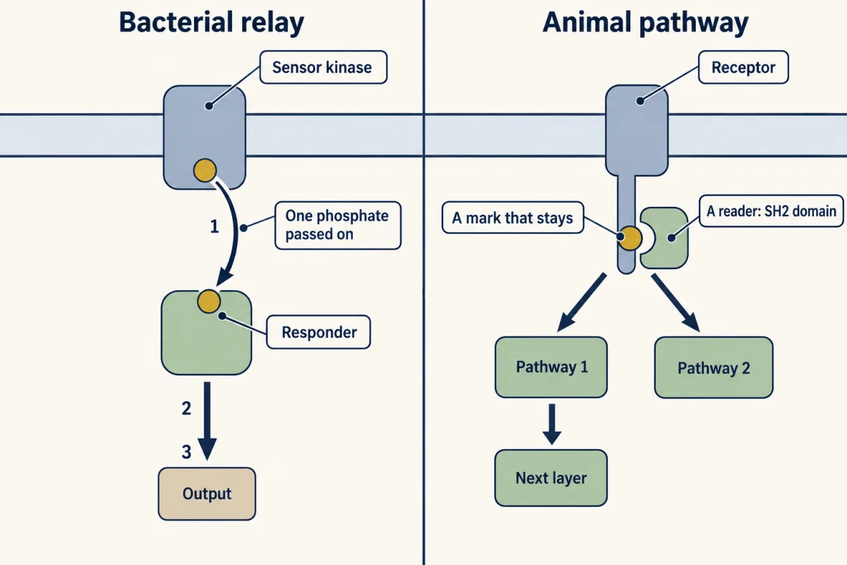
A bacterium senses most of its surroundings with two-component systems. Component one is a sensor kinase, usually set in the membrane. When it detects its signal, it takes the end phosphate from ATP. It puts that phosphate on one of its own histidines. Component two is the responder. It moves that phosphate onto one of its own aspartates. The change in shape that follows switches it on. Most responders are transcription factors, so the whole pathway is a sensor and a gene switch with one phosphate passed between them. Set this beside an animal pathway. A phosphate on aspartate is labile. How long it lasts differs from one responder to another, from seconds for CheY, a swimming responder, to minutes or more for others.
A phosphate on serine, threonine or tyrosine stays until a phosphatase removes it. So the cell controls off separately from on. Because that mark stays, another protein can bind it. An SH2 domain reads a phosphotyrosine. In the bacterial relay, the phosphate changes the responder that carries it, and no separate domain reads it. Most sensor kinases pass their phosphate to one partner and largely ignore the rest. An animal receptor recruits several pathways at once, and each runs through tiers of kinases.
Many bacteria sense much of their surroundings with two-component systems, though single proteins that carry both a sensing domain and an output domain are even more common. Component one is a sensor kinase, usually set in the membrane. When it detects its signal, it takes the end phosphate from ATP and puts it on one of its own histidines. Component two is the responder (its formal name is response regulator). The responder moves that phosphate onto one of its own aspartates, and the change in shape that follows switches it on. Most responders are transcription factors, so the whole pathway is a sensor and a gene switch with one phosphate passed between them.
ATP → histidine on the sensor → aspartate on the responder → output
E. coli has about thirty of these pairs, each for a different condition such as osmotic strength or nitrogen supply. Its swimming uses a variant: the kinase, CheA, is a separate protein that sits beneath the receptors, and the responder, CheY, binds the flagellar motor instead of DNA (the circuit built on it is in signaling circuits). Comparing sequences showed that CheY resembles regulators of sporulation and of membrane protein synthesis, so unrelated behaviours ran on one family of proteins, and experiments with purified chemotaxis proteins showed the phosphate passing directly from CheA to CheY.
Set this beside an animal pathway and you can list what the animal pathway adds.
A mark that stays. A phosphate on aspartate is labile, and how long it lasts differs from one responder to another, from seconds for CheY to minutes or more for others. CheY sheds its phosphate in well under a minute unaided, and a helper protein, CheZ, makes it faster still. A phosphate on serine, threonine or tyrosine stays until a phosphatase removes it (the phosphorylation switch), so how long the signal lasts is set by phosphatases rather than by the chemistry of the mark.
A reader. Because the mark stays, another protein can bind it. An SH2 domain reads a phosphotyrosine. In the bacterial relay the phosphate changes the responder that carries it, and no separate domain reads it.
Branches and layers. Most sensor kinases pass their phosphate to one partner responder and largely ignore the rest. An animal receptor tyrosine kinase recruits several pathways at once, and several of them run through tiers of kinases.
The enzymes differ too. The catalytic domain of a sensor kinase has a different fold from the serine, threonine and tyrosine kinases you know. Animals have no two-component systems, though plants and fungi do.
Copy, then change: three fates for a spare gene
Mistakes in copying DNA sometimes duplicate a single gene, and now and then a whole genome. Susumu Ohno argued in his 1970 book Evolution by Gene Duplication that this is the main source of new genes, because a spare copy can change while the original keeps doing the job. The genomes of jawed vertebrates, ours included, carry the traces of two whole-genome doublings early in their history, the likely route from one epidermal growth factor (EGF) receptor to four.
A spare copy has three main fates.
- It is lost. A mutation disables it, nothing selects against the loss, and it decays into a pseudogene (a gene-like sequence that no longer makes a working protein). This is the common outcome.
- The copies divide the old job. Each loses a different part of what the ancestor did, so both must be kept. This is sub-functionalization. If the old receptor worked in gut and in nerve, one copy may lose the regulatory DNA for gut and the other the regulatory DNA for nerve, so the animal needs both.
- One copy takes a new job, such as answering a new ligand. This is neo-functionalization.
A related word is co-option, the use of an existing gene or pathway for a new purpose, which need not involve copying it: Toll patterns the fly embryo and also fights infection (lesson).
Now carry the duplication through a whole pathway. Begin with one ancestral cascade of ligand, receptor and kinase chain, and double the receptor gene twice. In one outcome everything downstream is duplicated as well and the copies stay apart, which gives four vertical cascades like four separate wires. In the other, the four receptors go on sharing downstream parts and gain cross-connections. That gives a network in which many inputs funnel into a few heavily connected proteins called hubs. Ras is one. The comparison in the first section fits the second outcome better, because receptors multiplied faster than the core they feed. The core was copied too, though: humans have three Ras genes and, after adjusting for gene number, about 1.8 times as many MAP kinases as four invertebrate animals (see Going deeper). Gene counts alone cannot show how the parts are wired.
”The virtue of modularity”: new wiring from old parts
Now the second question: “How are new pathways coupled to receptors??” If every new connection needed a new enzyme, expansion would be slow. The way out is the virtue of modularity.
Signalling proteins are assembled from domains that fold and work on their own (interaction modules). That gives evolution cheap ways to make a connection.
Move a domain. A DNA rearrangement that joins the coding segment for an SH2 domain to another gene makes a protein with a new partner in one step. Adaptors such as Grb2 are nothing but binding domains (adaptors and scaffolds).
A module keeps its job when you move it. Each reader grips only when its target is there, and an enzyme with no reader is not brought anywhere. This is a sketch of the logic, not a prediction for a real construct.
Add a docking site. A new docking site is a few amino acids. The SH2 domain of Grb2 reads a phosphotyrosine followed two positions later by asparagine. The SH2 domains of PI 3-kinase read a phosphotyrosine followed three positions later by methionine. One or two point mutations in a receptor’s tail can make or remove such a site, and with it a line to Ras or to PI 3-kinase.
Change where and when a gene is on. Short stretches of regulatory DNA near a gene, or sometimes far from it, decide which cells make it and when. A receptor that turns up in a new tissue meets different partners and different target genes, so an unchanged pathway gives a new result.
The logic is the same at both levels. A gene gains a new expression pattern through a short DNA motif, and a protein gains a new partner through a short peptide motif.
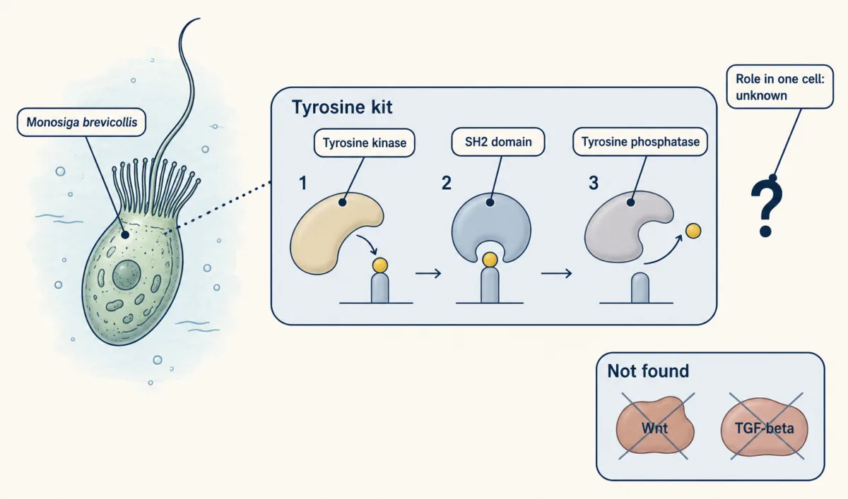
Genome counts agree. Many families of interaction domains grow in number from yeast to worm and fly to mouse and human, and the parts for tyrosine phosphorylation show it plainly. Budding yeast has one protein with an SH2 domain. Humans have more than a hundred, alongside 90 tyrosine kinases.
Where did that tyrosine kit come from? Choanoflagellates are the closest living single-celled relatives of animals. Nicole King and Sean Carroll reported a receptor tyrosine kinase gene in one of them, Monosiga brevicollis, in 2001. Its genome, published in 2008, encodes tyrosine kinases, tyrosine phosphatases and SH2 domains in numbers that match or exceed ours, yet it lacks recognisable Wnt and TGF-beta genes. So the kit of a kinase to write the mark, an SH2 domain to read it and a phosphatase to erase it predates animals. What it does in a single cell is still largely unknown.
Why a network and not a set of wires
A network gives up insulation, so what does it buy? Robustness: the output holds when a part fails or an input is noisy (signaling circuits). Remove one receptor and its sisters, which feed the same core, can cover part of the loss if they are made in the same cells. Negative feedback loops in the shared core can steady the output for every receptor that feeds it.
The same design has costs.
A hub is a single point of failure. Lock Ras on (GTPase switches) and the receptors above it lose control of everything Ras drives.
A shared core allows bypass. A lung tumour held in check by an inhibitor of the epidermal growth factor (EGF) receptor can escape by amplifying the gene for MET, a different receptor that feeds the same core.
And relatives resemble each other. Nearly all human kinases descend from one ancestral catalytic domain and keep a similar ATP pocket, so a drug shaped for one pocket often fits a relative. Imatinib, the drug for the BCR-ABL kinase of chronic myeloid leukaemia, also inhibits KIT and the platelet-derived growth factor (PDGF) receptors. KIT and one of the PDGF receptors are sister genes that sit side by side on chromosome 4. That cross-reaction is why imatinib treats a gut tumour driven by KIT, and it is why new kinase drugs are profiled against hundreds of kinases. The lesson on drugs in this unit starts from there.
Design problem
The gene for a receptor tyrosine kinase is copied by accident, so the animal now has two. Describe three different fates for the extra copy. For the fate in which the copy comes to carry a new message, say what must change in the copy, and what must change so that the new message does not leak into the old pathway.
Saved in this browser only.
One way to do it
Lost. The original still does the work, so a damaged spare is not removed by selection and decays into a pseudogene.
Shared. Suppose the old receptor worked in gut and in nerve. If one copy loses the regulatory DNA for gut and the other loses it for nerve, the animal needs both. No new message exists. One job is now held by two genes.
New message. The outside of the copy must come to bind a different ligand, often a drifting duplicate of the old one, or the copy must come to be made in cells where the old receptor was absent. Its tail may change too: a different set of tyrosines recruits a different mix of SH2 proteins.
Stopping the leak. A receptor tyrosine kinase signals as a pair (lesson), so the first leak is an old receptor pairing with a new one. The surfaces that hold the pair together must diverge, or the two receptors must be kept in different cells. The old ligand must also stop fitting the new receptor. Downstream, both receptors still reach Ras, so the new one must differ in its docking sites, in how long it signals, or in the scaffold that holds its kinases. Evolution rarely finishes this job, and the leaks that remain are the cross-connections of a network.
Going deeper
The 2007 genome comparison
A 2007 review of sequenced genomes (Amit, Wides and Yarden, Molecular Systems Biology 3:151) compared the human genome with four invertebrate animals, a sea squirt, a sea urchin, the fruit fly and a nematode worm, after adjusting for the number of genes in each genome. By that count humans have about 2.9 times as many receptor tyrosine kinases (RTKs), 2.5 times as many epidermal growth factor (EGF) receptors and 1.8 times as many MAP kinases. By the same adjustment the total number of protein kinases is only about 1.2 times higher, a difference too small to be significant.
Who showed that the relay is shared
In 1985 Ann Stock, with Daniel Koshland and Jeffry Stock, reported that the sequence of CheY resembled regulators of sporulation and of membrane protein synthesis, so unrelated behaviours ran on one family of proteins. In 1988 Melvin Simon’s laboratory mixed purified chemotaxis proteins and followed the phosphate from CheA to CheY, and Jeffry Stock’s group at Princeton reported the same transfer that year.
ErbB2 and ErbB3: a partnership, not a split
The EGF receptor family shows a division of labour that resembles sub-functionalization: ErbB2 has no known ligand, ErbB3 has a feeble kinase, and the two signal strongly as a pair. They are not sister copies of one gene, though (ErbB2’s closest relative is the EGF receptor, and ErbB3’s is ErbB4), so this is a partnership between relatives rather than a textbook split of one gene’s job.
Your notes
Select any text in the lesson to highlight it or add a note to it. Your highlights and notes are saved in this browser and collected in your notebook.
This browser is not letting the site save anything (a private window or strict privacy settings), so highlights and notes will be lost when you leave the page.
How we measure it
Kinome cataloguing by sequence search
Start from the conserved motifs of the kinase catalytic domain, build a statistical profile of them, and scan a whole genome for every gene that fits. Sorting the hits by similarity gives families, and comparing families across species shows which are old and which grew.
In vitro phosphotransfer with labelled ATP
Mix purified proteins with ATP whose terminal phosphate is radioactive, stop the reaction at intervals, separate the proteins on a gel and see which one carries the label. Adding the proteins in different orders shows the direction in which the phosphate travels.
Gene trees: orthologs and paralogs
Align related genes from several species and build a tree from the differences. Genes that split when two species split are orthologs, and genes that split by duplication inside one lineage are paralogs. The tree dates each duplication against the branching of species.
Domain swapping and chimeric proteins
Join a domain from one protein onto another and ask whether the hybrid carries the borrowed function. If a binding domain or the outside of a receptor still works in its new setting, it is a true module, and the swap shows what that module contributes.
Practice questions on this lesson (3) → MCAT-style, original, each with an explanation
Where this lesson fits
Before thisReview first
- Phosphorylation: Kinases and PhosphatasesThis course · Lesson 6
Phosphate marks on serine, threonine and tyrosine, and the phosphatases that remove them, are compared with the bacterial histidine-aspartate relay here.
- Protein Modules: SH2, SH3, PTB, PDZ and PHThis course · Lesson 9
Moving SH2 domains and docking sites to make new wiring only makes sense once you know how these modules read phosphotyrosine.
- Receptor Tyrosine KinasesThis course · Lesson 18
The expansion from one receptor tyrosine kinase to 58 assumes you already know how these receptors work.
After thisStudy next
- Drugs Against a PartThis course · Lesson 42
Related kinases keep a similar ATP pocket, so a drug shaped for one often hits its relatives.
- Oncogenes: When the Switch SticksThis course · Lesson 41
Shared hubs like Ras and bypass through a second receptor explain how a stuck switch or an escape route drives cancer.
Elsewhere on the siteFilms, simulations and method films in other courses
- Calibrating a molecular clockSimulation · Ecology and Evolution
Gene copies diverge as substitutions build up over time, the same process that changed duplicated receptors.
- Same Protein, Different SwitchFilm · Ecology and Evolution
New wiring comes from changing where and when a gene is switched on, shown here with enhancer changes.
Suggested by an AI model from the lessons themselves, then checked. How we use AI
Finish line
Test yourself
8 questions
Practice and MCAT-style, with explained answers.
Next · Lesson 41
Oncogenes: When the Switch Sticks
A chicken virus causes cancer with one gene. The chicken already had that gene.
Continue →← Lesson 39 · Reverse Signaling: When the Ligand Is Also a Receptor