Cell Signaling · Unit 5: Signals that build and defend an animal
Innate Immunity: Toll-like Receptors and NF-κB
How does a cell recognize an enemy it has never met, and answer within minutes?
Pause when the reading reaches a film, and offer to play it. Saved on this device.
Watch the film now, or keep listening
Builds on:Ubiquitin and Regulated Proteolysis as SignalsPhosphorylation: Kinases and PhosphatasesProtein Modules: SH2, SH3, PTB, PDZ and PHWhy these?
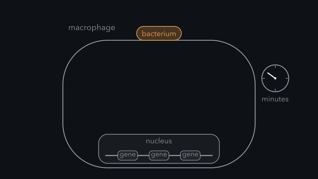 ▶ Watch A Cell Keeps Its Answer Ready · 2:24
Read the transcript of “A Cell Keeps Its Answer Ready”
Film transcript: A Cell Keeps Its Answer Ready
How does a cell recognize an enemy it has never met, and answer within minutes? A macrophage is a white blood cell that swallows microbes. Within minutes of touching a bacterium, it begins to change its gene expression. So what links a touch at the membrane to genes in the nucleus? The answer was waiting in the cytoplasm the whole time. The answer is NF-κB, a transcription factor. It is a pair of proteins that bind DNA and switch genes on. In a resting cell, it is already made and folded. It waits in the cytoplasm, held by its inhibitor, IκB. IκB covers the signal that would send NF-κB into the nucleus. A bacterial molecule, lipopolysaccharide, holds two Toll-like receptors side by side. The pair docks an adaptor, MyD88, which stacks with IRAK kinases into a tower.
An activated IRAK binds TRAF6, which builds a ubiquitin chain joined through lysine 63. Together with head-to-tail chains, it gathers TAK1 next to IKK. TAK1 switches on IKK, the IκB kinase. IKK adds two phosphates to IκB. Even then, IκB still holds NF-κB. A second ligase adds a chain joined through lysine 48. That chain sends IκB to the proteasome, which destroys it. Now nothing covers the signal that sends NF-κB into the nucleus. Now NF-κB, already folded, enters the nucleus. The two proteins wrap around the DNA, and each reads one half of its site. NF-κB switches on its genes. Nothing had to be transcribed or translated first. So NF-κB reaches its genes within minutes.
Take a cell line with no NF-κB activity, and block its protein synthesis. Lipopolysaccharide still makes the activity appear. So the protein must already have been there, inhibited. Here is the rule. Finish the activator, hold it outside the nucleus, and remove its inhibitor when the signal comes.
How does a cell recognize an enemy it has never met? Toll-like receptors read molecules that microbes cannot do without, and within minutes the cell switches on inflammatory genes, such as the gene for tumor necrosis factor (TNF), by freeing a transcription factor, NF-κB, that was waiting in the cytoplasm. Antibodies take days to appear, and a fruit fly makes none at all. Yet a fly survives on rotting fruit, and your macrophages, the white blood cells that swallow microbes, begin to change their gene expression within minutes of touching a bacterium. The same alarm, if it runs too long or too hard, injures the host. This lesson follows the fast answer from a receptor at the membrane to NF-κB, a protein that binds DNA and switches genes on, then shows how the response is ended and what goes wrong when it signals too little or too much.
The idea: detect what a microbe cannot give up
In 1989 the Yale immunologist Charles Janeway pointed at a puzzle in his own field. A purified protein injected alone provokes little immunity unless it is mixed with an adjuvant, an additive that provokes the immune system, and nobody knew why. His answer was a prediction: animals must carry receptors for “certain characteristics or patterns common on infectious agents but absent from the host”. The microbial features became known as pathogen-associated molecular patterns, and he called the proteins that bind them pattern-recognition receptors. The receptors he had in mind had not yet been identified as a class.
A receptor for adrenaline fits adrenaline and its close relative noradrenaline. A pattern receptor fits a whole class, chosen because the microbe cannot do without it: the lipid coat of a bacterium, or the double-stranded RNA of a virus. A bacterium cannot discard its outer membrane. It can, though, alter the part the receptor reads: at body temperature Yersinia pestis makes a lipid A that TLR4 barely detects, and some flagellated bacteria carry a flagellin that TLR5 does not recognize. That is why a short list of receptors can do work for which antibodies need billions of variants.
A fly gene for back and belly
The evidence came from an animal with no antibodies. In 1996 Jules Hoffmann’s laboratory showed that Toll, a fruit fly gene known for setting the back-to-belly axis of the embryo, is also needed to switch on an antifungal gene in adult flies, through a relay that ends at fly relatives of NF-κB. A year later Ruslan Medzhitov and Janeway reported a human relative of Toll which, locked in the ‘on’ state, switched on NF-κB and then inflammatory genes. Fly Toll does not bind the microbe itself; binding the microbial molecule directly is the mammalian innovation. What the human receptor detects was still unknown.
The receptor for endotoxin
That answer came from mice. Since the 1960s geneticists had known strains that barely react to lipopolysaccharide (LPS, or endotoxin), the lipid-and-sugar molecule that coats Gram-negative bacteria, the ones with an outer membrane. A dose that sends a normal mouse into shock leaves these strains nearly unaffected, and one gene makes the difference. In 1998 Bruce Beutler’s laboratory showed by positional cloning, which means reading the genes in the stretch of chromosome that always travels with the trait, that the gene was Tlr4, a mouse relative of Toll. Akira’s laboratory and others then matched most of the rest of the family, ten receptors in humans, to their targets, using gene deletions together with binding assays and structures.
| Receptor | What it detects | Where it sits |
|---|---|---|
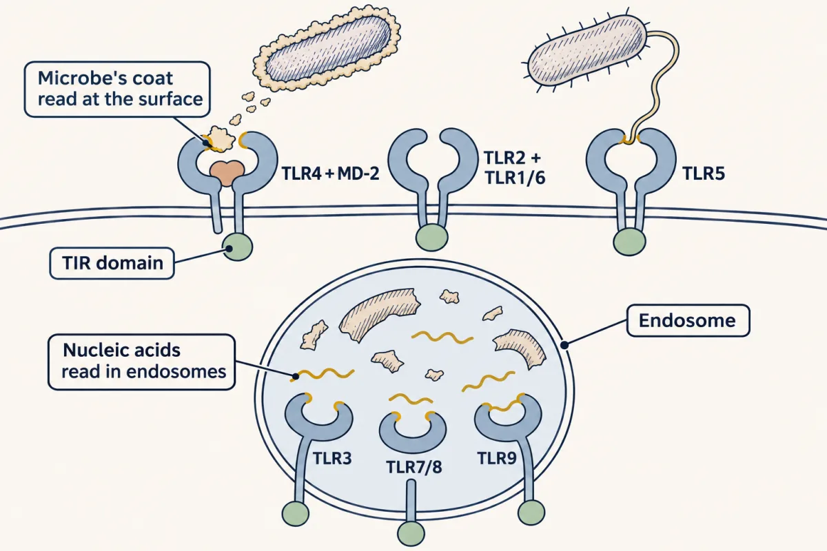
| TLR4, with its partner MD-2 | lipopolysaccharide | cell surface |
| TLR2, paired with TLR1 or TLR6 | bacterial lipoproteins | cell surface |
| TLR5 | flagellin, the protein of bacterial flagella | cell surface |
| TLR3 | double-stranded RNA | endosome |
| TLR7 and TLR8 | single-stranded RNA | endosome |
| TLR9 | DNA with unmethylated CG sequences | endosome |
Molecules from the outside of a microbe are read at the cell surface. Nucleic acids are exposed once what a cell has swallowed, whether a microbe or dying host material, is broken open, so their receptors sit in endosomes, the compartments that hold what a cell has swallowed.
From receptor to gene: an answer held ready
A Toll-like receptor has a horseshoe of leucine-rich repeats outside the cell and a TIR domain inside, named for Toll and the interleukin-1 receptor, which share it. Ligand binding holds two receptors side by side and so pairs their TIR domains. The paired domains are a docking site for the adaptor MyD88. Every Toll-like receptor uses MyD88 except TLR3, which uses a different adaptor, TRIF, on a route that leads to interferon genes. At TLR4 and TLR2 a sorting adaptor, TIRAP, helps bring MyD88 to the paired TIR domains; TLR4 then switches to TRIF, through a second sorting adaptor TRAM, once it has been taken into an endosome.
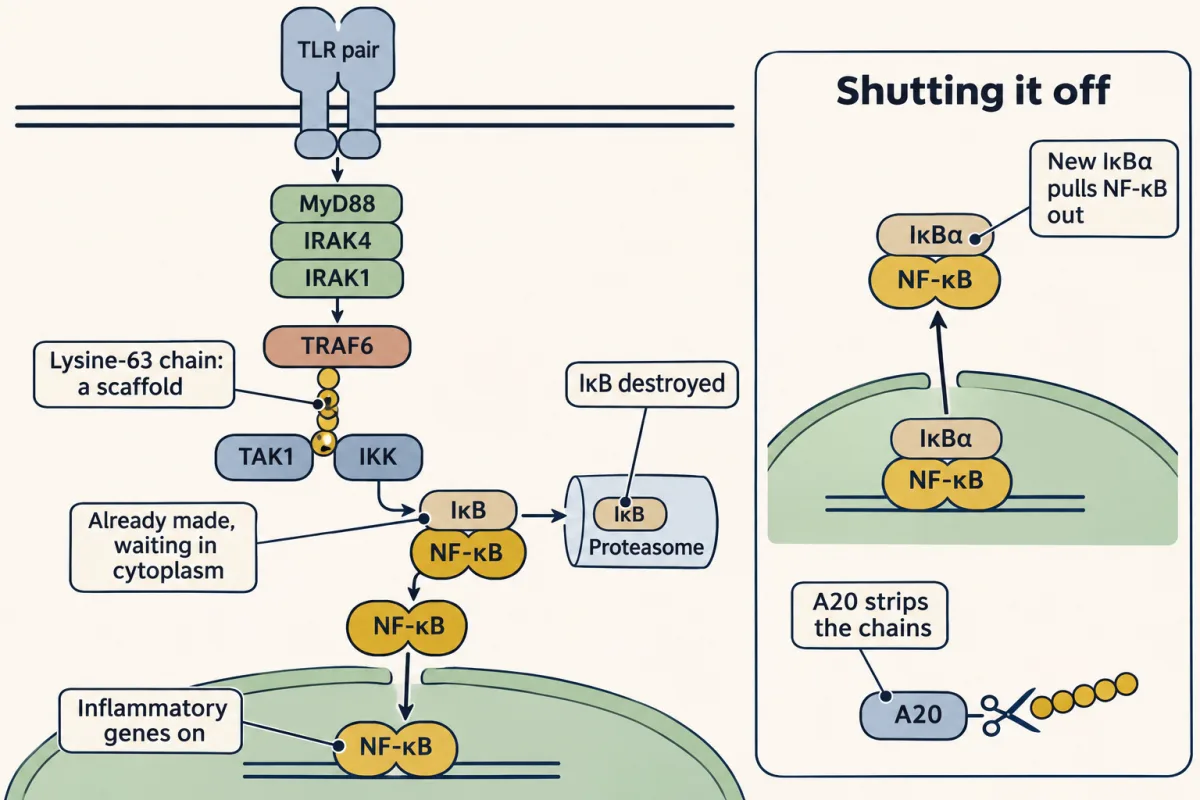
MyD88 has a TIR domain at one end and, at the other, a death domain, a small module that binds others of its kind. Those of MyD88 stack with those of the kinases IRAK4 and IRAK1 or IRAK2 into a helical tower. Inside it the IRAK4 kinases are close enough to phosphorylate one another and then their IRAK partners, and an activated IRAK binds the next protein, TRAF6.
TRAF6 is an E3 ubiquitin ligase. You know the rule from ubiquitin and proteolysis: a chain joined through lysine 48 sends a protein to the proteasome, and a chain joined through lysine 63 is a scaffold. In 2000 Zhijian Chen’s laboratory worked out this step in a test tube, purifying a missing factor from cell extracts. TRAF6, with a two-part enzyme called Ubc13/Uev1A, makes lysine-63 chains (in cells the related Pellino ligases can make them too), and those chains switch on the IκB kinase complex, IKK. The chain is a platform: the TAB2 and TAB3 partners of a kinase called TAK1 read it, while the IKK subunit NEMO is held by chains of a second kind, linked head to tail. With both kinds present, TAK1 phosphorylates the activation loop of IKK.
TLR pair → MyD88 → IRAK4 and IRAK1 → TRAF6 → lysine-63 chain → TAK1 → IKK → IκB destroyed → NF-κB enters the nucleus
NF-κB and its inhibitor
The target of IKK had been found years earlier in David Baltimore’s laboratory. In 1986 Ranjan Sen found a nuclear protein that bound a ten-base sequence in a control region of an antibody gene, the κ light chain, and the two named it NF-κB. In a cell line that lacked the activity, LPS made it appear, even with protein synthesis blocked. The protein must already have been there, inhibited. In 1988 Patrick Baeuerle found the inactive form in the cytoplasm, released it with a detergent, and named the inhibitor IκB.
NF-κB is a pair of Rel-family proteins. IκB proteins are built of ankyrin repeats, a common protein-binding fold, and they cover the signal that would send NF-κB into the nucleus. IKK adds two phosphates to IκB, a second E3 ligase adds a lysine-48 chain, and the proteasome destroys it. NF-κB, already folded, enters the nucleus. Nothing has to be transcribed or translated first, so it reaches its genes within minutes.
Ending the response
One gene that NF-κB switches on encodes IκBα, its own inhibitor. New IκBα enters the nucleus, takes NF-κB off the DNA and carries it back out. NF-κB also switches on A20, an enzyme that strips the lysine-63 chains and so shuts the pathway off further upstream. This is the negative feedback loop of signaling circuits, and under a lasting stimulus NF-κB can enter and leave the nucleus in waves.
”The anti-death, death pathway”: one receptor says die and stay alive
Read the transcript of “One Receptor, Two Messages”
Film transcript: One Receptor, Two Messages
Here is a loop. When NF-κB switches on, one of its target genes encodes tumor necrosis factor, or TNF. TNF is a cytokine, which is a signaling protein that immune cells secrete. Where does it go next? TNF binds TNF receptor 1 on the same cell, and on its neighbors. That receptor also signals to IKK and NF-κB. So the alarm spreads. But this receptor carries a second message. TNF receptor 1 is a death receptor. It can instruct a cell to kill itself. Usually it does not. NF-κB switches on genes whose products block the death machinery. So the receptor says die and stay alive at once. The second message normally wins. Now take cultured cells whose NF-κB arm is blocked, for example by an IκB that cannot be phosphorylated.
The same TNF kills. The death arm belongs to a later lesson. Keep this rule. NF-κB normally makes the stay-alive message win.
NF-κB earns this name from what the pathway does next. One of its target genes encodes tumor necrosis factor (TNF), a cytokine, which is a signaling protein that immune cells secrete. TNF binds TNF receptor 1 on the same cell and on its neighbors, and that receptor also signals to IKK and NF-κB, so the alarm spreads.
TNF receptor 1 is a death receptor: it can instruct a cell to kill itself. Usually it does not. Two checkpoints hold the death machinery back: at the receptor itself, ubiquitination and phosphorylation of a kinase called RIPK1 keep it from acting, and this needs no new transcription; then NF-κB switches on genes whose products block the death machinery. The receptor says “die” and “stay alive” at once, and the second message normally wins. In cultured cells whose NF-κB arm is blocked, for example by an IκB that cannot be phosphorylated, the same TNF kills. The death arm belongs to a later lesson.
A second alarm: nucleic acid in the wrong place
A virus has no cell wall to detect. It does have nucleic acid, and so does the cell. Cells watch for it in two places: inside endosomes, and in the cytosol, where the sensors RIG-I and MDA5 catch viral RNA and pass the signal through MAVS to interferon genes, and where a DNA sensor described below does the same for DNA.
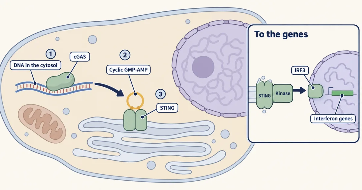
The sensor that links cytosolic DNA to interferon was purified in Zhijian Chen’s laboratory and reported in 2013. The enzyme, cGAS, binds double-stranded DNA and then joins one GTP and one ATP into a small ring, cyclic GMP-AMP, a second messenger of the kind you met with cAMP. The ring binds STING, a protein in the membrane of the endoplasmic reticulum. Activated STING then moves from the ER toward the Golgi and recruits the kinase TBK1, which activates the transcription factor IRF3, and IRF3 switches on interferon genes. Interferon then warns neighboring cells through the JAK-STAT pathway.
Too little and too much
Children born without working MyD88 or IRAK4 get severe, repeated infections with pus-forming bacteria such as Streptococcus pneumoniae and Staphylococcus aureus, often with little fever. They resist most other microbes, and the infections become rarer as they grow up.
Septic shock is the failure at the other end. In some Gram-negative infections LPS circulates and macrophages and other cells throughout the body meet it, and the tumor necrosis factor (TNF) and other mediators they release make blood vessels widen and leak, so blood pressure falls and organs fail. Human sepsis is broader than this picture: it often has no Gram-negative cause, it combines inflammatory injury with immune suppression, and drugs that block TLR4 or TNF have not improved survival in patients. Much of the damage is done by the host’s own response. The LPS-blind mice show both sides: endotoxin does not shock them, and infection with Gram-negative bacteria kills them far more easily.
In small, controlled amounts the alarm is useful. Monophosphoryl lipid A, a modified fragment of LPS that activates TLR4 with far less toxicity, has been used as part of the adjuvant in licensed vaccines against human papillomavirus and shingles. It is one answer to the question Janeway asked in 1989.
Design problem
A virus is built from the same four nucleotides as the cell that it infects. Design a way for a cell to detect the nucleic acid of a virus and ignore its own, using only where you place the receptor and one or two chemical differences. Then say what you would expect to go wrong in a person whose own DNA leaks into the wrong compartment.
Saved in this browser only.
One way to do it
Start with location. Put receptors inside endosomes, facing inward. A cell keeps its own DNA in the nucleus and mitochondria, while a swallowed virus is taken apart in the endosome and its nucleic acid is exposed there. TLR3, TLR7, TLR8 and TLR9 sit here.
Then add chemistry. TLR9 binds DNA in which the sequence CG is unmethylated. That is common in bacterial DNA and in many viral DNAs and rare in yours, where CG is scarce and usually methylated. TLR3 binds long double-stranded RNA, which a healthy cell makes little of and many viruses make as they copy their genomes.
In the cytosol, exposure does most of the work: long, free double-stranded DNA does not belong there. It is not location alone, since much of the sensor sits in the nucleus held on chromatin, and DNA packaging, clearance and control of the sensor keep it quiet. The sensor described above, cGAS, binds DNA of any sequence.
The weak point is the assumption that your own DNA stays put. If it leaks into the cytosol, the sensor fires with no infection and the cell makes interferon without stopping. People who lack TREX1, a nuclease that clears DNA from the cytosol, develop an inflammatory disease, Aicardi-Goutières syndrome, that resembles a viral infection present from birth.
Going deeper
Toll in the fly, in full
The evidence came from an animal with no antibodies. Toll was known in the fruit fly as a gene the mother uses to set the back-to-belly axis of her embryo. In 1996, in Jules Hoffmann’s laboratory in Strasbourg, Bruno Lemaitre tested adult flies with a disabled Toll pathway. They could not switch on the gene for drosomycin, a peptide that kills fungi, and they died after infection with the mold Aspergillus fumigatus. The relay inside the cell was mostly the one the embryo uses, and it ends at fly relatives of the mammalian transcription factor NF-κB. Fly Toll does not bind the fungus itself: it binds Spatzle, a fly protein cut into its active form by a protease cascade that other fly proteins set off when they detect fungal and bacterial cell walls. Binding the microbial molecule directly is the mammalian innovation.
A year later Ruslan Medzhitov and Janeway reported a human relative of Toll, which they had cloned, locked in the “on” state and expressed in human cells. NF-κB switched on, and inflammatory genes followed. What the human receptor detects was still unknown.
Cloning Tlr4, in full
That answer came from mice. Since the 1960s geneticists had known strains that barely react to lipopolysaccharide (LPS, or endotoxin), the lipid-and-sugar molecule that coats Gram-negative bacteria, the ones with an outer membrane. A dose that sends a normal mouse into shock leaves these strains nearly unaffected, and one gene makes the difference. Bruce Beutler’s laboratory found it by positional cloning, which means reading the genes in the stretch of chromosome that always travels with the trait. In 1998, with Alexander Poltorak as first author, they reported that the gene was Tlr4, a mouse relative of Toll. One resistant strain had a single amino acid changed in the part of the receptor inside the cell, and a second had no Tlr4 at all. Danielle Malo’s group reported the same gene early in 1999, and that year Shizuo Akira’s laboratory showed that mice with Tlr4 deleted barely respond to LPS. Akira’s laboratory and others then matched most of the rest of the family, ten receptors in humans, to their targets, using gene deletions together with binding assays and structures.
The Rel family
The family: Dorsal, Dif and Relish in the fly; RelA, RelB and c-Rel in mammals, along with p50 and p52.
Your notes
Select any text in the lesson to highlight it or add a note to it. Your highlights and notes are saved in this browser and collected in your notebook.
This browser is not letting the site save anything (a private window or strict privacy settings), so highlights and notes will be lost when you leave the page.
How we measure it
Positional cloning
Cross a mutant strain to a normal one, follow which stretch of chromosome is always inherited with the trait, narrow that interval, and sequence the genes in it. It finds a gene from its effect alone, with no guess about what the protein is. This is how Tlr4 was identified as the gene behind lipopolysaccharide resistance in mice.
Gene knockout in mice
Delete one gene in a mouse and ask what the animal can no longer do. A mouse lacking a single Toll-like receptor stops responding to one class of microbial molecule, which shows that the response needs that receptor. Binding assays and structures then show how the ligand is recognized.
Electrophoretic mobility shift assay (gel shift)
Mix a short labeled piece of DNA with a cell extract and run it on a gel. DNA with a protein bound to it moves more slowly. The assay reports whether an active DNA-binding protein is present, and it is how NF-κB was detected and its switching-on followed.
Reconstitution from purified components
Rebuild a reaction in a test tube from purified proteins, then leave out one at a time. If the reaction runs, those parts are sufficient under the conditions tested. The need for TRAF6, Ubc13/Uev1A and lysine-63 chains in activating IKK was shown this way, once the factors had been purified from cell extracts.
Method films · the methods library
- Binding assays: immunoprecipitation, pull-down and gel shift 3:43
- Cell-free extracts and reconstitution 4:01
- Engineered mutants: site-directed, dominant-negative and constitutively active 4:25
- Family linkage and positional cloning 3:52
- Gene cloning: vectors, libraries and screening 4:04
- Gene targeting: knockout and knock-in mice 4:13
- Pharmacological blockade and inhibitors 4:10
- Protein purification and column chromatography 4:14
Practice questions on this lesson (3) → MCAT-style, original, each with an explanation
Where this lesson fits
Before thisReview first
- Ubiquitin and Regulated Proteolysis as SignalsThis course · Lesson 8
You need the difference between lysine-48 chains that destroy and lysine-63 chains that scaffold to follow TRAF6 and IκB.
- Phosphorylation: Kinases and PhosphatasesThis course · Lesson 6
IRAK, TAK1 and IKK all pass the signal on by phosphorylating the next kinase or inhibitor.
- Protein Modules: SH2, SH3, PTB, PDZ and PHThis course · Lesson 9
MyD88 and the IRAKs assemble through TIR and death domains, which are protein-binding modules like those taught there.
After thisStudy next
- Antigen Receptors and CheckpointsThis course · Lesson 29
T-cell and B-cell receptors also switch on NF-κB, through a different relay, to drive adaptive immune responses.
- Death Receptors, BCL-2 and CaspasesThis course · Lesson 34
It takes up the death arm of TNF receptor 1 that this lesson leaves for later.
- Stress Signals: p38 and JNK, p53, Unfolded Proteins and OxidantsThis course · Lesson 33
It covers another fast stress response where a transcription factor is held ready and then released.
Suggested by an AI model from the lessons themselves, then checked. How we use AI
Finish line
Test yourself
8 questions
Practice and MCAT-style, with explained answers.
Next · Lesson 29
Antigen Receptors and Checkpoints
The T-cell receptor has no enzyme and almost no tail. How does it tell the cell to kill?
Continue →