Cell Signaling · Unit 4: Receptors with enzymes attached
Receptor Tyrosine Kinases
One helix crosses the membrane. So how does a growth factor outside switch on a kinase inside?
Pause when the reading reaches a film, and offer to play it. Saved on this device.
Watch the film now, or keep listening
Builds on:A Field Guide to ReceptorsPhosphorylation: Kinases and PhosphatasesProtein Modules: SH2, SH3, PTB, PDZ and PHWhy these?
 ▶ Watch The EGF receptor kinase switches on when two copies touch · 2:33
Read the transcript of “The EGF receptor kinase switches on when two copies touch”
Film transcript: The EGF receptor kinase switches on when two copies touch
The year is 2006. You work in John Kuriyan's laboratory at Berkeley. You are studying the receptor for EGF (epidermal growth factor). Three facts about it are known. 1. The receptor has three parts: an outer part that binds EGF, one pass through the membrane, and an inner part that is a kinase. This is the kinase that Cohen found. On its own, the kinase is off. 2. EGF is one small protein. One EGF molecule binds one receptor. It cannot hold two receptors. 3. But the receptor responds only in pairs. When you add EGF, receptors come together two at a time, each with its own EGF bound, and the kinases switch on. A receptor with EGF bound but no partner stays off.
Answer three questions. 1. EGF cannot join two receptors. So what has EGF binding physically done to the receptor, so that receptors now hold on to each other? 2. Once two receptors are held together, what switches the kinase on? Say what one kinase must do to the other. 3. Predict: cut the inner kinase free from the membrane, the outer part and EGF. Then crowd many copies of it together in a tube, so that they touch each other. Will the kinase be on or off? Explain your prediction using your answer to question 2. You now have a kinase that stays off until another copy comes close. This page asks what the two copies do to each other when they meet, and whether both copies end up the same.
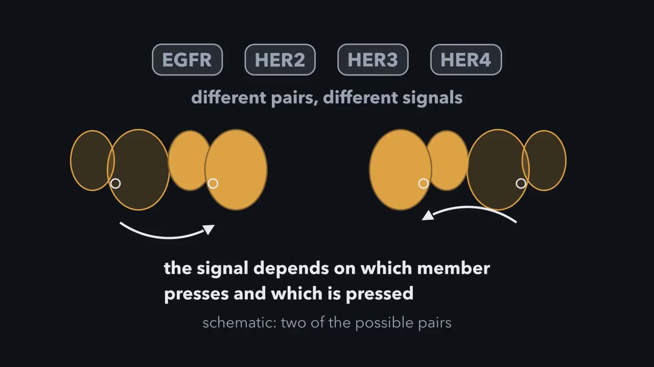
The EGFR kinase's off shape is one that two other enzymes also use. Src kinase and the cyclin-dependent kinases both fold into a resting shape that blocks their own activity. The isolated EGFR kinase, left alone, has the same kind of shape. Kuriyan's group solved the structure of the kinase pair. In the pair, the back lobe of one kinase presses against the front lobe of the other. So the two copies do different things. The pressing copy uses the shape of its back surface to push the other's active site into its working position. A cyclin protein switches on a cyclin-dependent kinase in the same way. The copy that is pressed is the one that switches on.
The same contact is used across the whole family of these receptors: EGFR, HER2, HER3 and HER4. So a cell can make different signals from the same set of parts by pairing different members. The signal depends on which member presses and which member is pressed.
Fifty-eight of your 518 protein kinases are built into the cell surface, with a ligand-binding half outside the cell and the kinase inside. Only one helix crosses the membrane between the two halves. So how does a growth factor that never enters the cell switch on an enzyme on the far side? The cell’s answer is to bring two receptors together.
The idea: one plan, built 58 times
A receptor tyrosine kinase (RTK) is a membrane receptor whose inner half is an enzyme that adds phosphate to tyrosine side chains. You met the architecture in the receptor field guide. Every RTK has three parts.
- The ectodomain is the part outside the cell. It binds the ligand.
- A single helix crosses the membrane. It anchors the receptor and joins its two halves.
- The kinase and its tail sit inside. The kinase is the two-lobed fold from the phosphorylation lesson, with ATP held in the cleft between the lobes. Past the kinase runs a flexible tail that carries tyrosines of its own.
The human genome encodes 518 protein kinases. About 90 are tyrosine kinases, and 58 of those are receptors, sorted into about 20 families that include the receptors for epidermal growth factor (EGF), insulin, nerve growth factor (NGF), platelet-derived growth factor (PDGF) and VEGF. The kinase is recognizably the same fold in each one. The ectodomain and the tail differ most, though each family also holds its resting kinase off in its own way.
The working unit is a pair. A lone receptor’s kinase stays off, or close to it, and the whole plan fits on one line:
two receptors side by side → kinases switch each other on → phosphotyrosines on the tails → SH2 proteins dock → several pathways
The dimerization question: three ways to make a pair
Growth factors are often dimeric, meaning built of two chains, and most receptors are monomeric, one chain each. How might ligands activate receptors? Cells use at least three answers.
A ligand that is itself a pair. Nerve growth factor (NGF) is two identical chains, and the pair binds two copies of its receptor, TrkA, one on each side. Platelet-derived growth factor (PDGF) and VEGF are pairs too and work the same way.
A ligand that makes each receptor expose an arm. Epidermal growth factor (EGF) is a single small chain, and one EGF binds one receptor, so EGF cannot be the bridge. At rest, the EGF receptor’s ectodomain is mostly folded over on itself, with one loop, the dimerization arm, tucked out of reach. EGF binds between two regions of the ectodomain and holds it extended with the arm out. Two extended receptors clasp each other by their arms, and in the crystal structure of the pair the two EGF molecules sit on the outer faces, far from the contact. Whatever holds the pair together belongs to the receptors themselves.
A receptor that is already a pair. The insulin receptor is built as two halves joined by disulfide bonds. Each half has an alpha chain outside and a beta chain that crosses the membrane and carries the kinase. Pairing cannot be its signal, because the pair is permanent. Insulin instead changes the shape of the pair, so that the two kinase-bearing legs, held apart at rest, come together.
All three routes end in the same place: two kinases held side by side. If contact between two kinases is the switch, contact supplied any other way should throw it too.
Rule to keep: a lone receptor’s kinase stays off, or close to it, until a partner is brought side by side; the insulin receptor works differently, because the pair is permanent, so pairing cannot be its signal, and insulin instead changes the shape of the pair so that the two kinase-bearing legs, held apart at rest, come together. Distances and which tails are marked are a sketch of the logic, not to scale.
”Ligand activates receptor…now what?” The two kinases switch each other on
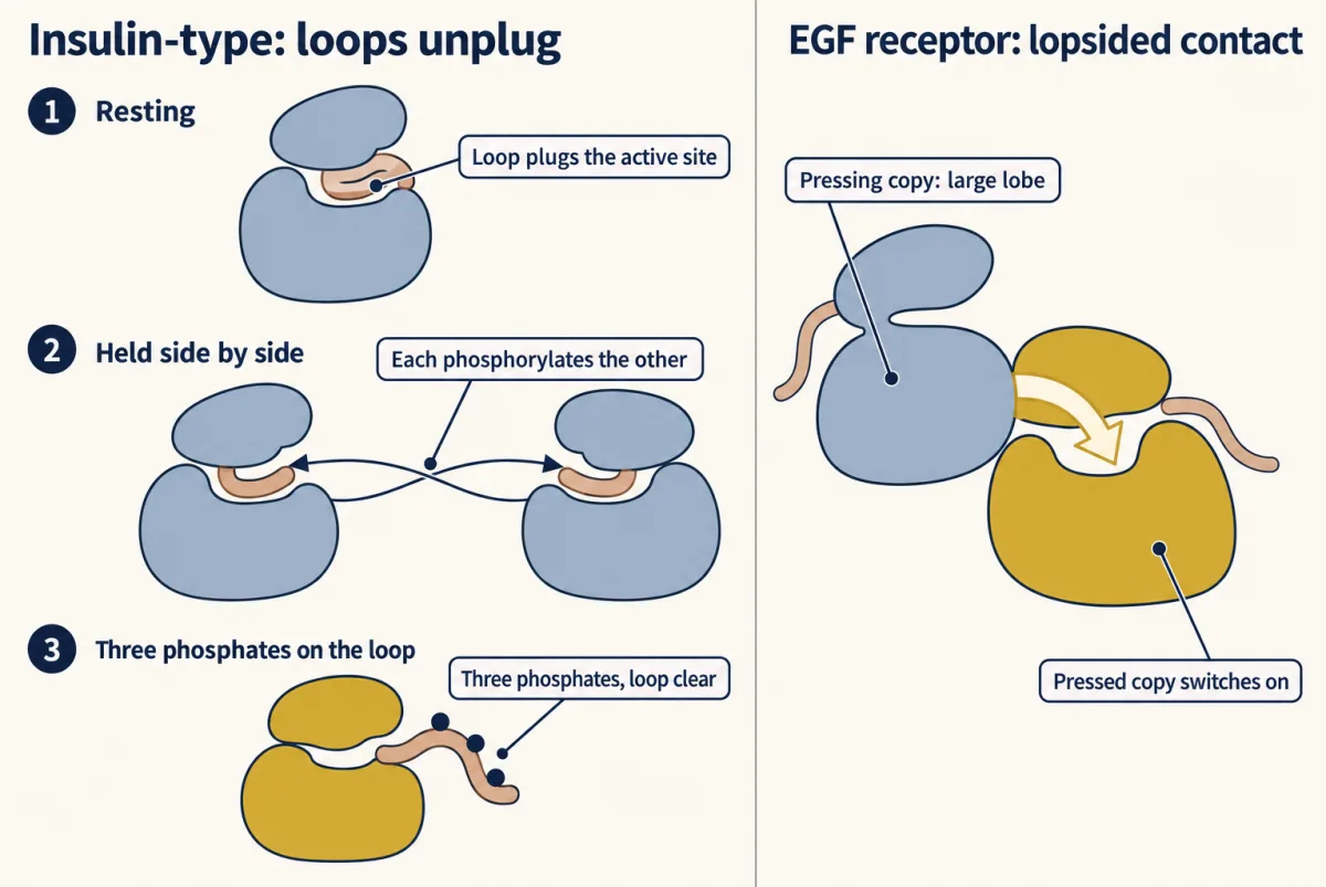
Recall the activation loop, the mobile strand beside a kinase’s active site. In the insulin receptor’s resting kinase the loop lies folded into the active site, and one of its own tyrosines sits where a substrate tyrosine should go. The kinase is plugged by a piece of itself.
When two kinases are held together, each phosphorylates the loop of the other. This is called autophosphorylation in trans: the receptor phosphorylates its own kind, but across the pair. With three phosphates on the insulin receptor’s loop, the loop stays clear of the active site, the two lobes close on ATP, and the kinase runs.
Most receptor tyrosine kinases (RTKs) use this scheme, in order: in a fibroblast growth factor (FGF) receptor followed step by step, a loop tyrosine was phosphorylated first, which raised the kinase’s rate, and the docking tyrosines followed.
The epidermal growth factor (EGF) receptor is the best-studied exception (RET is another), although at first it was assumed, by analogy with other receptors, to use the same mechanism. In 1987 Yosef Yarden and Joseph Schlessinger had presented evidence, from purified receptor, that EGF switches on the receptor’s kinase through contact between receptor molecules, and by 1989 there was evidence that one receptor adds phosphate to another, so a phosphate on the activation loop was the expected switch. Experiments from 1992 on had already found that this loop phosphate is not needed, but they did not say what does the switching. Two results in 2006 pointed to an answer.
First, changing one amino acid in the loop, a leucine to an arginine, makes the isolated kinase about 20 times more active than normal, and it does not need a phosphate on the loop. Clinicians call this change L858R, and it is found in many lung cancers whose EGF receptor is always on. Second, in John Kuriyan’s laboratory, Xuewu Zhang took the normal kinase alone, with no ectodomain and no growth factor, and crowded it by gathering it on the surface of lipid vesicles. It switched on, again without needing the loop phosphate.
So the resting kinase holds itself off, and contact with a second copy releases the hold. Crystal structures from Kuriyan’s group, published in 2006, showed the contact, and the pair is lopsided. The large lobe of one kinase presses against the small lobe of the other and pushes that kinase’s active site into its working shape. A cyclin, the protein that switches on the kinases that drive cell division, works on its kinase in much the same way. The pressed copy is the one that switches on. The pressing copy only has to supply a surface.
The EGF receptor has three relatives, HER2, HER3 and HER4, and all four use the same contact, so they can work in mixed pairs. HER3’s own kinase has almost no activity, yet a pair of HER2 and HER3 signals strongly, because HER3 can do the pressing.
”RTK gets activated…what next?” The tail becomes a set of docking sites
Read the transcript of “The tail's sequence picks the partner”
Film transcript: The tail's sequence picks the partner
The two kinases are on. They add phosphate to tyrosines outside the kinase core, mostly on the two flexible tails. Each of those phosphates is a docking site. So how does a tail pick its partner? An SH2 domain binds a phosphotyrosine. It also checks the few residues that follow. So the sequence decides which protein binds. On the EGF receptor tail, phosphotyrosine 1068 has an asparagine two residues later. That suits the SH2 domain of Grb2, which leads to Ras and the MAP kinase cascade.
Phosphotyrosine 992, on the same tail, binds PLC-gamma. That enzyme makes IP3 and DAG. Now look at HER3. Its tail has six tyrosines, and each has a methionine three residues later. That suits the SH2 domains of PI 3-kinase, which leads to Akt. So one activated pair starts several pathways at once. The choice of partner changes the mix. Does each site serve its own protein? The PDGF receptor carries sites for all three proteins on a single chain. Researchers tested the sites one at a time. They changed a tyrosine to phenylalanine, which cannot be phosphorylated. That protein failed to dock, and the others still did. PI 3-kinase uses two such tyrosines.
Once on, the kinases phosphorylate tyrosines outside the kinase core. In the epidermal growth factor (EGF) receptor most of them are on the two tails. Other receptors also use the segment just inside the membrane or an insert in the kinase, and the insulin receptor puts many of its docking sites on separate proteins, the IRS proteins. Each of these phosphates is a docking site.
You know the reader of these marks from the lesson on interaction modules: an SH2 domain binds a phosphotyrosine and checks the few residues that follow it. So the docking-site sequence largely decides which target binds. Take the EGF receptor family.
- Phosphotyrosine 1068 on the EGF receptor’s tail has an asparagine two residues later. That suits the SH2 domain of Grb2, which leads to Ras and the MAP kinase cascade.
- Phosphotyrosine 992 on the same tail binds PLC-gamma, the enzyme that makes inositol trisphosphate (IP3) and diacylglycerol (DAG).
- Six tyrosines on HER3’s tail each have a methionine three residues later. That suits the SH2 domains of PI 3-kinase, which leads to Akt.
One activated pair therefore starts several pathways at once, and the choice of partner changes the mix. The platelet-derived growth factor (PDGF) receptor carries sites for all three proteins on a single chain, and there the sites were tested one at a time: change the tyrosine that serves one protein to phenylalanine, which cannot be phosphorylated, and that protein fails to dock while the others still do. PI 3-kinase uses two such tyrosines. The next three lessons follow the branches.
The same marks arrange the receptor’s removal. A phosphotyrosine recruits Cbl, an E3 ubiquitin ligase, which tags the receptor with ubiquitin. The receptor is taken into the cell by endocytosis, the pinching-in of a patch of membrane as a vesicle, and the ubiquitin tag routes it to the lysosome, the compartment that digests proteins, and not to the proteasome. Meanwhile protein tyrosine phosphatases (PTPs) strip the phosphates, and there are plenty of them: the human genome has 107 PTP genes, of which 38 are classical enzymes that, with a few exceptions, act only on phosphotyrosine, set against about 90 protein tyrosine kinases (PTKs).
RTKs in cancer: three ways to stay on, two kinds of drug
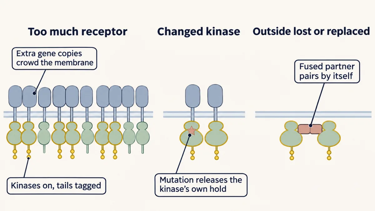
Many protein kinases have been linked to human disease, and receptor tyrosine kinases (RTKs) come up again and again in human tumours: the epidermal growth factor (EGF) receptor and its relatives, c-KIT, the VEGF receptors, c-MET and RET. If contact between kinases is the switch, a tumour can throw it without the ligand in at least three ways.
Make too much receptor. Gene amplification gives a cell extra copies of a gene. In 1987 Dennis Slamon and colleagues found the HER2 gene amplified in 30 percent of 189 breast tumours, and amplification predicted earlier relapse and shorter survival, most strongly in women whose cancer had reached the lymph nodes. HER2’s arm is exposed without any ligand, so crowding alone makes pairs.
Change the kinase. L858R sits in the kinase itself. The outside of the receptor is normal, but the kinase no longer waits for it.
Lose or replace the outside. In some thyroid cancers the RET gene breaks, and the half that encodes the kinase is joined to part of another gene whose protein forms pairs without help. The fused protein has no ectodomain at all.
Two kinds of drug answer these faults. An antibody is a large protein that binds one target tightly. It cannot cross the membrane, so it works on the ectodomain: trastuzumab (approved in 1998) binds HER2, and cetuximab (2004) binds the EGF receptor partly where EGF would and holds it folded. A small molecule can enter the cell and sit in the kinase’s ATP pocket. Gefitinib (2003) and erlotinib (2004) do this to the EGF receptor, and imatinib (2001), the Abl drug you have met, also blocks c-KIT. In 2004 three papers, by Lynch, Paez, Pao and their colleagues, reported that most lung cancers that shrank on gefitinib carried mutations in the EGF receptor’s kinase, L858R among them.
Why so many kinase drugs? “Highly Conserved— target for lots of drugs.” The ATP pocket is much the same across the kinase family, so one chemical strategy suits many kinases, and a molecule made for one pocket tends to fit others.
Design problem
A tumour cell must keep an RTK switched on with no growth factor present. Design three different ways to do it, each one changing a different part of the receptor or the amount of it. For each, predict whether an antibody against the outside of the receptor would still work, and whether a small molecule in the ATP pocket would.
Saved in this browser only.
One way to do it
- Change the amount. Amplify the gene, so that the membrane is crowded with normal receptors and pairs form without ligand. The ectodomain is present and normal, so an antibody can bind it, as trastuzumab binds HER2. The kinase is normal too, so a small molecule in the ATP pocket should also work.
- Change the kinase. Put in a mutation such as L858R that releases the kinase’s hold on itself. An antibody can still bind outside, and the mutant kinase still needs a partner, so cetuximab, which holds the receptor folded and unable to pair, can still act: it shrank mouse lung tumours driven by L858R. In patients, though, adding it to the kinase inhibitor afatinib as a first treatment did not help. A small molecule in the ATP pocket acts on the faulty part directly, as gefitinib and erlotinib do.
- Change the outside. Delete the ectodomain, or swap it for a piece of protein that pairs by itself, as in the RET fusions. The antibody has nothing to bind and fails. The small molecule works as long as the ATP pocket is unchanged.
The antibody needs the outside to be present and to matter. The small molecule needs only the pocket.
Your notes
Select any text in the lesson to highlight it or add a note to it. Your highlights and notes are saved in this browser and collected in your notebook.
This browser is not letting the site save anything (a private window or strict privacy settings), so highlights and notes will be lost when you leave the page.
How we measure it
X-ray crystallography of receptor fragments
A soluble piece of the receptor, such as the ectodomain or the kinase domain, is crystallized, and its atomic structure is worked out from the way the crystal scatters X-rays. For these receptors the outside and the inside were solved as separate pieces. How the molecules pack against each other in the crystal gave the clue to the EGF receptor's lopsided kinase pair.
Tyrosine-to-phenylalanine mutagenesis
Phenylalanine is tyrosine without its hydroxyl group, so it cannot accept a phosphate. Changing one tyrosine at a time, then asking which protein no longer docks or which response is lost, assigns a job to each phosphorylation site.
Kinase assay at controlled density
A purified kinase domain is mixed with ATP and a peptide substrate, and the rate of phosphate transfer is measured. Comparing the kinase free in solution with the same kinase gathered on the surface of lipid vesicles tests whether closeness alone switches it on.
Southern blotting for gene copy number
Tumour DNA is cut with a restriction enzyme, separated by size, and probed with a labelled piece of the gene of interest. The strength of the band, compared with a control gene present at the normal number, reports how many copies the tumour carries. The 1987 study of HER2 in breast tumours measured amplification this way.
Practice questions on this lesson (3) → MCAT-style, original, each with an explanation
Where this lesson fits
Before thisReview first
- A Field Guide to ReceptorsThis course · Lesson 4
It introduced the three-part receptor architecture that this lesson builds on, including the single-helix enzyme-linked receptors.
- Phosphorylation: Kinases and PhosphatasesThis course · Lesson 6
You need the kinase fold, the activation loop and the role of phosphatases from there to follow how the pair switches on.
- Protein Modules: SH2, SH3, PTB, PDZ and PHThis course · Lesson 9
The SH2 domain's reading of a phosphotyrosine and the residues after it explains which target docks on each tail site.
After thisStudy next
- Ras to MAPK: The Kinase CascadeThis course · Lesson 19
It follows the Grb2 branch from the activated receptor to Ras and the kinase cascade.
- PI 3-Kinase, Akt and InsulinThis course · Lesson 20
It takes the PI 3-kinase docking sites and the insulin receptor's IRS proteins further, through Akt.
- Oncogenes: When the Switch SticksThis course · Lesson 41
It takes further the idea that a stuck receptor kinase, such as an amplified or mutated one, drives cancer.
Elsewhere on the siteFilms, simulations and method films in other courses
- Receptor binding and dose-response analysisMethod film
Ligand-binding and dose-response fits are how receptor affinity and activation by a growth factor are measured.
- Gel electrophoresis, SDS-PAGE and western blotMethod film
Phosphotyrosine western blots show a receptor and its docking partners becoming phosphorylated after ligand.
Suggested by an AI model from the lessons themselves, then checked. How we use AI
Finish line
Test yourself
8 questions
Practice and MCAT-style, with explained answers.
Next · Lesson 19
Ras to MAPK: The Kinase Cascade
A growth factor at the cell surface switches on Ras, and a relay of three kinases carries the message to the cell's proteins and genes. The same relay, stuck on, is found in many cancers, and in PC12 cells how long it stays on seems to decide what the cell does.
Continue →← Lesson 17 · Sensing the World: Vision, Smell, Nitric Oxide and cGMP