Cell Signaling · Unit 7: Space, time and distance
Reverse Signaling: When the Ligand Is Also a Receptor
When a receptor grips a ligand that is stuck to another cell, which cell gets the message? Often both.
Pause when the reading reaches a film, and offer to play it. Saved on this device.
Watch the film now, or keep listening
Builds on:Protein Modules: SH2, SH3, PTB, PDZ and PHLipids as Anchors and AddressesPhosphorylation: Kinases and PhosphatasesWhy these?
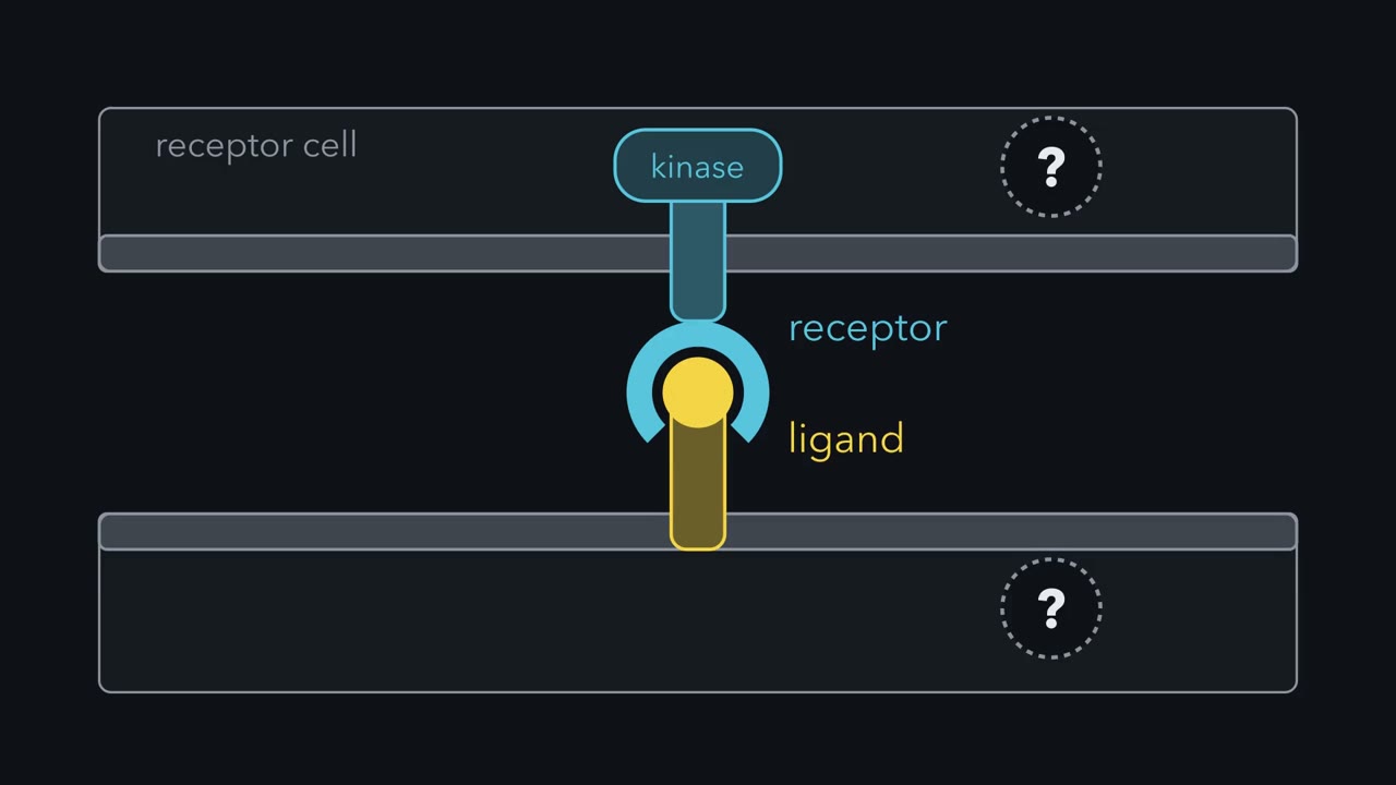 ▶ Watch A Grip That Signals Both Ways · 2:44
Read the transcript of “A Grip That Signals Both Ways”
Film transcript: A Grip That Signals Both Ways
When a receptor grips a ligand that is stuck to another cell, which cell gets the message? The receptor sits in the upper cell. Its ligand is held in the membrane of the lower cell. Does the message reach one of them, or both? Often, both. Many ligands are made as membrane proteins. Such a ligand can have a tail inside its own cell. A tail in the cytosol is what you expect of a receptor. When the two proteins bind, the receptor signals into its own cell. That is the forward signal. A second message passes into the cell that carries the ligand. That is reverse signaling.
Ephrins are the clearest case of reverse signaling. A B -type efren crosses the membrane once and ends in a short tail. The tail contains no enzyme. Yet when the efren meets the outer part of its receptor, its tail gains a phosphate on a tyrosine. That outer part has no kinase, and neither does the efren, So a kinase in the ephrin's own cell must do the work. Which kinase? SARC family kinases gather where B -type ephrins cluster, and they help to put phosphate on the tail. An adaptor then binds the phosphorylated tyrosines. It connects the ephrin to proteins that reshape the actin skeleton. This is one route; PDZ proteins offer another.
Suppose you delete the gene for a B -type ephron, and an organ fails to form. You have removed the forward signal, the reverse signal, and the grip that held the cells together. The knockout cannot tell you which loss caused the defect. So change the ephrin instead. Keep its outer part and its membrane segment, and remove only the tail. Check that it reaches the surface, then measure the forward signal. Reverse signaling through the tail is gone.
Other ligands work this way too. Membrane bound TNF acts as a receptor, and so does a fly semaphorin. In each pair, the protein named receptor also gives a signal. The protein named ligand also receives one. The names were fixed by history. So keep this rule: a membrane ligand can receive a signal as well as give one.
A ligand held in a cell’s membrane can receive a signal as well as give one. When a receptor on a neighbouring cell binds it, a message also passes into the cell that carries the ligand. This is reverse signaling. Epidermal growth factor shows why it is surprising that ligands are built this way. EGF is 53 amino acids long, and the protein it is cut from is 1,207 amino acids long and spans the membrane. Why would a cell build something that large to release something that small? For EGF the answer is still incomplete. For several other ligands it is known, and it changes what the word “ligand” means.
A curiosity: the ligand starts out as a large membrane protein
The epidermal growth factor (EGF) that Stanley Cohen purified (see the lesson on finding a signal) is a small peptide. When its messenger RNA was cloned from the mouse in 1983, the sequence predicted a protein more than twenty times longer. The human version has 1,207 amino acids. It begins with a signal peptide, the short stretch that sends a new protein into the secretory pathway. After that comes a long outer part. One segment crosses the membrane, and a tail lies in the cytosol. The 53 amino acids of EGF sit just outside the membrane, and proteases cut them free.
EGF is not a special case. Tumor necrosis factor (TNF), Fas ligand and the Notch ligand Delta are made as membrane proteins too. So a “ligand” can have a tail inside its own cell, and a tail in the cytosol is what you expect of a receptor.
What is Reverse Signaling? Both cells get a message
Picture two cells that touch. One carries a receptor. The other carries that receptor’s ligand, held in its membrane. When the two proteins bind, the receptor signals into its cell. Call this the forward signal. Reverse signaling is a second message, passing into the cell that carries the ligand. When both happen, the exchange is called bidirectional signaling.
A handshake is a fair picture. You cannot grip someone’s hand without your own being gripped. In biology the names were fixed by history. They record the role each protein was first seen to play, not which directions it can signal in.
A reverse signal needs two things. The ligand must stay attached to its cell, and it must have a route to the cytosol, through its own tail or through a neighbor in the same membrane. A useful hint when you meet a new ligand: if it stays in the membrane and its tail has changed little between species, suspect that the tail does something.
The processes affected make a long list: cell migration and proliferation, axon guidance, angiogenesis (the growth of new blood vessels), the immune response, apoptosis and development. Notch also needs two touching cells, but in the account you met in the Notch lesson the message runs one way, into the cell that carries Notch.
Ephrins: a tail that borrows a kinase
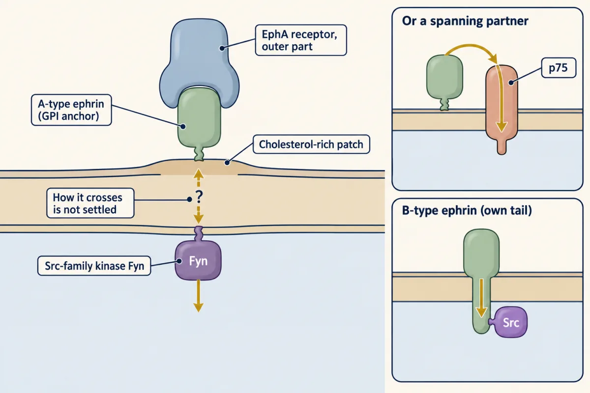
Ephrins are the clearest case. Their receptors, the Eph receptors, are the largest family of receptor tyrosine kinases, with 14 members in humans. Every ephrin is made attached to a membrane, in one of two ways. The five A-type ephrins are held by a GPI anchor, a lipid on the outer face of the membrane (see the lesson on lipid anchors). The three B-type ephrins cross the membrane once and end in a tail of 83 to 93 amino acids in the cytosol. The receptors fall into two matching classes, EphA and EphB, named for the ephrins they prefer, though some pairs cross the line: EphA4 also binds B-type ephrins.
The B-ephrin tail contains no enzyme. It has a few tyrosines whose positions are the same in all three B-type ephrins, and its last residues fit PDZ domains (see the lesson on protein interaction modules).
In the mid 1990s two laboratories reported the key observation. When cells carrying a B-type ephrin met the outer part of an EphB receptor, the ephrin’s tail became phosphorylated on tyrosine. The receptor fragment had no kinase domain and the ephrin has none, so a kinase inside the ephrin’s own cell had to be responsible.
In 2002 Rüdiger Klein’s laboratory in Heidelberg named it. Src-family kinases gather where B-type ephrins cluster, and they act as positive regulators of the tail’s phosphorylation. The same study found an off switch: a tyrosine phosphatase, PTP-BL, binds the PDZ-binding end of the tail and removes the phosphates after a delay. Mark Henkemeyer’s laboratory found that the adaptor Grb4 binds the phosphorylated tyrosines through its SH2 domain and connects the ephrin to proteins that reshape the actin skeleton.
The take-away from these studies fits in one line: “Ephrins use Src-family kinases to carry out reverse signaling.” It is not the whole story. When Klein’s laboratory changed the ephrin-B2 tail in mice, removing the PDZ-binding end caused major defects in lymphatic vessels, while changing all five conserved tyrosines to phenylalanine caused only a mild defect. In that tissue the PDZ route mattered more than the tyrosines.
”How could you signal via a GPI anchored protein?”
An A-type ephrin has no tail. It sits in the outer leaflet and never touches the cytosol. Try the question before you read on.
The answer the evidence supports is a partner. A-type ephrins collect in small patches of membrane rich in cholesterol, and Src-family kinases sit on the inner face of such patches, held there by their own lipid anchors. In 1999 Davy and colleagues reported that clustering ephrin-A5 with the outer part of an EphA receptor drew one of these kinases, Fyn, into the patches and changed how the cells adhered to their surroundings, a change that needed Fyn. Later work found partners that span the membrane. In the axons of the retina, ephrin-A associates with p75, a receptor for neurotrophins, and the reverse signal there needs p75.
Other GPI-anchored proteins solve the problem the same way. GFR-alpha-1 binds the growth factor GDNF and passes the signal mainly to Ret, a kinase that spans the membrane. It can also switch on Src-family kinases without Ret, which raises the same question as ephrin-A. Which partner an A-type ephrin uses seems to depend on the cell, and how a protein in the outer leaflet switches on a kinase at the inner leaflet is not settled.
TNF and semaphorins: other ligands that listen
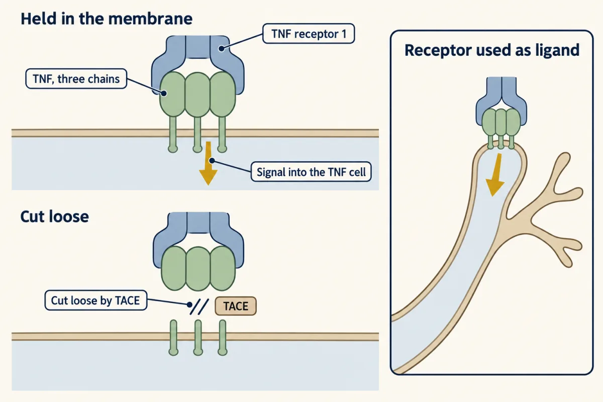
You met tumor necrosis factor (TNF) as a ligand in the lessons on NF-kappa-B and on cell death. It is made as a membrane protein, three chains together, with a short tail of 35 amino acids in the cytosol. A protease called TACE can cut the outer part free. Uncut TNF that stays in the membrane can act as a receptor: when a TNF receptor on another cell binds it, a signal passes into the TNF-bearing cell.
The tail has no enzyme activity. What the tails of several TNF-family ligands share is a short sequence of the kind that casein kinase 1 (CK1), a serine/threonine kinase, prefers. In membrane TNF a serine in the tail carries phosphate, and a 1999 study found that the phosphate was lost when a soluble TNF receptor bound. The take-away is worded with care: the CK1 site in the tail is likely an essential part of reverse signaling in this family. Note the word “likely”. The site is a short motif that CK1 acts on, not a domain of the ligand, and the evidence comes mainly from one study of membrane TNF in mouse macrophage cells.
The clearest case in neurons comes from Alun Davies’s laboratory. Developing sympathetic neurons carry TNF on their axons, and the tissues those axons grow into make TNF receptor 1. In culture, a soluble form of that receptor made the axons grow and branch more, and the effect depended on TNF in the axon’s membrane. Mice lacking either TNF or the receptor had sparser sympathetic innervation in several tissues. Here the protein named “receptor” is the signal, and the protein named “ligand” receives it.
Semaphorins make the same point. They are a large family of axon-guidance proteins, some secreted, some spanning the membrane, and their main receptors are the plexins.
In the fruit fly, the membrane-spanning semaphorin Sema-1a was first known as a ligand for plexin A. It also works the other way round. On the axons of the fly’s developing eye, Sema-1a receives a signal from plexin A that helps the axons hold to one another, and it needs its cytoplasmic part to do so. In motor axons, the tail of Sema-1a was found to bind two regulators of the small GTPase Rho, a guanine-nucleotide exchange factor (GEF) and a GAP (see the lesson on GTPase switches).
Reading a knockout: which direction did you remove?
Read the transcript of “Which Direction Did You Remove?”
Film transcript: Which Direction Did You Remove?
Picture two cells that touch. One carries a B-type ephrin, held in its membrane. The other carries its receptor. When the two proteins bind, the receptor signals into its own cell. That is the forward signal. A second message passes into the ephrin's cell. That is reverse signaling. Now suppose you delete the gene for the ephrin, and an organ fails to form. What did you take away? You have removed the forward signal into the receptor's cell. You have also removed the reverse signal into the ephrin's cell. And you have removed the plain grip that held the two cells together.
Deleting the receptor has the same problem. Its outer part was a ligand for the other cell. So here too, one deletion cannot say which loss mattered. The knockout cannot tell you which loss caused the defect. The failed organ does not say which. So here is a design problem for you. How could we disrupt reverse signaling while leaving forward signaling intact? Design the mouse, or the reagent, for an ephrin of the B class.
Suppose you delete the gene for a B-type ephrin and an organ fails to form. You have removed the forward signal into the receptor’s cell. You have also removed the reverse signal into the ephrin’s cell, and the plain grip that held the two cells together. The knockout cannot tell you which loss caused the defect. Deleting the receptor has the same problem, because its outer part was a ligand for the other cell.
Design problem
“How could we disrupt reverse signaling while leaving forward signaling intact?” Design the mouse, or the reagent, for an ephrin of the B class. Then say how you would do the opposite. Last, say what you would conclude if an animal with no ephrin at all had a defect that neither of your two animals had.
Saved in this browser only.
One way to do it
Remove the reverse signal. Change the ephrin’s gene so that the protein keeps its outer part and its membrane segment and loses its tail. Henkemeyer’s laboratory did this for ephrin-B2, putting the enzyme beta-galactosidase where the tail had been. The outer part can still bind and cluster EphB receptors on the next cell, so the forward signal should survive. With no tail there is nothing for a Src-family kinase to phosphorylate and nothing for an adaptor to bind, so the reverse signal through the tail is gone. A finer version changes only the tail’s tyrosines to phenylalanine, or removes the last residue, which PDZ domains need. These two changes remove different routes, so each takes away only part of the reverse signal. One control is essential: show that the altered protein reaches the cell surface in normal amount. If it does not, you have weakened the forward signal as well. A second check is wise: measure the forward signal in the receptor’s cell directly, because a later study found that changing the ephrin-B2 tail can alter the forward signal there, in one case making it stronger.
The reagent. Add the outer part of an EphB receptor, fused to the Fc region of an antibody and clustered, to cells that carry the ephrin. This gives a reverse signal with no receptor cell present. The reagent asks whether a reverse signal is enough to cause an effect, and the mouse asks whether it is needed.
The opposite. Alter the receptor instead: keep its outer part and membrane segment, and remove or inactivate its kinase domain. Henkemeyer and colleagues made this mouse for EphB2 in 1996. Mice with no EphB2 had misrouted axons in a brain tract called the anterior commissure. Mice with the kinase-less receptor did not. The axons carried B-type ephrins and the cells beneath them carried the receptor, so the result pointed to a reverse signal into the axons. A caution came later: Eph receptors can cluster with one another, so a kinase-less receptor might still help other Eph receptors send a forward signal. Since one ephrin binds several EphB receptors, you may need to alter more than one receptor gene.
The third animal. If the full knockout has a defect that neither altered animal shows, losing the ephrin’s tail alone does not cause it, and neither does losing the receptor’s kinase alone. Either direction may be enough alone, or the cells may only need to be held together. A mouse carrying both alterations at once would tell these two apart.
Going deeper
How big is the EGF precursor, and which other ligands are made this way?
The precursor is about as large as the epidermal growth factor (EGF) receptor. The finished human EGF receptor has 1,186 amino acids, and the EGF precursor has 1,185 once its signal peptide is removed.
EGF’s relative TGF-alpha is 50 amino acids, cut from a membrane-spanning precursor of 160. Tumor necrosis factor (TNF) is made as a membrane protein of 233 amino acids, and a protease releases the outer 157.
Who first saw ephrin tails become phosphorylated
The two laboratories behind the observation that B-type ephrin tails become phosphorylated on tyrosine were Tony Pawson’s in Toronto and Rüdiger Klein’s in Heidelberg, who reported it in 1996 and 1997.
The semaphorin family and its sema domain
Semaphorins make the same point. The first was found in 1992 in the grasshopper embryo, as a membrane protein that guides growing axons. The family is now sorted into eight classes; some are secreted, some span the membrane, and one class is held by a GPI anchor. All share the sema domain, about 500 amino acids at the outer end: “Sema domain is highly conserved between all classes.” Their main receptors are the plexins.
Your notes
Select any text in the lesson to highlight it or add a note to it. Your highlights and notes are saved in this browser and collected in your notebook.
This browser is not letting the site save anything (a private window or strict privacy settings), so highlights and notes will be lost when you leave the page.
How we measure it
Receptor-Fc fusion protein (a receptor body)
The outer part of a receptor is joined to the Fc region of an antibody and made as a soluble protein, which is often clustered further with an antibody against Fc. Added to cells that carry the membrane-bound ligand, it binds the ligand with no receptor cell present. A response that Fc alone does not cause, and that needs the ligand, is a reverse signal.
Knock-in of a truncated protein
Gene targeting in the mouse replaces the normal gene with a version that lacks one part, such as the cytoplasmic tail, sometimes with beta-galactosidase in its place as a marker. The animal shows what the rest of the protein can do without the missing part. It needs a control showing that the altered protein reaches the cell surface.
Immunoprecipitation and phosphotyrosine blotting
The protein of interest is pulled out of a cell extract with an antibody, separated on a gel, and probed with an antibody that recognizes phosphotyrosine. A band shows that the protein carried phosphate on tyrosine when the cells were broken open, provided the band is at that protein's size and not a partner pulled down with it.
Compartmented neuron culture
Neurons are grown in a device whose barriers let axons through and keep the fluid around the axons apart from the fluid around the cell bodies. A reagent added to one side reaches only that part of the neuron, which shows where on the cell a signal is received.
Method films · the methods library
Practice questions on this lesson (3) → MCAT-style, original, each with an explanation
Where this lesson fits
Before thisReview first
- Protein Modules: SH2, SH3, PTB, PDZ and PHThis course · Lesson 9
The ephrin tail signals through PDZ and SH2 readers, which that lesson explains in detail.
- Lipids as Anchors and AddressesThis course · Lesson 11
You need to know how GPI anchors and lipid patches hold proteins in the outer leaflet to follow ephrin-A.
- Phosphorylation: Kinases and PhosphatasesThis course · Lesson 6
Reverse signaling through ephrin tails rests on Src-family kinases and phosphatases adding and removing phosphates.
After thisStudy next
- How Signaling EvolvedThis course · Lesson 40
It takes up how receptor and ligand families like Eph and ephrin arose and were reused.
Elsewhere on the siteFilms, simulations and method films in other courses
- A Rank Against a SlopeFilm · Developmental Neuroscience
Eph receptors and ephrin gradients are shown steering axons, the forward half of the signal studied here.
- The Cue Marks the PlaceFilm · Developmental Neuroscience
Semaphorins and their repulsive cues are introduced there, including the family whose transmembrane member signals back into its own cell.
- There, but Not WorkingFilm · Developmental Neuroscience
Contact-dependent adhesion molecules on opposing cells can signal into both cells, as in this lesson's two-way grip.
Suggested by an AI model from the lessons themselves, then checked. How we use AI
Finish line
Test yourself
8 questions
Practice and MCAT-style, with explained answers.
Next · Lesson 40
How Signaling Evolved
A human has 58 receptor tyrosine kinases and baker's yeast has none. Where did they come from?
Continue →← Lesson 38 · Exosomes and Extracellular Vesicles: Signals Between Cells