Cell Signaling · Unit 2: The parts — switches and wiring
Phosphorylation: Kinases and Phosphatases
Rewritten on : new text, flashcards and film. Your notes and highlights stay, but the text around them may have moved.
Under review Parts of this lesson are still being checked.
- One citation detail, a gel-shift result attributed to a 2003 BATF paper, is being checked against the original figure.
The text stays up while the check is done. If you spot a problem, report it. All lessons under review.
How can one small chemical mark change what a protein does without rebuilding it?
Pause when the reading reaches a film, and offer to play it. Saved on this device.
Watch the film now, or keep listening
Builds on:Circuits: Amplification, Feedback, Adaptation and SwitchesBinding: Affinity, Occupancy and Dose–ResponseWhy these?
 ▶ Watch The Charge That Changes the Odds · 3:35
Read the transcript of “The Charge That Changes the Odds”
Film transcript: The Charge That Changes the Odds
How can a small mark change what a protein does without rebuilding it? Watch this population of protein copies. Each copy flickers among shapes, and those shapes make different contacts. The cell will change the odds, not manufacture a replacement. That is faster than waiting to make new protein.
A kinase brings ATP beside a hydroxyl-bearing site on a protein. The kinase transfers the end phosphate, leaving ADP. In animal signaling, the best-studied targets are serine, threonine, and tyrosine. The protein remains the same chain, but its local chemistry has changed. This attachment is a reversible covalent modification. Near neutral pH, the attached phosphate carries a strong negative charge. Put that charge among the protein's other charges. Attractions strengthen in some arrangements and weaken in others. The population shifts toward shapes and contacts that accommodate the new charge.
Partners can also read the phosphate's shape. Phosphate changes probabilities; it does not issue a universal command. Here is your prediction. If the chemical mark is always the same, must phosphorylation always activate its target protein?
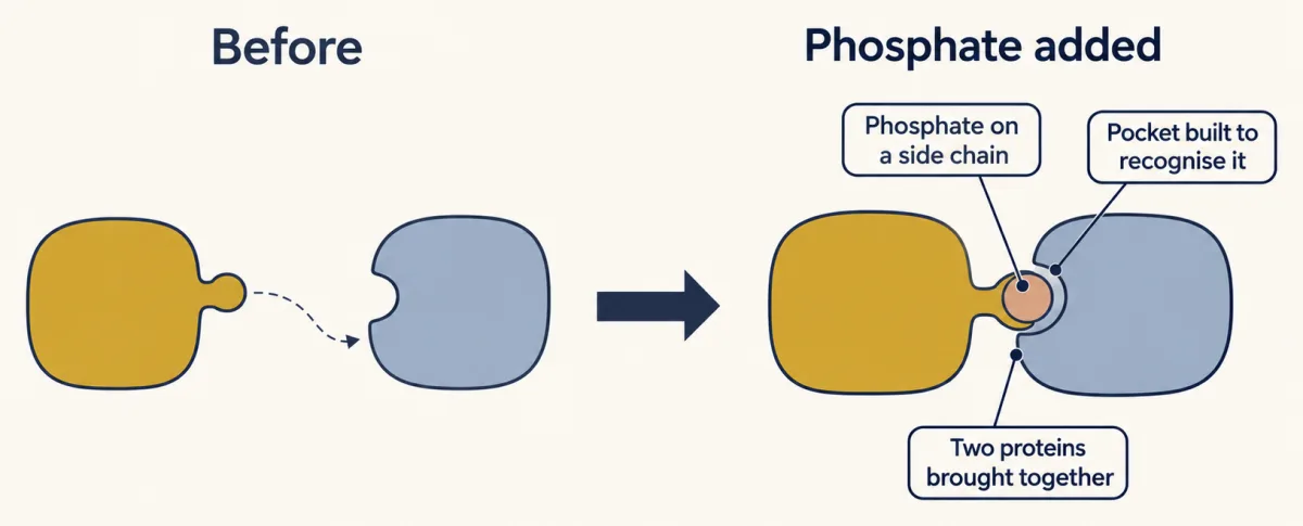
Phosphorylation shifts glycogen phosphorylase toward activity. It inhibits glycogen synthase, which builds glycogen. The direction depends on the protein and the site. The same charge can favor a different shape or disrupt a different contact. Sometimes the important change is a contact, not catalytic activity. A partner can recognize the phosphorylated surface and dock. Elsewhere, the mark can expose or hide a location signal. A phosphate can also prepare a nearby site for another kinase. These are different outcomes of the same physical change. The mark lasts until an enzyme removes it. A phosphatase uses water to release inorganic phosphate and restore the unmodified site. It does not reverse the kinase reaction or remake ATP.
Writing and erasing are separate reactions, controlled by separate enzymes. A complete trip spends an ATP molecule. That separation makes the mark both stable and reversible. ATP is plentiful enough that a kinase rarely waits for its donor. The phosphate persists without an eraser, yet can disappear quickly when a phosphatase acts. Cells can therefore alter existing proteins faster than rebuilding the whole population. Carry this rule into every case. Ask where the phosphate appears, which shapes or partners it favors, and what removal restores. The outcome may be activity, inhibition, docking, movement, or priming. The common cause is a stable phosphate, read through its charge and shape, that an enzyme can erase.
From the film
- A stable, removable phosphate changes protein behavior through its charge and shape.
- Kinases transfer phosphate from ATP to proteins, and phosphatases remove it with water.
- Phosphorylation may change activity, docking, location or a later modification.
- A phosphate can favor activity or inhibition; it is not an automatic on signal.
The first film shows the switch; this page adds the chemistry, numbers and evidence.
The one idea. A stable, removable phosphate changes protein behavior through its charge and shape.
Scope: regulatory phosphorylation of proteins, especially the well-studied serine, threonine and tyrosine sites in animal signaling. The charge is the main handle, but partners also read the phosphate’s shape and the residues around it, which is why aspartate is only a rough mimic. It may favor activity or inhibition; it is not an automatic on signal.
Write the mark, erase the mark
A cell can change what a protein does by making more of it, which is slow, by binding a small molecule to it, or by attaching a chemical group after the protein is made. That last route is a covalent modification, and signaling pathways use phosphate most. The first film’s two reactions:
protein-OH + ATP → protein-O-PO3(2−) + ADP (kinase)
protein-O-PO3(2−) + H2O → protein-OH + Pi (phosphatase)
A protein kinase moves the end, or gamma, phosphate of ATP onto the protein; a protein phosphatase removes it with water. ATP sits at millimolar concentration in the cell. Near pH 7 the attached phosphate carries close to two negative charges where there were none.
Most protein-bound phosphate sits on serine and threonine. Tyrosine carries well under 1% of it in a cell, about 2% of mapped sites, which kept phosphotyrosine hidden until 1979. Mammals also phosphorylate histidine, but the acid in standard methods strips that phosphate, so how common phosphohistidine is remains unclear.
Four consequences of one added phosphate
The phosphate has no activity of its own; the protein and its neighbours respond to it. The first film’s four outcomes, case by case:
Activity through shape. In muscle glycogen phosphorylase the phosphate lands on serine 14, near the chain’s start and far from the catalytic site, yet it favors the active form. The same mark inhibits glycogen synthase.
A docking site. A partner whose pocket recognises the phosphorylated side chain can dock; a later lesson covers those pockets.
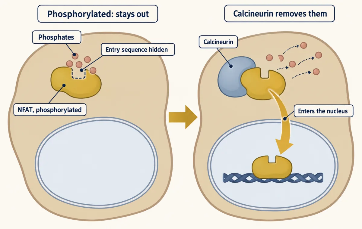
A new location. Nuclear factor of activated T cells (NFAT) is a transcription factor, a protein that switches genes on. It waits in the cytoplasm with many phosphates hiding its nuclear-entry sequence. When calcineurin removes them, NFAT enters the nucleus: the eraser carries the signal.
Predict. If calcineurin is inhibited while the kinases keep working, where should NFAT accumulate?
Check your prediction
In the cytoplasm. Its phosphates remain, so the sequence that sends NFAT into the nucleus stays hidden.
Priming another modification. Glycogen synthase kinase 3, for example, prefers a serine or threonine four residues before a site another kinase has phosphorylated. If the priming kinase answers a different signal, the substrate can wait for two inputs.
These outcomes concern single copies; the second film asks what sets the fraction of a population carrying the mark.
Read the transcript of “Two Enzymes Set the Level”
Film transcript: Two Enzymes Set the Level
Each outcome so far concerned one protein copy. But a cell controls a whole population. If a kinase writes the mark, why cannot kinase activity alone predict the level? Follow a fixed population while writing and erasing happen together.
Divide the population into an unphosphorylated pool and a phosphorylated pool. The kinase moves available copies toward the phosphorylated side. The phosphatase moves marked copies back. The total number of protein copies stays fixed while membership keeps changing. Use the simplest model: one site, a fixed pool, and effective first-order rates. Kinase flow depends on how many unmarked copies remain.
Phosphatase flow depends on how many marked copies exist. A steady level appears when those opposing flows match, although individual copies still cycle. Make a prediction. Keep kinase activity fixed but slow the phosphatase. Which way will the phosphorylated fraction move?
The fraction rises. Writing now outruns erasing at the old level, so marked copies accumulate. As that pool grows, phosphatase flow also grows. A new steady level appears when the flows match again. Inhibiting an eraser can therefore mimic activating a writer. The balance has a compact rule. At steady state, the kinase rate divided by the sum of both rates gives the phosphorylated fraction. Compute that rule across possible kinase rates. The curve rises smoothly and approaches a ceiling rather than jumping at a fixed threshold. The same enzymes also set response speed. Increase both rates by the same factor. The steady fraction stays put because their ratio is unchanged.
The population reaches that level faster because the opposing flows are larger. Faster cycling spends ATP faster. A signal can act at either side of the cycle. It may activate the kinase, inhibit the phosphatase, or later activate the phosphatase to end the response. Measuring only the writer misses those possibilities.
Measure the substrate's modification and both opposing activities. Keep the model's boundary visible. It assumes a fixed pool, a single site, and effective first-order rates. Saturated enzymes, several sites, feedback, or protein turnover can change the response. Start with the simple balance, then test whether the system obeys it. When phosphorylation rises, ask what changed on both sides. More kinase activity is only a possible answer. Less phosphatase activity can produce the same direction of change. The observed level belongs to the cycle, not to either enzyme alone. That is why the substrate measurement matters.
From the film
- Phosphorylation level depends on both the kinase and the phosphatase.
- At simple first-order rates, their ratio sets the steady phosphorylated fraction.
- Either activating the kinase or inhibiting the phosphatase can raise that fraction.
- Faster opposing rates make the level settle faster but spend more ATP.
How much protein carries the phosphate?
Predict. A kinase works at 0.6 per minute against a phosphatase at 0.2 per minute. A drug cuts only the kinase rate tenfold, to 0.06 per minute. Does the phosphorylated fraction fall tenfold?
The phosphorylated fraction, f, is the share of a protein pool carrying phosphate at the measured site. Take the second film’s simplest model: one site, a fixed total pool and effective first-order rates, with no saturated enzymes and no multisite network. Fraction (1 − f) is available to the kinase:
rate in = k(kinase) × (1 − f)
rate out = k(phosphatase) × f
At steady state the rates match, so:
f = k(kinase) / (k(kinase) + k(phosphatase))
Check your prediction
No. Before the drug, f = 0.6 / (0.6 + 0.2) = 0.75. After it, f = 0.06 / (0.06 + 0.2), about 0.23. A tenfold cut in kinase rate gives only about a threefold fall, because the phosphatase rate stays in the denominator.
So the phosphorylation level depends on both the kinase and the phosphatase, not on kinase activity alone. Because the summed rates set how fast the level settles, a pathway that must shut off in seconds needs a fast phosphatase; running it constantly costs ATP, so some pathways switch it on only when the signal ends.
How the enzymes find the right targets
The human genome holds about 518 protein-kinase genes. Most encode serine/threonine kinases; about 90 encode tyrosine kinases. The tyrosine-phosphatase family is about as large by count, but only about 38 members are classical enzymes that read only phosphotyrosine, and many erasers are built like membrane receptors.
Serine/threonine counts are lopsided: hundreds of kinases but only a few dozen genes for phosphatase catalytic subunits. Specificity comes from partners. PP1, a major catalytic subunit, is encoded by three genes yet pairs with well over a hundred regulatory subunits, which choose its location and substrates. The counts suggest that tyrosine dephosphorylation is steered more by enzyme choice, and serine/threonine dephosphorylation by company. That is an inference from counts, not proof.
Build the evidence: detect, map, test
Three claims need different evidence: the protein carries phosphate, a particular residue carries it, and changing that site changes function.
Predict. A stimulated sample shows an extra, slower band on an SDS gel. What control would tie that band to phosphate, and what would it still leave open?
Check your prediction
Treat half the sample with purified phosphatase. If the slow band collapses into the fast one and a phosphatase inhibitor prevents that, the shift depends on phosphate. The residue and the function remain open.
Detect it. A phosphorylated protein often runs more slowly on an SDS gel, which sorts proteins by apparent size; the phosphatase test above is the quick first check. Confirm that the protein carries phosphate by growing cells with radioactive phosphate (32P), pulling the protein out with an antibody and checking for label. A site found only in a test tube must still be confirmed in cells.
Map it. Break the labelled protein into amino acids and separate them to learn which kind of residue carries the label. Change candidate serine or threonine residues to alanine, or tyrosine to phenylalanine, removing the hydroxyl, and ask which change removes the label or shift. Mass spectrometry is faster: each phosphate adds 80 Da to a peptide, and peptide fragments usually locate the residue.
Predict. A peptide from the upper gel band is 240 Da heavier than the matching peptide below, and the band runs an apparent 3 kDa higher. How many phosphates does the peptide carry?
Check your prediction
Three, because 240 / 80 = 3. Three phosphates add only 0.24 kDa; the 3 kDa is apparent migration, not mass, so it cannot be converted into a phosphate count.
Test function. Compare an alanine mutant, which cannot be phosphorylated, with an aspartate mutant, whose negative charge roughly imitates a persistent phosphate. Ask which of the four consequences the mimic reproduces: activity, docking, location or priming.
Predict. A kinase activates enzyme E. The serine-to-alanine mutant no longer responds to the kinase, but the serine-to-aspartate mutant is no more active than the unstimulated normal enzyme. What can you conclude?
Check your prediction
The alanine result points to this serine as the site that matters. The silent mimic proves little: aspartate carries one charge where phosphate carries two, it is smaller, and a partner may not recognise it. Seek another line of evidence.
The normal protein should respond to kinase inhibition or activation; the alanine mutant should not. Inhibitors can narrow the writer list. Kevan Shokat’s analog-sensitive kinases are engineered to accept an ATP analog that normal kinases cannot use, so they label only direct targets.
Work this approach on a real case: the transcription factor in Deppmann and colleagues’ 2003 paper, which combined a phosphatase-sensitive gel shift, serine and threonine sites and a serine-to-aspartate mimic.
When the switch breaks: kinases in disease and the clinic
A lecture slide claims nearly every kinase has been linked to disease, a slight stretch: many have, but much of the kinome is poorly studied. A common cancer pattern is a gain-of-function mutation that leaves a kinase, often a receptor tyrosine kinase, active without a signal and telling the cell to grow. Losing a phosphatase can cause similar damage because the brake is gone.
A second slide calls kinases “highly conserved: target for lots of drugs.” The ATP cleft between the two lobes is snug and mostly greasy, so small drugs can fill it. Imatinib, approved in 2001 for chronic myeloid leukemia, binds BCR-ABL, the overactive fusion kinase made when a chromosome rearrangement joins two genes. Gefitinib and erlotinib, approved in the United States in 2003 and 2004, target the epidermal growth factor (EGF) receptor similarly.
Conservation makes selectivity hard: a molecule fitting one ATP pocket may fit some of the other 517, causing side effects, and it must compete with millimolar cellular ATP. Chemists find selectivity at the edges of the pocket, where kinases differ, or in inactive shapes that only some kinases adopt. Imatinib binds Abl in an inactive conformation, helping explain why it spares most other kinases.
Going deeper
How reversible protein phosphorylation emerged
1943. Carl and Gerty Cori and Arda Green in St. Louis crystallized glycogen phosphorylase, the muscle enzyme that releases glucose units from glycogen. Phosphorylase a was active alone; b required AMP. An enzyme turned a into b. They thought it removed bound AMP; it was a phosphatase.
1954. Burnett and Kennedy described a liver enzyme moving phosphate from ATP onto casein, the first protein kinase described.
1955. Edmond Fischer and Edwin Krebs in Seattle set out to make phosphorylase a by the Coris’ recipe. They kept getting b unless they filtered the extract through paper. The paper leached calcium, and standing extracts ran out of ATP. Over the next few years, conversion of b to a proved to be an enzyme moving one phosphate from ATP onto one serine on each chain. The overview lesson on signal transduction tells the full story.
1979 to 1980. Tony Hunter saw a radioactive spot between phosphoserine and phosphothreonine markers; his phosphotyrosine standard matched it. He and Bartholomew Sefton showed that Src, the cancer-causing protein of Rous sarcoma virus, phosphorylates tyrosine. Within the year, H. Ushiro and Stanley Cohen used tumour-cell membranes without living cells to show that epidermal growth factor (EGF) switches on the same activity. It was later traced to the cell’s own EGF receptor.
1988. Nicholas Tonks, working with Fischer, purified PTP1B, the first protein tyrosine phosphatase, from human placenta. Its sequence resembled part of CD45, a white-blood-cell surface protein, showing that a phosphatase could be receptor-like.
Pseudokinases and family counts
About one in ten human kinases, called pseudokinases, has lost catalytic activity and instead works by binding other proteins.
Of the roughly 90 human tyrosine kinases, 58 are receptors spanning the plasma membrane with the kinase inside. The tally “107 PTPs to counteract 90 protein tyrosine kinases (PTKs)” (protein tyrosine phosphatases and kinases) counts the whole phosphatase family: only about 38 are classical phosphotyrosine-specific enzymes, and relatives such as phosphatase and tensin homolog (PTEN) prefer substrates other than proteins.
Your notes
Select any text in the lesson to highlight it or add a note to it. Your highlights and notes are saved in this browser and collected in your notebook.
This browser is not letting the site save anything (a private window or strict privacy settings), so highlights and notes will be lost when you leave the page.
How we measure it
Radioactive labelling with gamma-32P ATP
You incubate a kinase and its substrate with ATP whose end phosphate is radioactive, or grow cells in radioactive phosphate, then separate the proteins on a gel and expose it to film. A dark band shows which protein received phosphate. Only a label in the gamma position works in the test tube, because that is the phosphate the kinase transfers.
Phosphatase-sensitive mobility shift
Many phosphorylated proteins run more slowly on an SDS gel. If treating the sample with a purified phosphatase, such as calf intestinal alkaline phosphatase, collapses the slow band into the fast one, the difference was phosphate. It is quick and needs no radioactivity, though some phosphates cause no shift at all.
Phosphoamino acid analysis
You break a 32P-labelled protein down to its amino acids in acid and separate them by electrophoresis on a thin-layer plate, beside phosphoserine, phosphothreonine and phosphotyrosine standards. The position of the radioactive spot tells you which kind of residue carries the phosphate. This is the method that revealed phosphotyrosine in 1979.
Site-directed mutagenesis: alanine and aspartate substitutions
You change the suspected residue to alanine, which cannot accept a phosphate, or to aspartate, whose negative charge imitates one. Loss of labelling in the alanine mutant points to the site, and the behaviour of the aspartate mutant suggests what the phosphate does. A carboxylate is only a rough copy of a phosphate, so the result needs a second line of evidence.
Method films · the methods library
- Analog-sensitive kinases (chemical genetics) 1:28
- Binding assays: immunoprecipitation, pull-down and gel shift 3:43
- Engineered mutants: site-directed, dominant-negative and constitutively active 4:25
- Gel electrophoresis, SDS-PAGE and western blot 4:01
- Protein purification and column chromatography 4:14
- Radioactive and stable isotope tracers 4:03
- X-ray crystallography 4:06
Practice questions on this lesson (3) → MCAT-style, original, each with an explanation
Where this lesson fits
Before thisReview first
- Circuits: Amplification, Feedback, Adaptation and SwitchesThis course · Lesson 5
You need the idea of switches and feedback from there to see why a kinase and phosphatase pair sets a signal's level.
- Binding: Affinity, Occupancy and Dose–ResponseThis course · Lesson 3
The binding and occupancy ideas there underlie how a drug competes with ATP in the kinase pocket.
After thisStudy next
- Protein Modules: SH2, SH3, PTB, PDZ and PHThis course · Lesson 9
These domains read the phosphate marks introduced here, turning a phosphate into a docking site.
- Receptor Tyrosine KinasesThis course · Lesson 18
This takes the idea of tyrosine phosphorylation to receptors that phosphorylate themselves and their partners.
- Ras to MAPK: The Kinase CascadeThis course · Lesson 19
A relay of kinases, each phosphorylating the next, shows the writer-and-eraser logic working in a pathway.
Suggested by an AI model from the lessons themselves, then checked. How we use AI
Finish line
Test yourself
8 questions
Practice and MCAT-style, with explained answers.
Next · Lesson 7
GTPase Switches: The Timer
After a receptor lets go, what decides how long an already-loaded G protein keeps signaling?
Continue →← Lesson 5 · Circuits: Amplification, Feedback, Adaptation and Switches