Cell Signaling · Unit 2: The parts — switches and wiring
Adaptors, Docking Proteins and Scaffolds
Rewritten on : new text, flashcards and film. Your notes and highlights stay, but the text around them may have moved.
A receptor activates Ras without ever touching it. How does the message cross the gap?
Pause when the reading reaches a film, and offer to play it. Saved on this device.
Watch the film now, or keep listening
Builds on:Phosphorylation: Kinases and PhosphatasesGTPase Switches: The TimerProtein Modules: SH2, SH3, PTB, PDZ and PHWhy these?
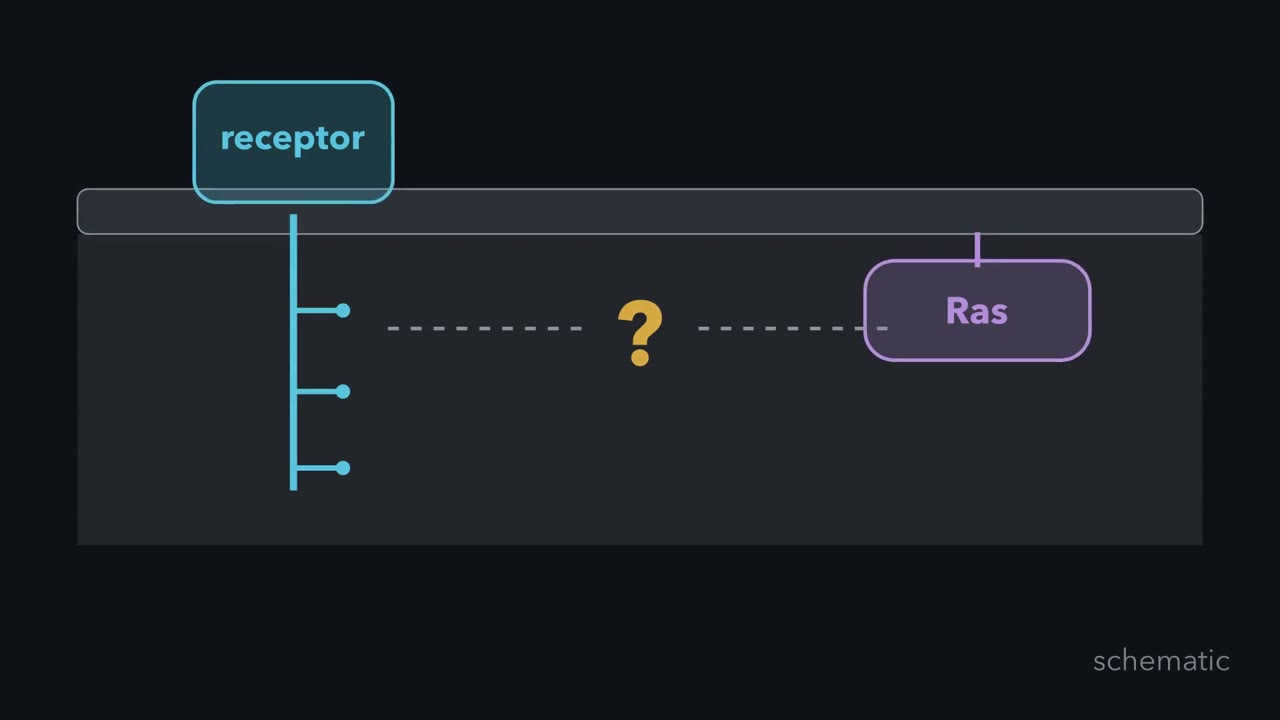 ▶ Watch How a Receptor Reaches Ras · 3:15
Read the transcript of “How a Receptor Reaches Ras”
Film transcript: How a Receptor Reaches Ras
A receptor activates Ras without ever touching it. How does the message cross the gap? Watch the blue receptor and purple Ras. Both sit at the membrane, but nothing yet connects them. A growth factor makes the receptor add phosphate marks to its tail. Each gold mark is a possible docking site. Yet a phosphate cannot activate Ras by itself. The receptor and Ras are separate proteins, each free to drift in the membrane. The missing part must bind the receptor without consuming its phosphate. It must also carry something that can act on Ras. The reader is Grb2, a green adaptor with no enzyme activity. Its central SH2 domain is a cup for phosphotyrosine.
Its SH3 domains are paired grips. Those grips already hold the orange exchange factor Sos. Which part should face the phosphorylated receptor: the SH2 cup or an SH3 grip? Keep your choice in mind while the two shapes move toward the gold phosphate.
The SH2 cup closes around the receptor phosphotyrosine. The SH3 grips keep holding Sos. The complete Grb2 and Sos unit moves to the receptor tail. The adaptor has connected a marked receptor to an enzyme. Membrane-bound Ras now meets the recruited Sos. Sos is a guanine-nucleotide exchange factor. It releases GDP from Ras, allowing GTP to bind. Purple Ras changes from off to on. Grb2 never catalyzes that exchange. Recruitment changes location, not chemical identity.
Sos was already attached to Grb2 in the cytoplasm. After the SH2 cup docks, the same Sos sits at the membrane, where it can meet Ras. Positioning has become a decisive signaling step. Sos also has another Ras-binding site, away from the exchange site. Ras with GTP can bind there and speed Sos. One activated Ras can therefore help Sos activate more, once recruitment starts. Now follow the route once. The receptor recruits Grb2. Grb2 brings Sos to the membrane, where Ras sits. Sos performs the exchange that activates Ras. Binding controls the encounter, while Sos performs the chemical work. The missing link from the opening is filled, but the receptor still never touches Ras. The adaptor changes who meets whom and where.
It lets the right enzyme reach its target without the adaptor being an enzyme itself.
From the film
- Adaptors route signals by linking partners, not by catalyzing reactions.
- Grb2 uses its SH2 domain to bind a receptor phosphotyrosine.
- Grb2 uses its SH3 domains to hold the exchange factor Sos.
- Recruiting Sos to the membrane lets it meet Ras and exchange GDP for GTP.
A growth-factor receptor can activate Ras without touching it. The bridge is assembly: an adaptor with no enzyme activity, Grb2, brings the exchange factor Sos to Ras.
The one idea. Binding proteins route signals by controlling which partners meet, where they meet, and when they can act.
Scope: this rule describes the adaptor, docking-protein and scaffold examples here. Binding alone may not activate a pathway, not every scaffold lacks enzyme activity, and more scaffold does not always mean more signal.
Three ways to organize a signal
A phosphotyrosine is a tyrosine carrying a phosphate. In a matching local sequence, it can become a docking site for an SH2 domain, a folded module that recognizes phosphotyrosine and nearby amino acids.
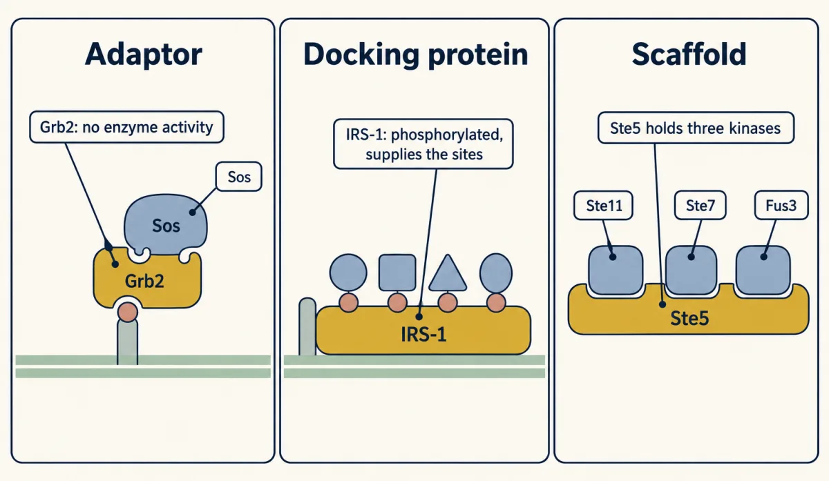
- An adaptor has no enzyme activity and supplies a link between partners. Grb2 is the model.
- A docking protein is itself phosphorylated at several sites and supplies a regulated platform for several partners. IRS-1 is the model.
- A scaffold organizes several successive members of one route. Ste5 in yeast is the model.
Partner count is only a shortcut: an adaptor typically bridges two partners, and a scaffold holds several, often three. The better test is what the protein supplies. KSR plays a scaffold-like role in mammals, but this pseudokinase changes its partners’ shapes and has some kinase activity of its own.
Quick check. A receptor phosphorylates many tyrosines on a non-enzyme protein, and different sites recruit different effectors. Which category fits best?
Check your prediction
A docking protein. It supplies several regulated phosphosites itself; holding several partners would not identify that role.
Grb2 routes a receptor signal to Ras
The first film follows the relay, receptor phosphotyrosine → Grb2 → Sos → Ras-GTP. Ras is a small switch protein anchored to the inner face of the plasma membrane, so bringing Sos to the membrane brings it within reach. Sos also has a second Ras-binding site, away from its exchange site; Ras-GTP bound there can speed Sos, so each Ras it activates helps it activate more.
Grb2’s two SH3 domains grip short proline-rich stretches, here the tail of Sos. Its SH2 domain reads more than the phosphate: nearby amino acids help decide which SH2 domain fits, so two phosphorylated tyrosines need not recruit the same SH2 protein.
Predict. Two receptor tails are phosphorylated equally, but only one has the local sequence recognized by Grb2’s SH2 domain. Which one recruits the Grb2–Sos complex?
Check your prediction
Only the matching one should recruit it efficiently. The phosphate is necessary for SH2 recognition here; the surrounding residues help provide specificity.
How experiments found the chain
A proposed order makes a genetic prediction: switching on a downstream part should bypass a missing upstream link.
Predict. Fly eye cells lacking a gene X never become the R7 photoreceptor. Suppose X acted downstream of Ras rather than upstream. What would always-active Ras do in those cells?
Check your prediction
Nothing: R7 would still fail, because a part after Ras would still be missing.
In the fly eye, the receptor tyrosine kinase sevenless decides whether one cell in each unit becomes R7. In a sensitized screen, Michael Simon and Gerald Rubin weakened sevenless signaling, then found mutations in fly ras and in Sos, a relative of the yeast Ras activator CDC25. In nematodes, genetics placed the EGF-receptor-like let-23 before let-60 Ras, with sem-5 on the same pathway.
Biochemistry supplied the physical link. In 1992 Eve Lowenstein and colleagues in Joseph Schlessinger’s laboratory isolated Grb2, showed that its SH2 domain associates with activated epidermal growth factor (EGF) and platelet-derived growth factor (PDGF) receptors, and found that Grb2 plus Ras, but neither alone, drove resting rat fibroblasts to copy DNA. In 1993 several groups, including Tony Pawson’s and David Bowtell’s, showed that Grb2’s SH3 domains bind mammalian Sos and that growth factor recruits the pair to the receptor.
Grb2, fly Drk and worm Sem-5 are orthologs, as are Ras, fly Ras1 and worm let-60; the chain predates the split between those lineages. What follows Ras differs: three of six cells beside the worm gonad become vulval tissue, fly cells adopt R7 fate and fibroblasts copy DNA.
IRS-1 turns one input into several branches
A docking protein expands the available surface. Insulin receptor substrate 1, IRS-1, reaches the membrane through a PH domain that binds membrane lipids and a PTB domain that recognizes a receptor phosphotyrosine motif. The insulin receptor then phosphorylates many tyrosines on IRS-1. The amino acids beside each phosphotyrosine decide which SH2 protein binds there, so different sites recruit PI 3-kinase, Grb2, the phosphatase Shp-2 and the adaptor Nck, sending the signal down several branches at once. Two of them:
Grb2 → Sos → Ras
PI 3-kinase → PDK1 → AKT
AKT helps regulate metabolism.
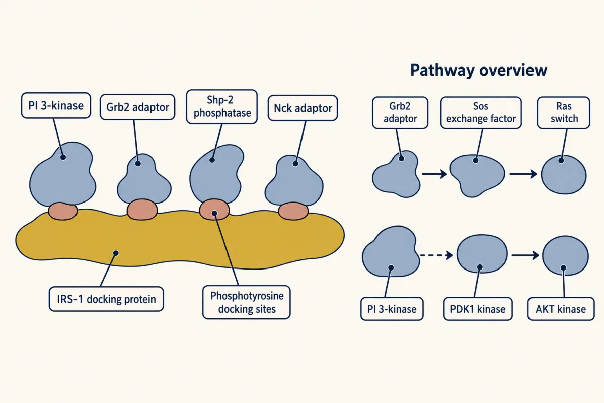
Grb2 now sits in two routes that both lead to Ras. Harder is a kinase shared by routes that must reach different outputs, the problem the second film takes into a yeast scaffold.
Read the transcript of “How a Scaffold Keeps Signals on Route”
Film transcript: How a Scaffold Keeps Signals on Route
The first film used Grb2 to route a receptor signal toward Ras. But what if two pathways share the same kinase? Follow the same colors as one orange kinase faces two possible purple outputs. Yeast mating uses a kinase route ending in Fus3. Some upstream kinases also serve other responses. A pheromone input must reach the mating output without making the cell follow every route that shares a part. A shared kinase does not carry a label saying which response should follow. Its meaning depends on the partners around it. The cell therefore needs an organizer that assembles the mating route under the mating input. Ste5 is a green scaffold with no enzyme activity.
Different regions bind Ste11, Ste7 and Fus3, so one Ste5 can hold all three. This assembly can make productive encounters more likely. Within the assembled route, Ste11 activates Ste7, and Ste7 activates Fus3. Fus3 helps prepare the cell for mating. The scaffold does not perform those kinase reactions. It organizes the proteins that do. Proximity is only the first model. Ste5 responds to pheromone-linked recruitment at the membrane. Its shape opens there. A Ste5 domain also helps Ste7 activate Fus3, which is otherwise a poor substrate. The scaffold therefore helps select an output. It controls partners, place and activation, rather than locking every shared kinase away. The same shared parts can behave differently inside a different organized context.
The filament-growth route uses Ste11 and Ste7 but ends at Kss1. One salt-stress branch uses Ste11, then a different middle kinase, and ends at Hog1. Only the mating route has Ste5, so only the mating route switches on Fus3. Suppose the cell keeps adding empty Ste5 scaffolds. Must the number of complete kinase assemblies keep rising? Watch where each kinase might land, then make your prediction.
No. In the simplest model, all three kinases must share one scaffold to signal. With scaffold in excess, they spread across different scaffold molecules. Fewer scaffolds are complete, so predicted output falls. This model effect is called combinatorial inhibition. Experiments support both layers. Ste5 binds the mating kinases on different regions. Later work showed that a Ste5 domain makes Fus3 easier for Ste7 to activate. Assembly and selective activation work together. Return to the fork. A shared kinase does not choose an output alone. The organizer around it changes which partners meet, where the complex forms and whether the last step can proceed.
From the film
- Scaffolds route shared parts by controlling partners, place and activation.
- Ste5 organizes Ste11, Ste7 and Fus3 in the yeast mating pathway.
- Ste5 helps Ste7 activate Fus3; proximity alone does not explain specificity.
- In the simplest model, excess scaffold splits partners across incomplete complexes and lowers output.
Ste5 routes shared kinases
The second film shows Ste5 controlling partners, place and activation on the mating route. The second film traced the shared parts to Kss1 and Hog1; of the two branches of the salt-stress cascade, Ste11 feeds only one. Fus3 helps halt the cell cycle for fusion, and without Ste5 a yeast cell cannot mate.
In 1994 Choi, Satterberg, Lyons and Elaine Elion showed that the three kinases bind different regions of the non-enzyme protein Ste5.
Scaffolds are often pictured as passive assembly platforms. A kinase and its target normally meet by random drift through a crowded cytoplasm, so holding both can increase productive encounters. That model is a useful start, but incomplete.
The first specificity idea was sequestration: a pathway-dedicated scaffold keeps kinase modules from talking to each other. Wendell Lim’s laboratory later found that Ste5 does not insulate by locking its kinases away; membrane gating and Fus3 unlocking do the work. Restraint also runs the other way: Hog1 helps keep high salt from activating the mating route.
Place has clearer cases elsewhere. InaD uses PDZ domains, modules that grip the last few amino acids of a partner, to gather light-response proteins in a fly photoreceptor. Paxillin gathers Ras-pathway proteins at focal adhesions, where a cell grips its surroundings.
More scaffold can mean less signal
The second film’s excess-scaffold result comes from a model. Levchenko, Bruck and Sternberg modeled a generic scaffolded kinase cascade in 2000, found that output rose and then fell as scaffold increased, and called the effect combinatorial inhibition. The model assumes fixed kinase amounts and signaling only when all three share one scaffold; cooperative assembly or other regulation can change the result.
Predict. A Ste5 fragment promotes Fus3 autophosphorylation at one of its two activating sites. Must that raise the pathway’s final output?
Check your prediction
No. The network effect must be measured. In the 2006 experiment from Wendell Lim’s laboratory, Fus3 made this way was only partly active and lowered pathway output, so Ste5 tuned the signal rather than simply accelerating it.
Ste5 has no human counterpart. KSR in the Ras–mitogen-activated protein kinase (MAPK) pathway and JIP-1 organize cascade kinases comparably but are not descended from Ste5; sequence similarity to Ste5 would not have found them.
Local signals and rewiring
A kinase reads a messenger, and an enzyme destroys it. What holds the two close enough for a local pool of messenger to exist? An anchoring protein. In John Scott’s laboratory, Dodge and colleagues showed that muscle-selective A-kinase anchoring protein, mAKAP, holds protein kinase A and PDE4, a phosphodiesterase that destroys cyclic AMP. The kinase therefore responds especially to cyclic AMP made nearby, before it spreads away.
Separate binding surfaces also make scaffolds rewritable: swap one surface for another that recognizes a different partner, and an input can reach a new output. Wendell Lim’s laboratory has used that logic to build signaling proteins no organism has had.
Design problem
Two pathways share kinase K. Input A must give only output A, and input B only output B. Design two ways to keep the messages apart.
Saved in this browser only.
One way to do it
Separate assemblies. Scaffold A holds K with A’s activator and target; scaffold B holds K with B’s partners. This is the original sequestration proposal for Ste5; a stronger design also gates each scaffold by its input and helps activate the final kinase. Its weak point is dose: excess scaffold can split partners across incomplete complexes.
Separate places. Anchor K at two locations. Input A makes the messenger at site A, where K sits beside target A; input B makes it at site B, beside target B. Each anchor also holds an enzyme that clears the messenger before it reaches the other site. This is the mAKAP logic. Its weak point is local clearance: remove the clearing enzyme and the signal can grow and spread.
Other answers can work. A good design states what enforces specificity and what failure would break it.
Going deeper
Discovery order
The fly screen appeared in November 1991; Utpal Banerjee’s laboratory had isolated Sos earlier that year. The worm ordering came from Paul Sternberg’s group; Stuart Clark, Michael Stern and Robert Horvitz cloned sem-5 in March 1992, and Lowenstein’s Grb2 paper linked Grb2 to it in August. Across about nine months, a fly screen, worm clone and rat-cell experiment converged on one receptor-to-Ras chain.
The 1994 Ste5 result surprised because a protein essential for mating appeared to catalyze nothing. Elion’s group combined two-hybrid mapping with co-purification; Michael Wigler’s group (PNAS, 2 August) and George Sprague’s (Genetics, November) reached the same result with the then-new yeast two-hybrid method. Marcus and colleagues proposed that a dedicated scaffold might sequester a kinase module.
A debated cardiac target
The mAKAP complex also holds a target. Marx and colleagues reported that protein kinase A phosphorylates the cardiac ryanodine receptor, the calcium-release channel for each heartbeat, causing FKBP12.6 to leave. They also reported defective regulation in failing hearts. Both findings remain debated, and some groups have not reproduced FKBP12.6 loss.
Your notes
Select any text in the lesson to highlight it or add a note to it. Your highlights and notes are saved in this browser and collected in your notebook.
This browser is not letting the site save anything (a private window or strict privacy settings), so highlights and notes will be lost when you leave the page.
How we measure it
Sensitized genetic screen
You weaken one pathway so that it is only just working, then look for mutations that matter only against that weak background. A mutation that does nothing in a normal animal can now tip the pathway over. Simon and Rubin used it in the fly eye and found ras and Sos.
Binding-based cloning and microinjection
You find a gene by what its protein sticks to, for example a phosphorylated growth-factor receptor, then inject the purified protein into resting cells and score whether they copy their DNA. It tells you that a protein binds a receptor and that it feeds a pathway that drives growth.
Co-purification of protein complexes
You pull one protein out of a cell extract and see what comes with it. Elion's group used it to show that Ste5 copurifies with Ste11, Ste7 and Fus3.
Yeast two-hybrid assay
You fuse two proteins to the two halves of a transcription factor in yeast. If the proteins bind each other, the halves come together and switch on a reporter gene. Printen and Sprague used it to show that Ste5 interacts with each kinase of the mating cascade.
Practice questions on this lesson (3) → MCAT-style, original, each with an explanation
Where this lesson fits
Before thisReview first
- Phosphorylation: Kinases and PhosphatasesThis course · Lesson 6
You need to know how tyrosine phosphates are added and removed, since adaptors read those phosphotyrosine marks.
- GTPase Switches: The TimerThis course · Lesson 7
Sos is an exchange factor that turns Ras on, so you need how GDP-GTP switches work.
- Protein Modules: SH2, SH3, PTB, PDZ and PHThis course · Lesson 9
Grb2 works through its SH2 and SH3 domains, which that lesson explains.
After thisStudy next
- Receptor Tyrosine KinasesThis course · Lesson 18
It shows how an activated receptor creates the phosphotyrosine sites that Grb2 and docking proteins grab.
- Ras to MAPK: The Kinase CascadeThis course · Lesson 19
It follows Ras-GTP into the three-kinase cascade, where scaffolds like KSR hold the kinases together.
- PI 3-Kinase, Akt and InsulinThis course · Lesson 20
It takes the IRS-1 docking platform further, following the PI 3-kinase to AKT branch.
Suggested by an AI model from the lessons themselves, then checked. How we use AI
Finish line
Test yourself
8 questions
Practice and MCAT-style, with explained answers.
Next · Lesson 11
Lipids as Anchors and Addresses
How does a protein with no membrane-spanning segment end up stuck to a membrane, and how does a lipid label tell it where?
Continue →