Cell Signaling · Unit 2: The parts — switches and wiring
Protein Modules: SH2, SH3, PTB, PDZ and PH
Rewritten on : new text, flashcards and film. Your notes and highlights stay, but the text around them may have moved.
A receptor has just phosphorylated its own tyrosines. What reads the marks, and how does each reader pick its mark?
Pause when the reading reaches a film, and offer to play it. Saved on this device.
Watch the film now, or keep listening
Builds on:Phosphorylation: Kinases and PhosphatasesA Field Guide to ReceptorsBinding: Affinity, Occupancy and Dose–ResponseWhy these?
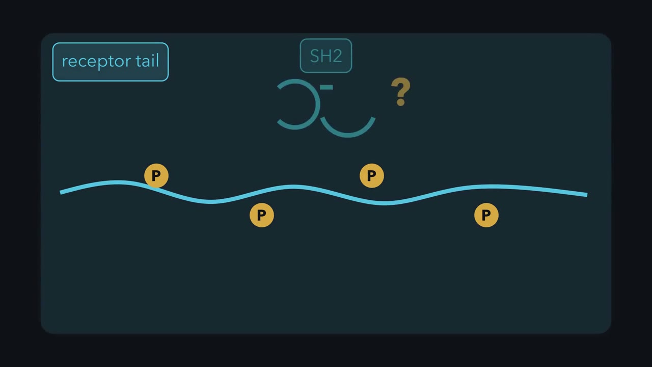 ▶ Watch How One Mark Finds Its Reader · 3:41
Read the transcript of “How One Mark Finds Its Reader”
Film transcript: How One Mark Finds Its Reader
A receptor writes phosphotyrosine marks across its tail. How can one protein find the right mark among all the others? Phosphate changes a small surface patch, but the mark usually does little alone. It becomes useful when a reader binds there. The reader brings its host protein into the growing signaling complex. A kinase writes the mark, and a phosphatase can erase it. An SH2 domain is one such reader. Its first binding pocket fits phosphotyrosine. Positively charged side chains help grip the phosphate. Unmodified tyrosine lacks that phosphate, so the pocket cannot make the same contact. That first pocket answers whether tyrosine is phosphorylated.
It cannot identify which phosphorylated site this is. A neighbouring surface reads residues beside the phosphotyrosine and supplies another layer of preference. Different SH2 domains carry differently shaped neighbouring surfaces. Now the reader tests both features together. Several tails offer the same gold phosphate. Their neighbouring patterns differ. Matching blocks settle into the groove; poor fits make the reader release.
Src-family readers favour one pattern, while other readers favour others. Here is the rule. Reusable binding domains turn recognized motifs or lipids into protein connections. For SH2, phosphorylation enables the contact, while sequence context helps choose among possible partners. The domain binds; it does not catalyse a reaction. Predict before the reader moves. Both tails carry phosphotyrosine. Only the lower neighbouring pattern fits this reader's groove. Which tail will retain the reader longer? Watch both parts of the binding surface.
The lower tail makes both contacts, so it retains this reader more strongly in this comparison. The upper tail still has phosphate. Its neighbouring pattern simply provides a poorer fit for this domain. Another SH2 domain could prefer a different pattern. The domain is modular because it keeps this binding activity when moved. Detach the reader from one host. Attach it to a different host, and its preferred target remains the same. The new host can bring a different cellular activity to that site. Preference is not destiny. Location, abundance, timing, and the rest of the host protein also shape contacts in cells. The domain narrows the candidates; cellular context narrows them further.
A preferred motif cannot recruit a reader that never reaches it. Other modules apply the same logic to other targets. SH3 reads proline-rich motifs, while some PH domains read membrane lipids. The target changes, but recognition still creates a connection. For PH, that connection is to the membrane.
From the film
- Reusable binding domains turn recognized motifs or lipids into protein connections.
- An SH2 domain grips phosphotyrosine and also reads the residues beside it.
- Sequence preference narrows possible partners; location, abundance and timing narrow them further.
A receptor tyrosine kinase has phosphorylated its own tail. What reads those marks, and how does one reader choose among them?
A phosphotyrosine on a receptor tail is mostly a mark that changes little until another protein recognises it. What the cell needs is many proteins arriving at the receptor together, and the phosphate is how that request is written. A motif is a short amino-acid sequence, a few residues long, that such a reader can recognise.
The one idea. Reusable binding domains turn recognized motifs or lipids into protein connections.
Scope: a connection here means protein to protein or protein to membrane. A motif alone does not determine binding in a cell, not every PH domain binds a phosphoinositide, and not every contact requires phosphorylation.
Why a mark needs a reader
A phosphate on tyrosine adds two negative charges to a small patch of a protein’s surface, and phosphatases can remove it within seconds. The cell can therefore switch a docking site on and off without making or destroying a protein. The first film gives the reader’s two jobs; here is where readers came from.
A module keeps its binding job when it moves
A modular interaction domain is a protein segment, from a few dozen to roughly two hundred amino acids, that folds by itself, recognises a motif or lipid, and keeps that binding activity in another protein.
In 1986 Ivan Sadowski, a graduate student in Tony Pawson’s laboratory in Toronto, altered about a hundred amino acids just before a sarcoma-virus kinase domain. The same region sits in the same place in Src, Abl and Fps, so it probably mattered, but nobody knew for what. The mutant, made in bacteria, phosphorylated tyrosine in a test tube about as well as the original. In rat cells, however, its kinase activity was low and it no longer made the cells cancerous.
That mismatch points to a job other than catalysis, holding other proteins and steering the kinase beside them, which a test-tube assay of phosphate transfer cannot see.
The lab named the region Src homology 2, or SH2, after the second conserved block in Src. By 1990, SH2 domains isolated from a phospholipase, a GTPase-activating protein and Src itself each bound activated growth-factor receptors in vitro.
Crk showed that readers can organize signaling without an enzyme of their own. Bruce Mayer, in Hidesaburo Hanafusa’s laboratory at Rockefeller University, cloned this cancer gene from an avian sarcoma virus collected in 1931. Its protein is viral Gag joined to an SH2 and an SH3 domain. Crk does not phosphorylate tyrosine, yet transformed cells carry more phosphotyrosine than normal cells: it reads phosphotyrosines and gathers the cell’s own enzymes outside their usual arrangement.
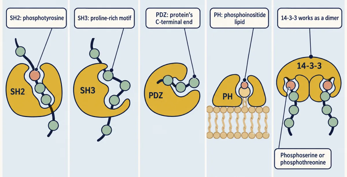
| Module | What it binds |
|---|---|
| SH2 (about 100 residues) | a phosphotyrosine and the residues just after it |
| PTB (phosphotyrosine-binding) | an NPXY motif, which some PTB domains need to be phosphorylated and others bind unphosphorylated; the Shc type reads the residues before the tyrosine |
| SH3 (about 60 residues) | a proline-rich motif with a PxxP core |
| WW | a proline-rich motif such as PPxY |
| PDZ | the last few residues at a protein’s C-terminal end, including its free carboxyl group |
| PH (about 120 residues) | phosphoinositides, the phosphorylated lipids of the membrane, for some PH domains |
| 14-3-3 | phosphoserine or phosphothreonine motifs; a whole small protein that works as a dimer |
Predict. A PDZ-binding tail gains one extra residue at its carboxyl end. The old final residues are still present. Can a PDZ domain read them as the same terminal motif?
Check your prediction
No. A PDZ domain reads the last residues together with the free carboxyl end. Adding a residue moves that end, so the old sequence is no longer the same C-terminal target.
Many PH domains bind lipid weakly or not at all, and only a minority bind a particular phosphoinositide with real specificity. The one that matters for the design problem below is the PH domain of Akt, which binds phosphatidylinositol 3,4,5-trisphosphate (PIP3) and also PI(3,4)P2, a second lipid that appears when PI 3-kinase is active.
SH2 specificity: two parts of one binding surface
Here is the evidence behind the first film’s two grips. Many tyrosines are phosphorylated in a stimulated cell, and around a hundred human proteins carry SH2 domains, so the phosphate grip alone cannot choose a partner.
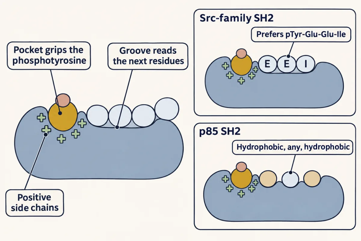
In 1992 Gabriel Waksman, a postdoctoral researcher in John Kuriyan’s laboratory at Rockefeller, crystallized the SH2 domain of v-Src with a short phosphopeptide. That structure and a 1993 follow-up with a tighter-binding peptide showed lysine and arginine side chains in one pocket stacking against the phosphotyrosine ring and bonding to the phosphate, and a neighbouring groove holding the residues just after it.
One structure cannot show whether the groove produces selectivity. In 1993 Zhou Songyang, in Lewis Cantley’s laboratory in Boston, tested that with a peptide library: phosphotyrosine at a fixed position, random residues after it, sampled by purified SH2 domains from at least ten signaling proteins one at a time.
Src, Fyn, Lck and Fgr all selected phosphotyrosine–glutamate–glutamate–isoleucine. Domains from p85, the regulatory subunit of PI 3-kinase, phospholipase C-gamma and the phosphatase SHPTP2 preferred phosphotyrosine–hydrophobic–any–hydrophobic. Most other domains selected patterns of their own, so the groove sets much of an SH2 domain’s sequence preference.
Predict. A Src-family SH2 domain has the arginine in its phosphate pocket mutated, but its groove is intact. Will it still pull phosphotyrosine–glutamate–glutamate–isoleucine peptides out of the library?
Check your prediction
Not well. The groove adds preference among phosphopeptides that the pocket already holds; it cannot hold them alone. That is why a pocket-arginine mutation is the standard way to abolish SH2 recruitment, as in the design problem below.
How much is bound?
For one independent binding site at equilibrium, occupancy follows:
fraction of domain bound = [ligand] / (Kd + [ligand])
Kd is the free-ligand concentration at which half the domains are occupied. A lower Kd means less ligand is needed for the same occupancy.
Predict. Substitute a ligand concentration equal to Kd. What fraction of the domains is bound?
Check your prediction
One-half: the numerator is Kd and the denominator is Kd plus Kd, or two Kd.
The equation describes occupancy, not residence time or multivalent binding. Reversible binding matters here: while an SH2 domain is bound it shields phosphotyrosine, but when it releases, a phosphatase can reach the site. Once the phosphate is gone, that contact cannot re-form.
Weak, reversible grips have a second use: a protein with several modules can grip parts of itself, or two targets at once. The second film shows both.
Read the transcript of “Two Grips, One Decision”
Film transcript: Two Grips, One Decision
The first film tracked one domain to its target. What changes when one protein carries several binding modules? Those modules can now influence one another's effects. Start with cellular Src as an open strip. It carries an SH3 domain, an SH2 domain, a kinase, and a short tail. Each block keeps a distinct job. Flexible connections let those blocks meet within one protein. A kinase called Csk places phosphate on a tail tyrosine. The tail bends back. Src's own SH2 pocket catches that phosphotyrosine, bringing the tail against the protein. This contact uses the reader internally. The SH3 domain catches the linker at the same time. These internal contacts settle both domains against the kinase.
Together they stabilize in an inactive shape. Neither module performs the kinase reaction itself. Now offer the SH2 domain a better external phosphotyrosine target. Predict what happens. Can Src's own tail still complete the same grip?
Compare the two targets competing for one pocket. No. The external target can occupy SH2 and pull it from the tail. Losing one internal contact loosens the clamp. Other inputs can also disturb the SH3 contact. Opening makes kinase activation more accessible. Viral Src has lost the tail and its regulatory tyrosine, along with other mutations.
Without that tail contact, the full internal clamp cannot form, so the kinase is always on. The missing module target removes an off-switch. The same modular logic can build a conditional detector. Give one protein an SH2 domain for a phosphorylated receptor and a PH domain for a membrane lipid. Join them with a flexible linker. Suppose either contact alone is too weak to retain the detector. With only the receptor mark, it visits and leaves. With only the lipid, it also visits and leaves. One input produces only a brief encounter. When both targets are close, both domains bind at once. Each contact helps keep the other within reach. The detector remains recruited only where the two inputs coincide.
Removing either target again releases it. This joint retention is avidity: two weak contacts together can behave like one much tighter interaction at that location. That is the second rule. Combining modules can make binding conditional on more than one signal. In Src, two internal contacts together hold the kinase off. In the detector, two external contacts together hold the protein in place.
From the film
- Combining modules can make binding conditional on more than one signal.
- Src's SH2 and SH3 domains help hold its own kinase in an inactive shape.
- A multi-domain detector stays recruited only while its required targets are together.
Src: a protein that reads itself
Bishop and Varmus won the 1989 Nobel Prize for showing that the cancer gene of a chicken sarcoma virus, v-Src, is a captured copy of a normal cell gene, c-Src. The two proteins differ in how well they stay quiet.
From its amino end, c-Src carries myristate, a fatty acid attached to its second residue that anchors it to the inner plasma-membrane face. Then come a short unique region, SH3, SH2, a linker, the kinase domain and a short tail. Csk phosphorylates tyrosine 527 in the tail, six residues from the end in chicken Src. Crystal structures confirm the clamp the second film draws.
An outside ligand wins because the tail after tyrosine 527 fits SH2 less well than phosphotyrosine–glutamate–glutamate–isoleucine, so the internal contacts are weak enough to be displaced. Once the clamp opens, one Src molecule can phosphorylate tyrosine 416 in another Src’s activation loop, which helps keep the kinase active until a phosphatase removes the phosphate.
v-Src’s missing tail, as the second film showed, leaves it with no clamp. Changing tyrosine 527 to phenylalanine in c-Src has the same activating effect and makes it transform cells.
Predict. If c-Src loses its amino-terminal myristate but keeps an intact phosphorylated tail, which property changes most directly: membrane anchoring or the SH2-tail grip?
Check your prediction
Membrane anchoring. The fatty acid is at the amino end, whereas the SH2-binding phosphotyrosine is in the carboxy-terminal tail. The mutation separates localization from the internal clamp.
The virtue of modularity: new pathways from old parts
The human genome encodes 518 protein kinases, including 58 receptor tyrosine kinases. A pathway can gain a connection without new chemistry: join a reader to an enzyme that already works, and a receptor can recruit that enzyme. Phospholipase C-gamma, a lipid-cutting enzyme, also carries two SH2 domains, an SH3 domain, a PH domain and a C2 domain. The SH2 domains are what let an activated receptor recruit it. Grb2, which the next lesson takes up, is little more than an SH3, an SH2 and a second SH3 in a row. Humans have around a hundred SH2-containing proteins; yeast, with no dedicated tyrosine kinases, has almost none.
Combining modules can make binding conditional on more than one signal. The design problem asks you to build such a detector.
Design problem
Design a protein that binds phosphatidylinositol 3,4,5-trisphosphate (PIP3) and an activated receptor tyrosine kinase. Then make it respond to calcium as well. PIP3 is a lipid made on the inner face of the membrane when PI 3-kinase phosphorylates phosphatidylinositol 4,5-bisphosphate (PIP2). Say which modules you use, how you join them, and one experiment that shows each part is doing its job.
Try your design in the mixer before you open the answer.
A module keeps its job when you move it. Each reader grips only when its target is there, and an enzyme with no reader is not brought anywhere. This is a sketch of the logic, not a prediction for a real construct.
Saved in this browser only.
One way to do it
Fuse a PH domain that binds PIP3 to an SH2 domain whose preferred sequence matches the phosphotyrosine motif on your receptor. Join them with a flexible linker long enough that both can reach the membrane at once. Like the second film’s coincidence detector, it stays only where a receptor tail and PIP3 are both within reach. The Akt PH domain would work, but it also binds PI(3,4)P2, so for a strict PIP3 detector choose a PH domain known to prefer PIP3.
For calcium, add a Ca2+-dependent C2 domain, which binds acidic membrane lipids only when it has Ca2+ bound. If you also weaken the PH and SH2 grips so that no two alone are enough, all three inputs are needed before the protein stays at the membrane. A second route is to hide one of the grips behind an inhibitory segment that Ca2+ and calmodulin pull away, as a better ligand pulls the SH2 domain off the Src tail.
Test each part. Each of these should abolish recruitment: a mutation in the arginine of the SH2 pocket, or an unstimulated receptor; a PI 3-kinase inhibitor, or a mutation in the basic pocket of the PH domain; chelating intracellular Ca2+ with a cell-permeant chelator such as BAPTA-AM. If removing one input, verified as truly removed, still leaves the protein at the membrane, the other grips are too strong and the design needs a poorer SH2 or PH domain.
Going deeper
More than the motif
SH2 domains commonly bind phosphopeptides with sub-micromolar to low-micromolar dissociation constants, a range that permits exchange. Motif preference narrows the candidate set but does not uniquely specify an in-cell partner. Beyond the location, abundance and timing shown in the first film, scaffold-imposed proximity, competition among related domains and the cell’s kinase and phosphatase complement all contribute.
The avidity in the second film comes from rebinding: while one contact holds, the other can re-form before the proteins separate.
Several routes can open Src
A phosphorylated platelet-derived growth factor (PDGF) receptor can pull Src’s SH2 domain from its tail. Focal adhesion kinase (FAK) can do so after integrins activate it. A proline-rich partner such as beta-arrestin can take the SH3 domain, a proposed route from an activated beta-adrenergic receptor to Src, and G-protein alpha subunits can also activate Src directly. These routes refine the clamp model; they do not replace it.
Your notes
Select any text in the lesson to highlight it or add a note to it. Your highlights and notes are saved in this browser and collected in your notebook.
This browser is not letting the site save anything (a private window or strict privacy settings), so highlights and notes will be lost when you leave the page.
How we measure it
Kinase assay paired with a transformation assay
You measure how fast a purified kinase puts phosphate on a substrate in a tube, and in a separate experiment whether the same protein turns cultured cells cancerous. If the first number is normal and the second is lost, the mutated region does something other than catalysis.
X-ray crystallography of domain-peptide complexes
A domain and a short peptide are crystallized together and the diffraction pattern is turned into an atomic model. It shows which side chains touch the phosphate and which pockets hold the neighbouring residues.
Degenerate phosphopeptide library screening
A mixture of many different peptides, each with a phosphotyrosine at a fixed position and random residues after it, is offered to a purified domain. The peptides that stick are sequenced, and the residues that are over-represented are the ones the domain prefers.
Binding assays with isolated domains
A single domain is produced on its own and tested for binding to a phosphorylated receptor or a peptide. If the isolated domain binds, the binding does not depend on the rest of the protein.
Method films · the methods library
- Binding assays: immunoprecipitation, pull-down and gel shift 3:43
- Engineered mutants: site-directed, dominant-negative and constitutively active 4:25
- Gene cloning: vectors, libraries and screening 4:04
- Pharmacological blockade and inhibitors 4:10
- Protein purification and column chromatography 4:14
- X-ray crystallography 4:06
Practice questions on this lesson (3) → MCAT-style, original, each with an explanation
Where this lesson fits
Before thisReview first
- Phosphorylation: Kinases and PhosphatasesThis course · Lesson 6
You need to know how kinases and phosphatases add and remove the phosphate that these domains read.
- A Field Guide to ReceptorsThis course · Lesson 4
It introduces receptor tyrosine kinases and their self-phosphorylated tails, which create the marks read here.
- Binding: Affinity, Occupancy and Dose–ResponseThis course · Lesson 3
The Kd and fraction-bound equation used here for domain grip is taught there.
After thisStudy next
- Adaptors, Docking Proteins and ScaffoldsThis course · Lesson 10
Adaptors like Grb2 are built from SH2 and SH3 modules strung together, as shown here.
- Receptor Tyrosine KinasesThis course · Lesson 18
It shows which SH2 and PTB readers dock on the activated receptor's phosphotyrosines.
- PI 3-Kinase, Akt and InsulinThis course · Lesson 20
It uses the PH domain of Akt reading PIP3 and the p85 SH2 domain docking.
Suggested by an AI model from the lessons themselves, then checked. How we use AI
Finish line
Test yourself
8 questions
Practice and MCAT-style, with explained answers.
Next · Lesson 10
Adaptors, Docking Proteins and Scaffolds
A receptor activates Ras without ever touching it. How does the message cross the gap?
Continue →