Physiology · Unit 9: Reproduction
Sexual Differentiation and the Testis
Rewritten on : new text, figures and flashcards. Your notes and highlights stay, but the text around them may have moved.
An AMH-null male mouse can make sperm and retain a uterus. Which testicular signals have separated?
Pause when the reading reaches a film, and offer to play it. Saved on this device.
Watch the film now, or keep listening
Builds on:Endocrine Axes and How to Test ThemThe Adrenal Cortex and MedullaSmooth Muscle and the Myogenic ResponseWhy these?
An AMH-null male mouse can produce functional sperm and retain a uterus. How can those outcomes coexist?
To answer it you need two steps. First, what turns an embryonic gonad into a testis: one gene on the Y chromosome, SRY. Second, what the testis then sends: at least two hormones, each from its own cell to its own target. One is AMH (anti-Müllerian hormone), which removes the ducts that would become a uterus. The adult testis uses the same two cells, and with the feedback loop that explains why a man who injects testosterone usually makes fewer sperm.
The one idea to carry through this lesson. Making a male reproductive tract is a chain of separate links, each one a cell, a hormone, an enzyme or a receptor:
- SRY → SOX9 → Sertoli cells → AMH → Müllerian ducts (would-be uterus) removed.
- Sertoli cells help guide Leydig-cell development → Leydig testosterone → androgen receptor → Wolffian ducts (would-be sperm ducts) kept.
- In humans: testosterone → dihydrotestosterone (DHT) → the same receptor → outside fully masculinized.
Break one link and everything downstream of it fails, while the other developmental branches carry on. In the adult the same two cells divide the work again: Leydig cells make testosterone under luteinizing hormone (LH), and Sertoli cells support sperm under follicle-stimulating hormone (FSH) and local testosterone, so blood testosterone is not the same as testicular testosterone.
The rule describes a link lost completely, early in development; partial loss gives in-between results, and feedback loops (added later) can shift other branches (see Going deeper). Most causal experiments below used rabbits and mice; human findings are labelled.
Making a testis: SRY flips the supporting cells
Every embryo starts with a bipotential gonad, one that can develop into either a testis or an ovary. In most mammals, including humans, the Y-linked gene SRY starts testis determination by raising SOX9, a transcription factor, in the gonad’s supporting-cell precursors, which then become Sertoli cells. In the mouse, positive feedback helps keep SOX9 switched on, reinforcing the Sertoli fate.
The ovary is not what is left when SRY is missing: it has its own gene program that pushes the other way, and the two compete in the same supporting cells (genes named in Going deeper). Once Sertoli cells exist, they shape the signals that guide developing Leydig cells and germ cells. A gonad’s fate and its effects on other organs are separate causal steps.
For SRY’s identification by Sinclair and colleagues in 1990 and Koopman’s 1991 mouse work, see sex linkage and beyond.
Predict. An XY mouse embryo lacks a working Sry gene. What do its supporting cells become, and what happens to everything the chain builds after them?
Check your prediction
SOX9 is not raised, so the supporting cells follow the ovarian program and no testis forms. SRY is the first link, so everything downstream fails: no AMH, no Leydig testosterone, no DHT. One missing gene changes the gonad, the ducts and the outside.
Two ducts, two jobs, two signals
Every early mammalian embryo, XY or XX, builds two pairs of ducts side by side. The Wolffian ducts (also called mesonephric ducts) can become the epididymis, where sperm mature after leaving the testis, the vas deferens, which carries them onward, and the seminal vesicles. The Müllerian ducts (paramesonephric ducts) can become the oviducts, the uterus and the upper vagina. An adult normally keeps only one set, with at most small remnants of the other.
Which set stays depends on the testis. Alfred Jost showed this in the 1940s by removing the gonads of rabbit fetuses in utero, while still in the uterus, and inspecting their ducts later (Jost’s report).
Predict. Jost removes the testes from an XY rabbit fetus early, before the ducts have changed. Which ducts does the animal have later?
Check your prediction
It keeps its Müllerian ducts and loses most of its Wolffian ducts, so it develops a female-typical tract. Jost also operated on XX fetuses: removing their ovaries did not prevent a female-typical tract either. So the female-typical tract does not need an ovarian secretion; the male-typical tract needs the testis. (This is about the tract; the ovary itself is built by an active program.)
So, left alone, the Müllerian ducts stay and the Wolffian ducts fade. A testis must do two opposite things: remove one pair and rescue the other.
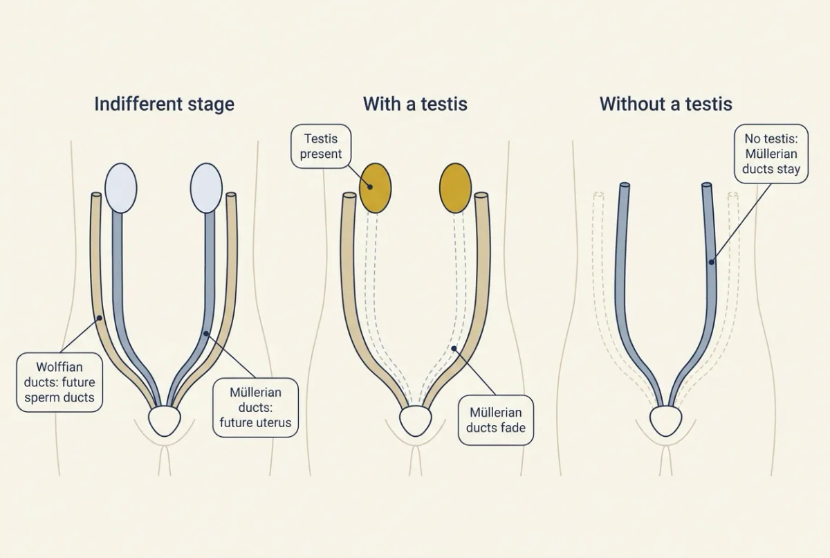
Could one hormone do both? Removing the whole gonad could not tell the signals apart, so Jost gave testosterone in place of a testis. The Wolffian ducts stayed, but so did the Müllerian ducts. If testosterone were the testis’s only signal, it should have removed those as well. It rescued one pair but could not remove the other, so he inferred a second signal, a Müllerian inhibitor.
Molecular work later identified it as anti-Müllerian hormone (AMH). It comes from Sertoli cells. Testosterone comes from a different cell, the Leydig cell, a steroid-making cell between the developing tubules (the human picture; see Going deeper for the mouse). Two cells, two hormones, two targets:
- Sertoli cell → AMH → Müllerian ducts regress.
- Leydig cell → testosterone → Wolffian ducts are kept.
Now you can answer the question this lesson opened with.
Predict. In 1994, Behringer, Finegold and Cate made male mice that lack only the gene for AMH (also called MIS, Müllerian-inhibiting substance). Their testes form. What do their ducts look like, and can they make sperm?
Check your prediction
They keep a uterus and oviducts, because nothing removes the Müllerian ducts, and they keep their male (Wolffian) derivatives, because testosterone is still made. Their testes make functional sperm, though most of these males are infertile because the retained organs get in the way of sperm transfer. One broken link changed one branch; the testosterone branch carried on.
The experiment shows that AMH is necessary for Müllerian regression in mice; it does not locate AMH’s receptor or measure a human response curve.
The outside needs DHT (in humans)
The external genitalia start from the same structures in every embryo. In humans, the genital tubercle contributes to the glans penis or clitoris, and the labioscrotal swellings become the scrotum or the labia majora. With androgen signalling, the urethral folds fuse and contribute to the penile urethra.
In humans, these tissues first convert testosterone into a stronger androgen. The enzyme 5-alpha-reductase type 2, encoded by the gene SRD5A2, converts testosterone to dihydrotestosterone (DHT). Human fetal DHT signalling is needed for full masculinization of the external genitalia and supports prostate development. Testosterone and DHT act through the same androgen receptor (AR), a hormone-responsive transcriptional regulator (see nuclear receptors). Why convert at all? DHT holds the receptor more tightly, so tissues that make DHT amplify the signal.
Predict. Use the chain. In an XY embryo whose testes form normally, what happens to the Müllerian ducts, the Wolffian ducts and the outside if (a) the androgen receptor does not work at all, or (b) SRD5A2 does not work?
Check your prediction
(a) No AR. AMH does not act through the androgen receptor, so the Müllerian ducts should still regress. But both androgen branches run through AR, so the Wolffian ducts are not kept and the outside is female-typical. One receptor, two outcomes changed.
(b) No SRD5A2 (human). Testosterone still acts on AR, so the Wolffian derivatives are generally kept. Only DHT is missing, so the outside is less masculinized.
Again, what fails lies downstream of the broken link: one branch (AMH, SRD5A2) or several (SRY, AR). Each row below removes one link from the start of development.
| Missing link and system | Müllerian outcome | Wolffian outcome | External outcome |
|---|---|---|---|
| Testis: early rabbit gonadectomy | Retained | Largely lost | Female-typical |
| AMH: male mouse null mutant | Uterus and oviducts retained | Male derivatives retained | Male-typical |
| AR: XY Tfm mouse | Regressed | Not maintained | Female-typical |
| SRD5A2: human mechanism | Usually absent | Generally retained | Reduced masculinization |
The Tfm mouse carries an X-linked AR mutation, named for the historical term “testicular feminization”; Lyon and Hawkes’s paper established its X linkage. In 46,XY humans, complete AR loss likewise leaves testis formation, and usually anti-Müllerian hormone (AMH) action, intact while androgen response is absent. Human SRD5A2 deficiency is autosomal recessive: affected people carry disease-causing variants in both copies, usually one from each parent, and some variants abolish enzyme activity while others reduce it. Real cases vary around these typical patterns (see Going deeper).
Predict each anatomical panel before you change a signal in the “Build the tract” model below; its enzyme toggle represents the human SRD5A2 mechanism.
In this sketch: an XY embryo with a bipotential gonad; every switch is a complete loss from the start of development with the others intact, as in the lesson’s table; the switches combine findings from rabbit, mouse and human, so two or more switches off at once is a prediction from these rules, not an observation; if the receptor is off the external genitalia are drawn female-typical whether or not the enzyme works; “reference” is a made-up baseline for LH and testosterone, with no numbers, concentrations or units; the LH and feedback arrows come from the lesson’s general statements, not from fetal data; the external-genitalia names (glans, scrotum, labia) are human structures.
A testis sends two separable signals: AMH removes the Müllerian ducts, and testosterone acting through the androgen receptor maintains the Wolffian ducts; in humans, conversion to DHT by 5-alpha-reductase type 2 also supports masculinization of the external genitalia.
The adult testis: the same two cells, two compartments
A man injects testosterone, so his blood testosterone rises. What happens to his sperm production? Many people would guess that it rises. Build the adult testis first, then decide.
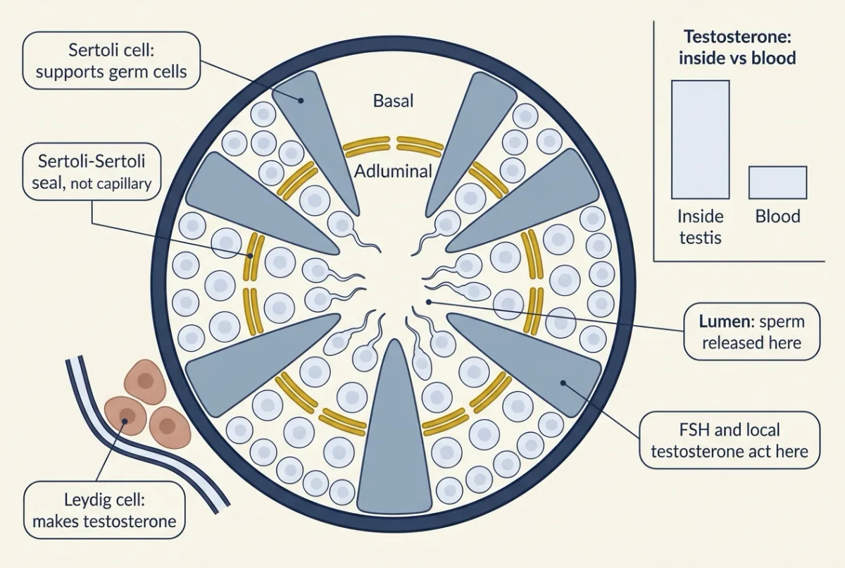
The seminiferous tubules are tubes in which germ cells develop into sperm; the interstitium is the tissue between them. The two fetal cell types split the adult work.
- Outside the tubules: Leydig cells make testosterone under the pituitary hormone luteinizing hormone (LH). LH-receptor signalling increases cyclic AMP and cholesterol delivery into mitochondria; the shared steroid machinery is covered in adrenal glands and the cascade in cAMP and PKA.
- Inside the tubules: Sertoli cells nourish the germ cells. They respond to the other pituitary gonadotropin (gonad-stimulating hormone), follicle-stimulating hormone (FSH), and to local testosterone. Both gonadotropin receptors use G-protein signalling.
Leydig testosterone reaches the tubules directly, so intratesticular testosterone is far higher than circulating testosterone.
Inside each tubule, the blood-testis barrier is a junctional seal between neighbouring Sertoli cells, not capillary endothelium. It separates a basal compartment, which holds spermatogonia (mitotic germ-cell precursors) and early spermatocytes (cells entering meiosis), from an adluminal compartment, toward the lumen, which holds later meiotic cells and spermatids (haploid products of meiosis). Mouse evidence shows the junctions coming apart and re-forming as spermatocytes cross; see epithelia.
Last, feedback. Testosterone restrains hypothalamic-pituitary LH and FSH output, partly after conversion to estradiol, and Sertoli cells make inhibin B, which selectively inhibits FSH. Apply the loop logic from endocrine axes; the ovarian axis is next.
Predict. Back to the man injecting testosterone. What happens to his LH, to the testosterone inside his testes, and to his sperm production?
Check your prediction
His LH and FSH fall, because the injected testosterone feeds back on the pituitary. With little LH, his Leydig cells make little testosterone, so testosterone inside the testis falls steeply while blood testosterone is high. The germ cells lose local androgen and FSH support, so sperm production falls. That is why testosterone therapy and anabolic steroids can suppress sperm production, sometimes to the point of infertility.
A 2005 trial in healthy men confirmed the hormone side of this and tested a workaround, low-dose hCG (see Going deeper).
Mouse experiments show that Sertoli cells need androgen even in the adult (see Going deeper).
A sperm ejaculated today began developing about two and a half months ago
Why care about a number? Anything that changes sperm production, such as a drug, a fever or the testosterone injections above, shows up in semen weeks to months later.
In spermatogenesis, spermatogonia divide by mitosis; a spermatocyte’s two meiotic divisions yield four haploid products; spermiogenesis reshapes spermatids into spermatozoa; and spermiation releases them from the tubule’s lining into the lumen. The cell sequence is in animal reproduction and the chromosome steps in meiosis.
Picture a conveyor belt. Stand at one spot in a tubule. Every 16 days (the conventional human value), a new batch of cells starts its journey there, so the same mix of stages comes round again every 16 days. That repeat is called an epithelial cycle. Any one batch needs about 74 days, the textbook human total from a differentiating spermatogonium to spermiation, to finish. So several batches are on the belt at once. Assuming steady production and a constant cycle length:
Cycles per lineage = 74 days / 16 days per cycle = 4.625, about 4.6 cycles.
Check: 4.625 × 16 = 74 days, already about 2.4 months. Add the time sperm then spend passing through the epididymis, and a sperm ejaculated today began developing about two and a half months ago.
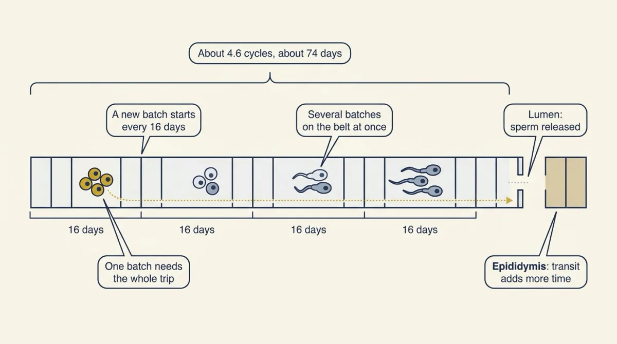
A tracer labels cells partway along the belt, so its lag depends on where it enters: count the cycles left from the labelled stage, multiply by 16 days, then add epididymal transit. The one human deuterium-labelling study, which followed label all the way to ejaculated sperm, measured about two months (worked example in Going deeper).
Some books give 64 days because they leave out the early spermatogonial divisions, so check which starting cell a number uses.
Erection and ejaculation: three phases, three outflows
The testis makes sperm; delivering them takes three more steps, each on its own nerves.
Erection is filling and stiffening of erectile tissue as inflow increases and venous drainage is restricted. It depends mainly on pelvic parasympathetic pathways, including nitrergic nerves that signal with nitric oxide (NO); endothelium also supplies NO. NO activates guanylyl cyclase, raising cyclic GMP (cGMP), and protein kinase G links cGMP to lower cytosolic calcium and smooth-muscle relaxation. PDE5 breaks down cGMP, so PDE5 inhibitors keep cGMP high; see cGMP signalling.
Cavernosal smooth muscle surrounds blood-filled spaces called sinusoids. When it and the arterial smooth muscle relax, the tissue fills and compresses the veins beneath the tunica, its fibrous covering. See smooth-muscle mechanics and autonomic outflow.
Emission moves sperm and glandular secretions into the urethra, mainly under sympathetic control of the vas deferens and accessory glands, with the bladder neck closing. Expulsion is the rhythmic outward movement driven by somatic pudendal motor output to muscles including bulbospongiosus. Ejaculation combines these coordinated events.
Three phases, three separate outflows, so they can fail separately: sympathetic nerve damage or an alpha-blocker can impair emission, while erection, which depends mainly on parasympathetic and nitrergic pathways, is largely preserved.
Design problem
A colleague claims that testosterone alone is enough to give an XY embryo a male-typical tract. Design experiments in fetal rabbits or mice that test the claim.
Saved in this browser only.
One way to do it
Replace the testis with testosterone. Remove the testes from XY rabbit fetuses early and give testosterone. If the claim were right, the tract would be fully male-typical. Instead both duct pairs are kept, so the claim fails for the Müllerian ducts. This is Jost’s result.
Run the converse in mice. Delete AMH or its receptor and leave testosterone intact: the Müllerian ducts persist. Delete AR and leave AMH intact: the Müllerian ducts regress but the Wolffian ducts are lost. Together they show two signals with separate targets.
Controls. Use littermates, confirm each deletion before reading the anatomy, and sample at matched ages inside the duct-response window.
Going deeper
The ovarian program and SOX9 enhancers
The opposing ovarian network includes WNT4, RSPO1 and beta-catenin, with FOXL2 contributing to ovarian identity.
An enhancer is regulatory DNA that increases a gene’s expression in a particular cellular context. Croft and colleagues’ 2018 human SOX9 work implicates enhancer duplication or deletion in altered testis determination, extending the mechanism beyond coding sequences. Their reporter assays tested cloned fragments in transfected cells; such activity does not establish a whole-gonad expression threshold. Croft’s study
What Jost’s timing showed
Jost’s 1947 report describes males castrated on fetal day 20 that retained female genital tracts yet still had rudimentary prostatic buds; later operations preserved more established male structures. His results support a requirement for fetal testicular influence during particular developmental windows in rabbits, rather than continuing hormone dependence of every structure. In experiments that left one testis in place, its influence could be stronger on nearby ducts than across the fetus, a sign that these signals act locally.
Mouse versus human steroid steps
“Leydig testosterone” is the human picture. In the fetal mouse, Leydig cells make mostly androstenedione, and fetal Sertoli cells convert it to testosterone, so even this link is split between the two cell types. The dihydrotestosterone (DHT) branch also differs: mouse Srd5a2 loss gives a much milder phenotype than the human deficiency.
When a link is only partly broken
Some people with complete androgen receptor (AR) loss keep Wolffian remnants. In campomelic dysplasia, one working copy of SOX9 is lost, and most, but not all, XY patients show sex reversal. In 5-alpha-reductase type 2 deficiency, the high testosterone of puberty partly masculinizes the external genitalia, so the DHT branch is a matter of potency, not an absolute switch. Partial-function variants give graded outcomes, and each target responds only during its developmental window.
LH and testosterone after receptor loss
The chain leaves out feedback. Loss of AR-mediated feedback predicts increased luteinizing hormone (LH) drive, but testosterone concentrations depend on species and testis function; normal or high testosterone is a human AR-loss pattern, not a guaranteed Tfm-mouse value.
The testosterone-injection trial
In Coviello and colleagues’ three-week study of healthy men in 2005, testosterone injections given with a placebo suppressed LH and follicle-stimulating hormone (FSH), and intratesticular testosterone fell by about 94%. Men who also received low-dose hCG, which acts on the LH receptor, kept intratesticular testosterone in the normal range.
Sertoli androgen receptor deletions
Larose and colleagues’ 2020 SCARKO mice lacked AR specifically in Sertoli cells: surviving spermatocytes could synapse and recombine chromosomes, yet failed to acquire competence for meiotic division. Cytology and single-cell RNA sequencing showed that mutant germ cells stayed in the gene-expression state of early prophase (leptotene and zygotene), a transcriptional block. That supports a Sertoli-mediated requirement without implying that every early meiotic event requires androgen, or that humans arrest identically.
A second design problem. Mice lacking Sertoli-cell AR from fetal life make almost no round spermatids and no elongated spermatids. A colleague argues that this reflects failed Sertoli maturation rather than an ongoing adult requirement. Design an experiment that separates those explanations.
One way to do it
Delete AR only after Sertoli maturation, in adult mice carrying a conditional AR allele: receptor DNA sits between recognition sites for the enzyme Cre. Target Cre to Sertoli cells to remove that DNA; verify AR loss in Sertoli cells and retention in Leydig and peritubular cells. Include conditional-allele mice without Cre, Cre delivery to wild-type testes, and a contralateral control, with matched handling.
Collect a time series scaled to mouse development, about 8.6 days per epithelial cycle and roughly 35 days for spermatogenesis, so that cells already beyond a block and stored sperm can clear. Score stage-specific meiotic markers and epididymal sperm; test barrier integrity with a tracer. Measure serum LH and testosterone: a relatively unchanged Leydig compartment is a prediction to test, rather than an assumption guaranteed by Sertoli specificity.
If adult signalling is required, predict meiotic arrest and eventual depletion of epididymal sperm. Willems and colleagues reported adult Sertoli AR deletion with meiotic arrest and no mature epididymal sperm at 40 days, supporting that prediction. Their adult-deletion experiment
Quantify mosaic deletion and control for delivery injury: persistent sperm after incomplete deletion would not establish receptor independence, and one arrest phenotype cannot identify the responsible androgen-regulated genes. The inference remains specific to adult mouse Sertoli function.
Timing: 64 versus 74 days, and the tracer lag
Heller and Clermont’s original 1963 labelling study estimated about 64 days for the whole process. Their 1964 analysis gave about 74 days once spermatogonial divisions were counted, and Amann’s 2008 review recommends 74. Their timing work is described further in animal reproduction.
A third number measures something else again. Misell and colleagues in 2006 labelled DNA with deuterated water in 11 men with normal sperm concentrations, who drank it daily for three weeks while semen was sampled every two weeks for up to 90 days. Label appeared in ejaculated sperm after 64 ± 8 days (mean and standard deviation), with a 42–76-day range. This label-to-ejaculate lag includes post-testicular transit and storage, so its start and endpoint differ from the textbook total.
Worked problem: count cycles, then add transit. Simplify: label incorporation occurs only during the last premeiotic S phase, the DNA-replication phase in preleptotene spermatocytes. Because the water was taken for weeks, real label availability can mark other dividing precursors, such as spermatogonia, too. In this made-up example, assume 3.5 cycles remain to spermiation and epididymal transit takes 10 days, with no additional storage delay; neither assumption is a measured result from Misell’s study.
Remaining testicular interval = 3.5 cycles × 16 days per cycle = 56 days.
Predicted label-to-ejaculate lag = 56 days + 10 days = 66 days.
The measured mean plus or minus one standard deviation spans 64 − 8 = 56 days to 64 + 8 = 72 days. The 66-day prediction falls inside that interval, but agreement does not validate the assumed 3.5 cycles or the transit time: different component delays could give the same total. The observed range shows variation in the measured lag, not an exact universal clock. Because semen was sampled only every two weeks, part of that spread also reflects the sampling interval.
Two vascular details
In scrotal mammals, the testis is cooler than the core, with countercurrent heat exchange between incoming arterial blood and the surrounding pampiniform plexus, a network of testicular veins.
Rajfer and colleagues’ 1992 experiments linked electrically evoked relaxation of isolated human cavernosal strips to endogenous nitric oxide (NO) synthesis. These surgically obtained strips test local signalling, not the integrated neural initiation of erection.
Your notes
Select any text in the lesson to highlight it or add a note to it. Your highlights and notes are saved in this browser and collected in your notebook.
This browser is not letting the site save anything (a private window or strict privacy settings), so highlights and notes will be lost when you leave the page.
How we measure it
Timed fetal gonadectomy
Remove fetal gonads in an animal preparation and compare later anatomy with age-matched controls. Varying developmental timing tests when gonadal influence is required.
Targeted and cell-specific gene deletion
Disrupt a hormone gene or remove a receptor in a specified cell population. Compare littermate controls and verify the deletion before attributing an outcome to that signal.
Stable-isotope DNA labelling
Deuterated water labels newly synthesized DNA, and sperm DNA can be examined for label. Appearance in ejaculated sperm measures a lag that includes processes after testicular development.
Single-cell RNA sequencing with cytology
Measure transcriptional states in individual testicular cells and compare them with chromosome-stage observations. A transcriptional block and a chromosome phenotype need not identify the same point in meiosis.
Practice questions on this lesson (3) → MCAT-style, original, each with an explanation
Where this lesson fits
Before thisReview first
- Endocrine Axes and How to Test ThemThis course · Lesson 2
You need the feedback logic of hormone axes from there to see why injected testosterone lowers LH and sperm output.
- The Adrenal Cortex and MedullaThis course · Lesson 20
The shared steroid-making machinery and cholesterol handling used by Leydig cells are laid out there.
- Smooth Muscle and the Myogenic ResponseThis course · Lesson 4
Erection rests on smooth-muscle relaxation in blood vessels and erectile tissue, which that lesson explains.
After thisStudy next
- The Ovarian Cycle and FertilityThis course · Lesson 28
It applies the same gonadotropin feedback loop to the ovary, the other half of the reproductive axis.
Elsewhere on the siteFilms, simulations and method films in other courses
- Gene targeting: knockout and knock-in miceMethod film
The lesson's AMH-null and receptor-deletion mice rely on knockout gene targeting.
- Cre-lox and binary expression systemsMethod film
The Sertoli androgen receptor deletions use cell-specific Cre-lox deletion.
- Tissue grafting and transplantationMethod film
Jost's fetal rabbit gonadectomy is surgical removal that shows which testicular signal does which job.
Suggested by an AI model from the lessons themselves, then checked. How we use AI
Finish line
Test yourself
8 questions
Practice and MCAT-style, with explained answers.
Next · Lesson 28
The Ovarian Cycle and Fertility
In one 1984 study, single progesterone samples from normally cycling women in the mid-to-late luteal phase ranged from 2.3 to 40.1 ng per mL. What does one sample tell you?
Continue →