Neuroscience · Unit 4: Motor systems
Descending Control and the Basal Ganglia
Rewritten on : new text, figures, flashcards and film. Your notes and highlights stay, but the text around them may have moved.
A stroke deep in the left hemisphere weakens the right side. A cut in the right half of the neck weakens the right side too. Why?
Pause when the reading reaches a film, and offer to play it. Saved on this device.
Watch the film now, or keep listening
Builds on:Motor Units, Reflexes and LocomotionIntegration, Inhibition and NeuromodulationTransmitter Systems and DrugsWhy these?
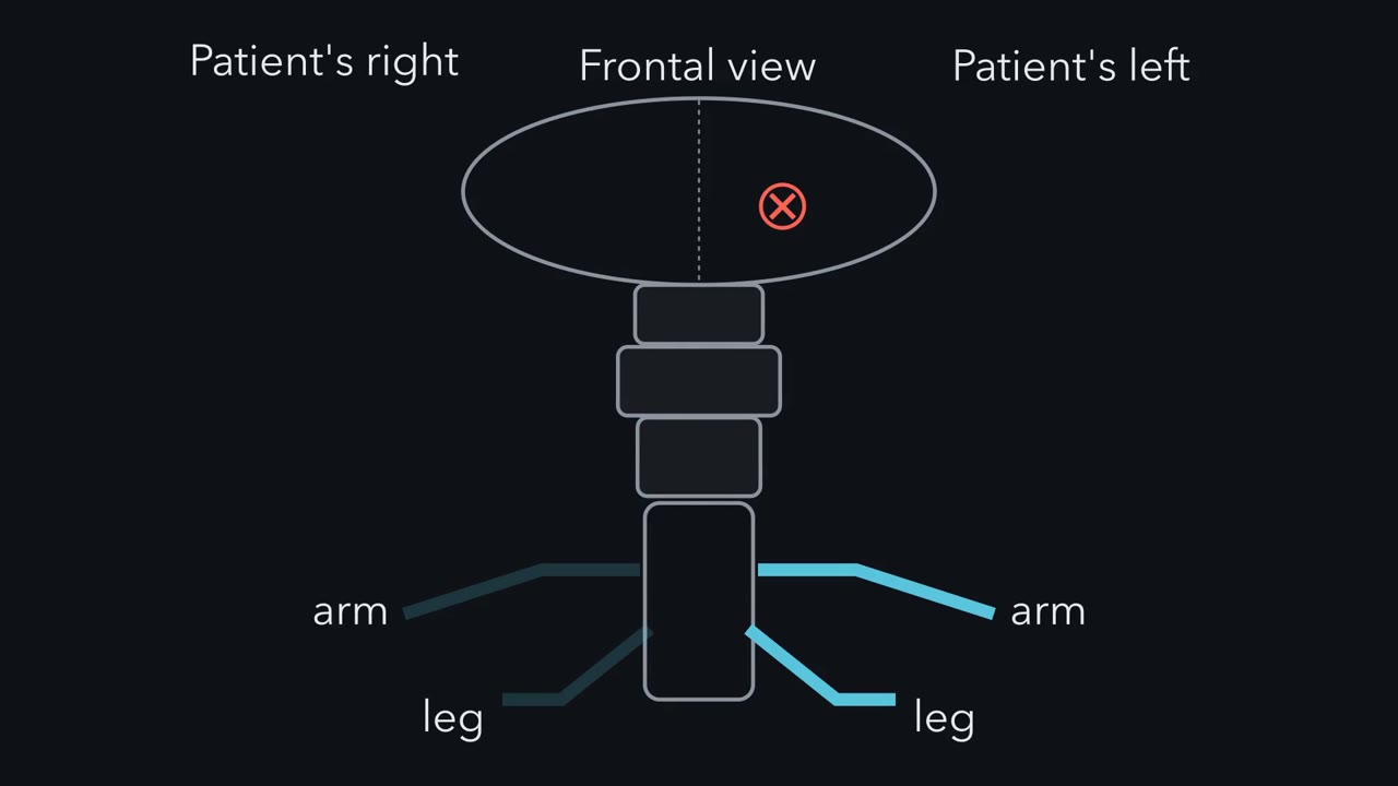 ▶ Watch Two Counts: Side and Sign · 2:41
Read the transcript of “Two Counts: Side and Sign”
Film transcript: Two Counts: Side and Sign
A stroke deep in the left side of the brain weakens the right side of the body. Why the opposite side? A cut through the right half of the cord in the neck weakens the right side too. Lesions on opposite sides, weakness on the same side. Follow the tract down from the motor cortex, through the internal capsule and the brainstem. In the caudal medulla, most of its fibres cross the midline. That crossing is the pyramidal decussation. Damage above the crossing shows up on the opposite side of the body. In the cord, most fibres have already crossed. So damage there shows up on the same side. So here is the first count. Count the crossings between the damage and the muscle.
One crossing flips the side. Try one. A lesion sits in the right pyramid, just above the crossing. Which limbs are weak? The left limbs. One crossing lies below the lesion, so the side flips. A second puzzle. The basal ganglia never reach the spinal cord, yet in the classic model they decide whether a movement goes ahead. Their trick rests on inhibition. Picture three neurons in a row. The first inhibits the second, and the second inhibits the third. Now the first neuron starts firing faster. What does the third one do?
The third fires faster too. The first holds the second down, so the second lets go of the third. Inhibiting an inhibitor releases its target. That is disinhibition. That is the second count. Each inhibitory link flips the sign. An even number keeps the direction. An odd number reverses it. Put them together. For a motor pathway, follow the chain from the lesion or drug to the muscle. Crossings set the side. Inhibitions set the sign. Some limits. The counts give the direction of a change along one chosen path, not its size. The sign count assumes each link keeps a fixed sign, and each neuron can still change its firing. When routes pull in opposite directions, the net effect depends on how strong each route is. And input from both sides breaks the crossing count: after a one-sided cortical lesion, the forehead is usually weakened less than the lower face.
Follow the chain, and count what flips.
From the film
- Count the midline crossings between a lesion and the muscle: each crossing flips the side of the effect.
- Each inhibitory link flips the sign, so an even number keeps the direction and an odd number reverses it, and inhibiting an inhibitor releases its target.
- The counts give the direction of a change along one chosen path, not its size, and they fail when routes pull in opposite directions.
A stroke deep in the left hemisphere weakens the right face, arm and leg. A cut through the right half of the spinal cord in the neck weakens the right arm and leg. Lesions on opposite sides, weakness on the same side.
Something about where each lesion sits decides which side of the body pays for it. The same kind of reasoning turns out to explain how a set of deep nuclei that never reach the spinal cord can still decide whether a movement goes ahead.
Three puzzles
Start with what has to be explained. Three observations about movement look unrelated.
- Side. Why do the two lesions above, on opposite sides, weaken the same limbs?
- Control without contact. The basal ganglia are a group of nuclei (a nucleus is a cluster of neuron cell bodies with shared connections) deep in the brain. They do not project to the spinal cord, yet in the classic model they decide whether a movement goes ahead. How?
- Opposite diseases. Losing the dopamine neurons of the substantia nigra makes movement slow and hard to start, as in Parkinson’s disease. Losing a different set of neurons early in Huntington’s disease is thought to contribute to extra, involuntary movements. Why do two losses in one circuit push movement in opposite directions?
The one idea to carry through this lesson. To predict what a lesion or a drug does to movement, follow the motor chain from the place it acts toward the muscle and count two things. Each crossing of the midline flips the side: an odd number of crossings puts the effect on the opposite side of the body. Each inhibitory link flips the sign: an even number of inhibitions passes a change on with its direction kept, and an odd number reverses it. The counts give the side and the direction of a change, not its size.
Each count has limits, and each is stated where the count is first used.
Counting crossings: the corticospinal tract
Picture a front view of the brain and spinal cord. Draw one line from the motor cortex of the left hemisphere down to the right hand, with one X low in the brainstem where the line crosses the midline. Now attach names to the picture.
- The line starts at an upper motor neuron: a neuron in the cortex or brainstem whose axon descends to control lower motor neurons, directly or through interneurons.
- It ends at a lower motor neuron: a motor neuron in the brainstem or the spinal cord’s ventral horn whose axon leaves the central nervous system and ends on muscle. You met it in motor units and reflexes.
- The descending axons travel together as a tract, a bundle of axons inside the central nervous system.
- The X is a decussation, a place where a tract crosses the midline.
- Ipsilateral means on the same side; contralateral means on the opposite side.
The corticospinal tract starts in layer V of primary motor cortex, with contributions from premotor, supplementary motor and parietal areas. On the way down its axons pass five stops:
- the corona radiata;
- the posterior limb of the internal capsule;
- the cerebral peduncle of the midbrain;
- the base of the pons;
- the medullary pyramids.
In the caudal medulla most of the fibres cross at the pyramidal decussation. That is the X in the picture. The crossed fibres run in the lateral column of the cord as the lateral corticospinal tract and end mainly on interneurons of the intermediate grey matter and on ventral horn motor neurons.
The X splits the pathway in two. Damage above it, with one crossing still to come, shows up on the opposite side of the body. Damage in the cord, below it, shows up on the same side.
Which neuron is damaged: the signs
The side tells you where along the tract to look. The signs tell you whether the damage is in the descending pathway or in the final motor neuron.
- A lower motor neuron lesion causes weakness with low tone and reduced or absent reflexes, because the motor limb of the reflex arc is cut. Later the muscle wastes and may show fasciculations, the visible twitches of single motor units firing on their own.
- An established upper motor neuron lesion causes weakness with raised tone (more resistance when the limb is moved passively), brisk stretch reflexes, clonus (rhythmic beats of contraction set off by a sudden stretch) and an extensor plantar response, in which stroking the sole makes the great toe extend. Independent finger movements are often lost.
“Established” matters. Right after a spinal cord injury the limbs below can be flaccid and areflexic (spinal shock), and spasticity appears later. And an upgoing toe is normal in infants, so in a young child it has to be read against age.
The face adds one more clue. Corticobulbar fibres to the motor neurons for the lower face come mainly from the opposite cortex, while the forehead receives input from both sides. An upper motor neuron lesion above the brainstem therefore weakens the contralateral lower face more than the forehead, which is relatively spared. This is the side count’s limit: the forehead, fed from both sides, is weakened less rather than flipped.
Two cases
Assume one focal lesion, examined weeks after onset so the upper motor neuron signs have developed.
Worked problem. Case 1. Weakness of the right lower face, right arm and right leg, with raised tone, brisk reflexes and an extensor plantar response on the right.
- Step 1: upper motor neuron signs put the lesion in the descending pathway, not the motor neurons or nerves.
- Step 2: the lower face is involved. Fibres for voluntary face movement end on facial motor neurons in the pons, so the lesion is above the facial nucleus: in the hemisphere, or occasionally in the pons above that level.
- Step 3: above the decussation the deficit is contralateral, so right-sided weakness means a left-sided lesion.
- Answer: most likely the left hemisphere, for example the posterior limb of the left internal capsule, where descending fibres for the face, arm and leg are packed into a small space.
For sensation, run the side count the other way: count the crossings between the body and the lesion. The dorsal column pathway for touch and vibration crosses in the medulla; the spinothalamic pathway for pain and temperature crosses within a segment or two of entering the cord (touch and pain).
Predict. Case 2. The same right arm and leg signs, but the face is normal. Vibration sense is lost on the right, and pain and temperature are lost on the left below about the C6 dermatome. Where is the lesion, and on which side?
Check your prediction
- Step 1: the face is spared, which fits a lesion below the facial nucleus, probably in the cord; the sensory findings in step 3 test this.
- Step 2: in the cord the corticospinal fibres have already crossed, so no crossing lies between the lesion and the muscle and the weakness is ipsilateral: a right-sided lesion.
- Step 3: check with sensation. The dorsal columns have not yet crossed in the cord, so right vibration loss points to the right cord. The spinothalamic fibres have already crossed, so right cord damage removes pain and temperature on the left, starting a little below the lesion.
- Answer: a right hemisection of the cord near C5, with lower motor neuron signs expected at the level itself, where ventral horn cells are destroyed.
Counting inhibitions: the basal ganglia brake
Read the transcript of “The Brake and Its Two Routes”
Film transcript: The Brake and Its Two Routes
How can nuclei that never reach the spinal cord decide whether a movement goes ahead? Start with a picture: a column of boxes. The cortex is at the top, then an input stage. An output stage feeds the thalamus, which sends an arrow back to the cortex, closing a loop. Now the names. The input stage is the striatum. The output stage is the internal globus pallidus, GPi. Its output neurons are inhibitory and fire steadily, even at rest. So the thalamus is held down, like a foot on a brake. In the classic model, movement needs the brake lifted. Two routes run from the striatum to the brake. Striatal projection neurons are inhibitory too. In the direct route, striatal neurons with D1 receptors inhibit the brake.
The brake inhibits the thalamus. Two inhibitions, an even number. More striatal firing releases the thalamus, toward movement. The indirect route takes a detour. Striatal neurons with D2 receptors inhibit the external globus pallidus, GPe. GPe inhibits the subthalamic nucleus, STN. And the STN excites the brake. Walk it through. D2 neurons fire more, so GPe fires less, and the freed STN fires more. The brake, excited, fires more. The thalamus is held down harder. Three inhibitions, an odd number. This route pushes away from movement. Now add dopamine, from the substantia nigra pars compacta. It acts on the first box of each route. Treat each action as one more link.
In the classic model, dopamine excites the D1 neurons and inhibits the D2 neurons. Acting through each receptor, does dopamine push toward movement or away from it? Toward movement, both ways. Through D1, dopamine adds no flip. Through D2, its own inhibition adds one flip to the route's three. Four flips, an even number. The two actions agree. So losing dopamine pushes both routes toward less movement. The direct route weakens, the indirect strengthens. So the brake fires more for two reasons: less inhibition from the striatum, more excitation from the STN. The thalamus is held down harder, and movement is hard to start: the model's prediction of akinesia.
The model rests on a measurement. In 1960, post-mortem brains from people with Parkinson's disease showed striatal dopamine greatly reduced. Inhibit an inhibitor, and you release its target.
From the film
- In the classic model, GPi fires steadily and holds the thalamus down like a brake, and movement needs the brake lifted.
- The direct route has two inhibitions and pushes toward movement, and the indirect route has three and pushes away from it.
- In the classic model, dopamine loss weakens the direct route and strengthens the indirect one, so the brake fires more and movement is hard to start.
The corticospinal tract carries the command. In the classic rate model, which treats each nucleus as one firing rate that rises or falls, the basal ganglia decide whether that command goes ahead. Start with a picture and add the names after.
Picture one hemisphere’s circuit as a column of boxes. At the top is the cortex. Below it is an input stage, and near the bottom an output stage, which feeds the thalamus. The thalamus sends an arrow back up to the cortex, closing a loop. Between input and output run two routes: a short one straight down, and a long one that detours through two more boxes. Off to one side sits one more box that sprays dopamine onto the input stage. Every arrow is either excitation or inhibition.
Now the names.
- The input stage is the striatum (caudate and putamen).
- The output stage is the internal segment of the globus pallidus, GPi, with a partner that works much the same way, the substantia nigra pars reticulata (SNr).
- The detour runs through the external segment of the globus pallidus, GPe, and the subthalamic nucleus (STN).
- The dopamine comes from the substantia nigra pars compacta, SNc.
Besides the thalamus, the output also reaches brainstem targets.
The brake
The output neurons of GPi and SNr are GABAergic, releasing the inhibitory transmitter GABA, and fire tonically, that is, steadily, even at rest. So the motor thalamus is held under steady inhibition, like a foot resting on a brake. In the classic model, movement needs the brake lifted: GPi must fire less.
How can a circuit made largely of inhibitory neurons lift anything? Try the smallest case.
Predict. Neuron A inhibits neuron B, and B inhibits neuron C. B is firing, and C has steady excitatory drive of its own. A starts firing faster. What does C do?
Check your prediction
A fires more, so B is inhibited more and fires less. B fires less, so C gets less inhibition and fires more. Inhibiting an inhibitor releases its target. This is disinhibition: two inhibitory links in a row pass the change on with its direction kept, as long as each cell can still change its firing (a B that is already silent cannot slow further). Removing inhibition only permits firing; C’s own drive does the exciting.
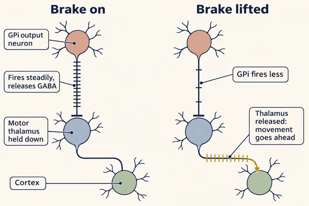
Two routes to the brake
Striatal projection neurons are themselves GABAergic, so every arrow leaving the striatum is an inhibition.
- In the direct pathway, striatal neurons that express D1 dopamine receptors inhibit GPi and SNr directly.
- In the indirect pathway, striatal neurons that express D2 receptors inhibit GPe; GPe, also GABAergic, inhibits STN; and STN, which is glutamatergic (it releases the excitatory transmitter glutamate), excites GPi.
Follow more striatal firing down each route and count the inhibitions from the striatum to the thalamus. A released thalamus means a push toward movement; a thalamus held down harder means a push away.
| Route | Chain from striatum to thalamus | Inhibitory links | More striatal firing means |
|---|---|---|---|
| Direct | striatum ⊣ GPi ⊣ thalamus | 2, even | thalamus released: toward movement |
| Indirect | striatum ⊣ GPe ⊣ STN → GPi ⊣ thalamus | 3, odd | thalamus held down harder: away from movement |
Read the indirect row one step at a time. D2 neurons fire more, so GPe fires less. STN, freed from GPe, fires more. GPi, excited by STN, fires more. The thalamus is held down harder. The links into the striatum from cortex and out of the thalamus to cortex are excitatory, so they add no flips.
Dopamine is one more link
In the classic model, SNc dopamine acts on the two kinds of striatal neuron in opposite ways: it excites D1 direct-pathway neurons and inhibits D2 indirect-pathway neurons. Dopamine is not a box in the loop; it acts on the first box of each route. So treat each action as one more link at the head of its route, and count again.
Predict. Acting through each receptor, does dopamine push toward movement or away from it? What should losing dopamine do?
Check your prediction
Through D1: an excitatory link, then the direct route’s two inhibitions. Two flips, an even number: more dopamine, more movement.
Through D2: dopamine’s inhibitory action is one flip, then the indirect route’s three. Four flips, an even number: more dopamine, more movement again.
Both actions agree, so the count can call the net effect. Losing dopamine pushes both routes toward less movement. The direct pathway weakens and the indirect pathway strengthens: GPe is more inhibited and STN is released. So GPi fires more for two reasons at once: less inhibition from the striatum, and more excitation from STN. The thalamus is held down harder and movement becomes hard to start, which is called akinesia. That is the classic model’s prediction for dopamine loss.
What the sign count can and cannot call. It assumes each link has one fixed sign, and it counts a lesion as a fall in activity. When several routes carry a change and agree in sign, the count predicts the net effect. When they disagree, it says only that they pull against each other; which one wins depends on how strong each route is, and signs cannot tell you that. “Where the box model breaks” returns to this.
How D1 and D2 receptors couple to cAMP belongs to G proteins and cAMP and PKA; where dopamine is released and cleared is in transmitter systems.
This counting is the logic of the rate model set out by Albin, Young and Penney in 1989 and by DeLong in 1990. It is a way to reason about the direction of a change, not a measured law of how firing rates add.
Use the simulation to watch the count: switch dopamine, the indirect-pathway neurons and the STN on and off, predict each box first, then check.
+ excitatory link, − inhibitory link. Each box shows its change from normal: ▲ up, ▼ down, ● no change, ? the two routes pull in opposite directions. Dashed box: removed in this state (cells lost, not a change in rate).
In this sketch: signs and directions only, no magnitudes. Direct and indirect neurons are both active around the start of an action in mice, so the pathways do not simply alternate; the hyperdirect cortex-to-STN route is not drawn; pattern and synchrony change as well as rate; and the model gives no account of rest tremor or of deep brain stimulation.
Rule to keep: give each excitatory link +1 and each inhibitory link −1 and multiply along a route. In this sketch signs and directions only, no magnitudes, after Albin, Young and Penney (1989) and DeLong (1990).
Both counts together: silencing the STN
Read the transcript of “The Test: Silence the STN”
Film transcript: The Test: Silence the STN
If dopamine loss makes the STN overactive, what happens when you silence it? Picture a monkey made parkinsonian with MPTP, a toxin that kills dopamine neurons of the substantia nigra. Now lesion its left STN. Which way should movement change? And on which side of the body?
Start with the sign. The STN excites the brake, and the brake inhibits the thalamus. That is one inhibition, an odd number. So when STN activity falls, the thalamus rises. The brake lifts, toward movement. Now the side. In the classic model, the left loop returns to the left cortex. Its commands then cross once, at the pyramidal decussation. So the change should appear on the right side. In 1990, Bergman, Wichmann and DeLong did this experiment in monkeys treated with MPTP. The parkinsonian signs improved on the side of the body opposite the lesion. The prediction held. This work helped make the STN a target for Parkinson's surgery, and for deep brain stimulation.
But the count has a hard limit. In mice, both pathways become active together around the start of an action. When two routes pull in opposite directions, counting cannot say which one wins. That depends on how strong each route is.
The model also gives no account of rest tremor. And how deep brain stimulation works is still unresolved. Count crossings for side and inhibitions for sign. Then check the count against the data.
From the film
- In the classic model, silencing the STN lifts the brake, so movement should improve on the side opposite the lesion.
- In MPTP-treated monkeys, lesioning the STN improved parkinsonian signs on the side opposite the lesion, as the count predicted.
- The count has limits: in mice both pathways become active together at action onset, and the model gives no account of rest tremor.
The dopamine count says that after dopamine loss the STN is overactive and drives GPi harder. If so, silencing the STN should help. This is where both counts work at once: crossings set the side, inhibitions set the sign. For the side, follow the loop. In the classic model each hemisphere’s basal ganglia loop returns to the cortex on its own side, and that cortex’s commands cross once, at the pyramidal decussation.
The test animal is a monkey made parkinsonian with MPTP, a toxin that preferentially kills SNc dopamine neurons (its discovery is in the anecdote “Parkinsonism from a contaminated drug”).
Predict. In a monkey treated with MPTP, you lesion the left STN. Which way should movement change, and on which side of the body?
Check your prediction
Sign. STN excites GPi, and GPi inhibits the thalamus: one inhibition, an odd number. So STN activity and thalamic activity move in opposite directions. A lesion makes STN activity fall, so the thalamus is released, toward movement.
Side. Left STN, left GPi, left thalamus, left cortex, then one crossing on the way to the cord: the right side of the body.
Prediction: the parkinsonian signs should ease on the right side.
Hagai Bergman, Thomas Wichmann and Mahlon DeLong did this experiment in MPTP-treated monkeys and reported in Science in 1990 that the parkinsonian signs improved on the side of the body opposite the lesion. The rate model had made a prediction a lesion could test, and the prediction held. The result made the STN a surgical target (DeLong’s profile has the award that followed).
The same count, with the same sign, gives a different outcome when dopamine is normal. A lesion in or near one STN, classically a small stroke in a person without Parkinson’s disease, can release the opposite limbs too far. The result is hemiballism, large flinging movements of one arm or leg.
Parkinson’s and Huntington’s through the circuit
Parkinson’s disease. The motor signs (slowness, small movements, rigidity, rest tremor) appear after substantial loss of SNc dopamine neurons; published threshold figures vary. The evidence that tied the disease to dopamine came from post-mortem tissue: Ehringer and Hornykiewicz found striatal dopamine greatly reduced in brains from patients (1960). The dopamine count above gives the model’s account of the slowness: GPi pressed harder from two directions, the thalamus held down.
Treatments follow the same logic. A dopamine precursor such as L-DOPA raises the dopamine available in the striatum. In mice with dopamine depleted by 6-hydroxydopamine, driving direct-pathway neurons with light rescued the motor deficits Kravitz and colleagues measured (2010). Why the dopamine neurons die is taken up in neurodegeneration.
Huntington’s disease has a different cause: an expanded CAG repeat in the huntingtin gene (the mutation type is in mutation), not a primary loss of dopamine.
Predict. Suppose the striatal neurons of the indirect pathway, the ones projecting to GPe, are affected first, so their output to GPe falls. Predict what GPe, STN and GPi do, and what happens to movement.
Check your prediction
Losing that output removes the inhibition of GPe, so GPe is released and fires more. STN is more inhibited and fires less. GPi gets less excitation and fires less, and the thalamus is released. In the model this pushes toward extra movement. That is thought to contribute to chorea, the brief, irregular involuntary movements of early disease.
What supports the premise? Enkephalin marks the striatal projection to GPe and substance P the one to GPi, and in post-mortem tissue the enkephalin terminals in GPe are the more affected in early and middle stages (Reiner and colleagues, 1988; the anecdote “Seventeen brains and two peptides” has the study). Staining alone cannot tell neuron death from terminal loss or reduced peptide. Later loss of direct-pathway output is associated with the akinesia of late disease.
Where the box model breaks
The rate model predicts the sign of several clinical effects. Four sets of results show its limits, and each meets a limit of the count stated with dopamine.
First, routes that disagree are active together. The cartoon has the pathways alternating on and off. Cui and colleagues (2013) found calcium signals from both D1-expressing (direct-pathway) and A2A-expressing (indirect-pathway) striatal populations in mice rising around the start of an action. Two routes of opposite sign active at once is exactly the case the count cannot call.
Second, there are more routes. In awake monkeys, Nambu and colleagues (2000) found that cortical stimulation evoked an early pallidal excitation that depended on STN and arrived faster than effects through the striatum: a hyperdirect pathway. Count it: cortex excites STN, STN excites GPi, GPi inhibits the thalamus. One inhibition, so it pushes away from movement, against the direct route. And in mice, direct-pathway neurons branch into GPe as well (Labouesse and colleagues, 2023). Counted with the simple signs, a branch through GPe and STN has three inhibitions and would push against the direct route. Yet inhibiting the direct-pathway terminals in GPe impaired the mice’s motor performance, the opposite of what that count predicts. The authors traced part of the effect to inhibition of Npas1-expressing GPe neurons, one of several GPe cell types with different targets. So the count cannot assign this branch a sign; the result shows only that the branch does real work.
Third, rate may not be the main variable. The count follows mean firing rates. In mice, dopamine depletion disrupted the clustered pattern of movement-related indirect-pathway activity (Parker and colleagues, 2018), a change in pattern rather than only in mean rate. Studies of parkinsonian animals and patients also report more bursting and synchronized oscillation in STN and GPi.
Fourth, some symptoms and treatments do not fit. Lesioning GPi relieves parkinsonism, as the model predicts. But the model also predicts that removing GPi output should cause extra movements, and pallidotomy usually does not cause them; it also eases dyskinesia and dystonia. The model gives no account of rest tremor, and deep brain stimulation of the STN relieves parkinsonian signs by a mechanism that is still unresolved. What the basal ganglia contribute to choosing actions by their value continues in motivation and reward.
Design problem
You have seen the result. Now design the experiment as if it had not been done, with its controls. The rate model predicts that the STN is overactive in parkinsonism and that silencing it should help. Design a primate experiment to test this, and say what result would count against the model.
Saved in this browser only.
One way to do it
Use monkeys made parkinsonian with MPTP. Record single units in STN and GPi before and after MPTP; the model predicts both rates rise. Then lesion or inactivate one STN and score akinesia and rigidity on the opposite side of the body, with the other side as a within-animal comparison. This is the lesion step Bergman, Wichmann and DeLong did.
Controls: sham surgery; histology for lesion position and SNc loss; scoring blind to side and condition; and GPi recording after the lesion to see whether its output fell.
Against the model: no motor improvement after an accurate STN lesion; or improvement with no fall in GPi activity, which would mean the benefit does not work by lowering GPi rate as the model says. Two cautions: pattern and synchrony change as well as rate, so record them too; and a lesion result does not explain deep brain stimulation.
Going deeper
How much of the tract crosses
Estimates of the fraction of corticospinal fibres that cross at the pyramidal decussation vary between sources, from roughly 75 to 90%. Most of the uncrossed minority forms the anterior corticospinal tract, whose fibres largely appear to cross segmentally, near the level where they end. How the fibres cross during development is in crossing the midline.
Direct contacts on motor neurons
In macaques some cortical cells contact digit motor neurons directly (Rathelot and Strick, 2006).
The count as a product of signs
The inhibition count can be written as arithmetic. Give each excitatory link +1 and each inhibitory link −1, and multiply along the route from cortex back to cortex. A positive product means activity entering the loop ends up releasing the thalamus and so favours movement; a negative product means it ends up holding the thalamus down.
Direct: cortex→striatum (+1) × striatum→GPi (−1) × GPi→thalamus (−1) × thalamus→cortex (+1) = +1
Indirect: cortex→striatum (+1) × striatum→GPe (−1) × GPe→STN (−1) × STN→GPi (+1) × GPi→thalamus (−1) × thalamus→cortex (+1) = −1
What the SNr subgroups did
Freeze and colleagues (2013) found that stimulating either pathway produced both excitatory and inhibitory responses among SNr neurons in mice, so neither pathway simply pushed all of SNr one way. Even so, movement initiation during direct-pathway stimulation tracked the inhibited SNr subgroup, and movement suppression during indirect-pathway stimulation was most strongly associated with the excited subgroup. These pathway-specific relationships fit the model despite the mixed responses.
Counting the branched cells
Of 148 direct-pathway neurons in the mouse dorsomedial striatum that carried the SNr tracer, 147 also carried the GPe tracer. That is 147/148 = 99.3% of the SNr-labelled cells counted, not of all striatal neurons. The anecdote “One cell, two targets” tells the story.
Your notes
Select any text in the lesson to highlight it or add a note to it. Your highlights and notes are saved in this browser and collected in your notebook.
This browser is not letting the site save anything (a private window or strict privacy settings), so highlights and notes will be lost when you leave the page.
How we measure it
The neurological examination as lesion localization
Strength, tone, stretch reflexes, the plantar response, wasting and fasciculations sort a weakness into upper or lower motor neuron patterns. Combined with which side and which body parts are affected, and with sensory findings, they place a lesion along a pathway whose crossings you know.
Single-unit recording in awake primates
A fine extracellular electrode records the spikes of one neuron while a monkey moves or rests. Comparing firing rates and patterns before and after MPTP treatment tested the rate model's predictions for the subthalamic nucleus and internal pallidum.
Post-mortem neurochemistry and immunohistochemistry
Measuring a transmitter in autopsy tissue, or staining terminal fields for peptides that mark one projection, shows what is lost in a human disease. It measures the end state of tissue, not living firing, and cannot by itself separate cell death from reduced marker.
Cre-dependent optogenetics
A light-gated channel is expressed only in neurons of one genetic type, here D1- or D2-expressing striatal neurons, and light then drives them. This tests whether driving those cells is sufficient to change a behaviour; it does not show how they act normally.
Practice questions on this lesson (3) → MCAT-style, original, each with an explanation
Where this lesson fits
Before thisReview first
- Motor Units, Reflexes and LocomotionThis course · Lesson 15
It teaches lower motor neurons and the reflex arc, which you need to tell upper from lower motor neuron signs.
- Integration, Inhibition and NeuromodulationThis course · Lesson 7
It explains how inhibitory inputs lower a cell's firing, which the inhibition count along each route relies on.
- Transmitter Systems and DrugsThis course · Lesson 6
It covers GABA, glutamate and dopamine receptors, which set the sign of each link in the circuit.
After thisStudy next
- The CerebellumThis course · Lesson 17
It adds the other big loop that shapes movement, and uses the same habit of following a motor chain by side.
- Feeding, Motivation and RewardThis course · Lesson 19
It takes the dopamine and striatum circuit further, from allowing movement to choosing actions by their value.
- When Mature Neurons Fail: NeurodegenerationThis course · Lesson 26
It asks why the dopamine neurons lost in Parkinson's disease die, which this lesson only assumes.
Elsewhere on the siteFilms, simulations and method films in other courses
- Adding up a reflexSimulation · Organismal Biology
The reflex route there lets you trace receptor, neuron and effector, the lower motor neuron end of the chain.
Suggested by an AI model from the lessons themselves, then checked. How we use AI
Finish line
Test yourself
8 questions
Practice and MCAT-style, with explained answers.
Next · Lesson 17
The Cerebellum
Most of the neurons in one count of the human brain sit in the cerebellum. Damage it and you can still move. What do you lose?
Continue →