Neuroscience · Skim version
The Map: How the Nervous System Is Organized
Read the full lesson → Skim the whole course →
The guiding idea
Inject a protein tracer into a mouse's blood and, in the cerebral cortex, it goes no farther than the cells lining the capillaries. Why there?
The central nervous system is one hollow tube bent once: its walls became the brain regions and spinal cord, and its canal became the ventricles.
From the film: One Tube, Bent Once
- Picture the central nervous system as one hollow tube, bent once.
- The tube's walls became the brain regions and spinal cord, and its canal became the ventricles.
- Dorsal and ventral are set by the tube's own axis, so the words change direction where the human tube bends.
From the film: Follow the Fluid, Find the Seal
- Cerebrospinal fluid is made mainly by the choroid plexus and flows from the lateral ventricles through the third and fourth ventricles to the space around the brain and cord.
- Replacing a fluid volume does not empty it, and a lasting block of the aqueduct tends to enlarge the ventricles upstream of it.
- The blood-brain barrier is at the capillary endothelial cells and the blood-CSF barrier is at the choroid plexus epithelium.
From the film: From Where to What
- Each kind of evidence licenses a different claim about where a function lives.
- A lesion shows contribution, a recording shows covariation, and imaging shows a blood signal rather than which neural signal.
- Stimulation in area MT shifted choices toward the neurons' preferred direction, which shows a causal contribution in that task, not that MT is the seat of motion.
-
A map and its puzzles
-
One tube, bent once
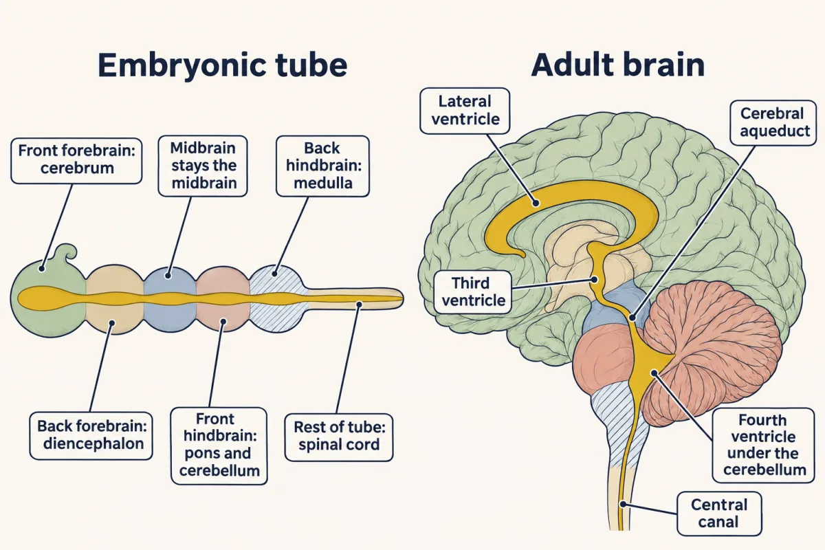
A working picture, not literal adult anatomy: each bulge of the embryonic tube becomes a region of the adult brain, and the tube's canal becomes the ventricles, from the lateral ventricles down to the central canal. AI-generated illustration -
Naming places on a bent tube
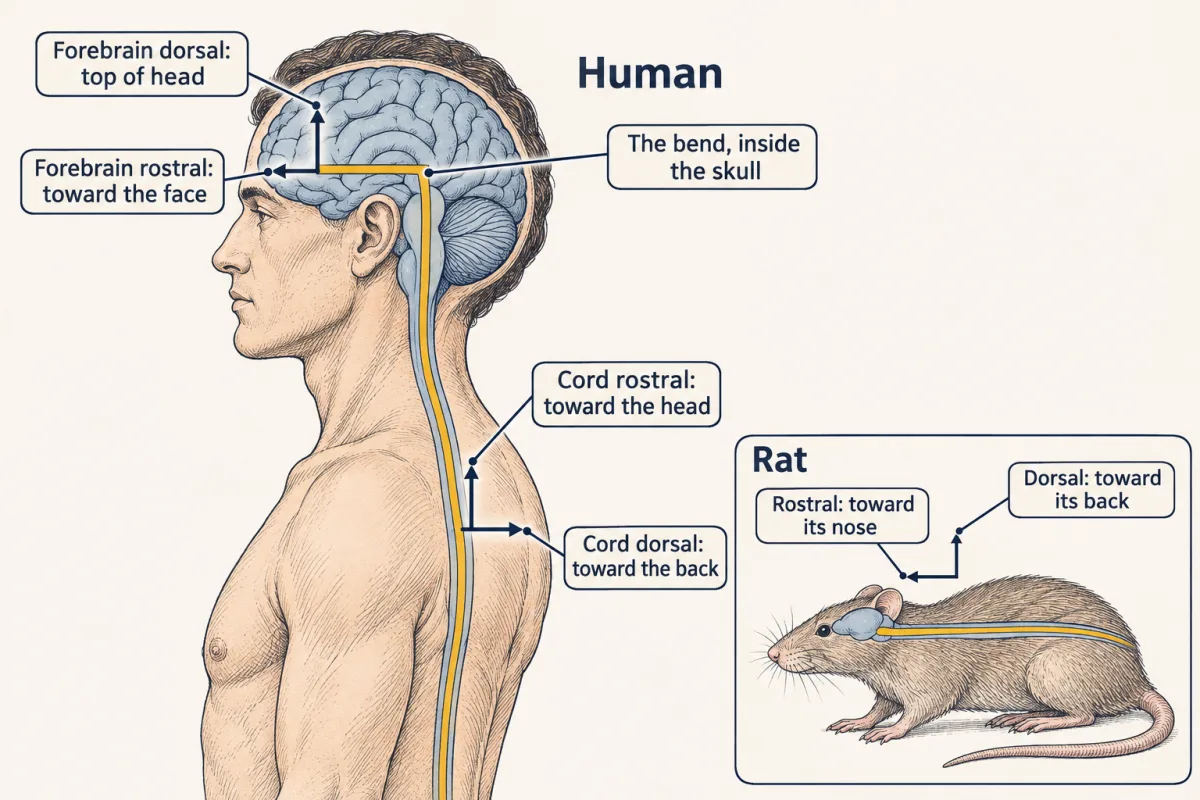
The human neuraxis bends inside the skull, so dorsal means the top of the head in the forebrain but the back of the body in the spinal cord; in a rat the axis runs nearly straight. AI-generated illustration -
The fluid in the tube
-
Where is the seal?
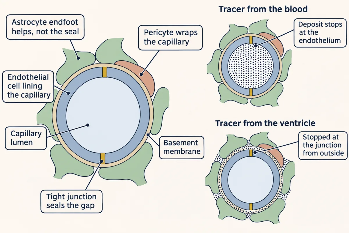
Peroxidase from the blood and peroxidase from the ventricles both stopped at the capillary's endothelial cells, where tight junctions were proposed as the seal, at least for that tracer and time window. AI-generated illustration -
From where to what: four kinds of evidence
-
Going deeper
Where this lesson fits
Before thisReview first
- Tissues, Organs and HomeostasisOrganismal Biology · Starting college
It introduces tissues, organs and the control systems that this lesson's map of nervous system divisions assumes.
- Nervous Control: A First LookOrganismal Biology · Starting college
It first traces a reflex route through dorsal and ventral roots, which this lesson explains by the tube's layout.
After thisStudy next
- Neurons and Glia at WorkThis course · Lesson 2
It takes the cells that fill the tube's walls, including the astrocytes that help maintain the barrier, further.
- Touch and PainThis course · Lesson 13
It traces where touch and pain pathways cross the midline, using the sensory dorsal root plan from this lesson.
- Memory SystemsThis course · Lesson 22
It tells the Scoville and Milner lesion story, using the limits of lesion evidence set out here.
Elsewhere on the siteFilms, simulations and method films in other courses
- Narrow First, Then FoldFilm · Developmental Neuroscience
It shows how the embryonic neural tube forms and what happens when part of it stays open.
- Two Segments, Two JobsFilm · Developmental Neuroscience
It shows how the neural tube is divided head to tail into the hindbrain segments that this map names.
Suggested by an AI model from the lessons themselves, then checked. How we use AI