Neuroscience · Unit 6: Cognition and learning
Memory Systems
Rewritten on : new text, figures, flashcards and film. Your notes and highlights stay, but the text around them may have moved.
H.M. got better at mirror drawing over three days and did not remember ever having done it. What kind of memory improved?
Pause when the reading reaches a film, and offer to play it. Saved on this device.
Watch the film now, or keep listening
Builds on:Descending Control and the Basal GangliaThe CerebellumThe Map: How the Nervous System Is OrganizedWhy these?
 ▶ Watch Two Ways of Remembering · 2:47
Read the transcript of “Two Ways of Remembering”
Film transcript: Two Ways of Remembering
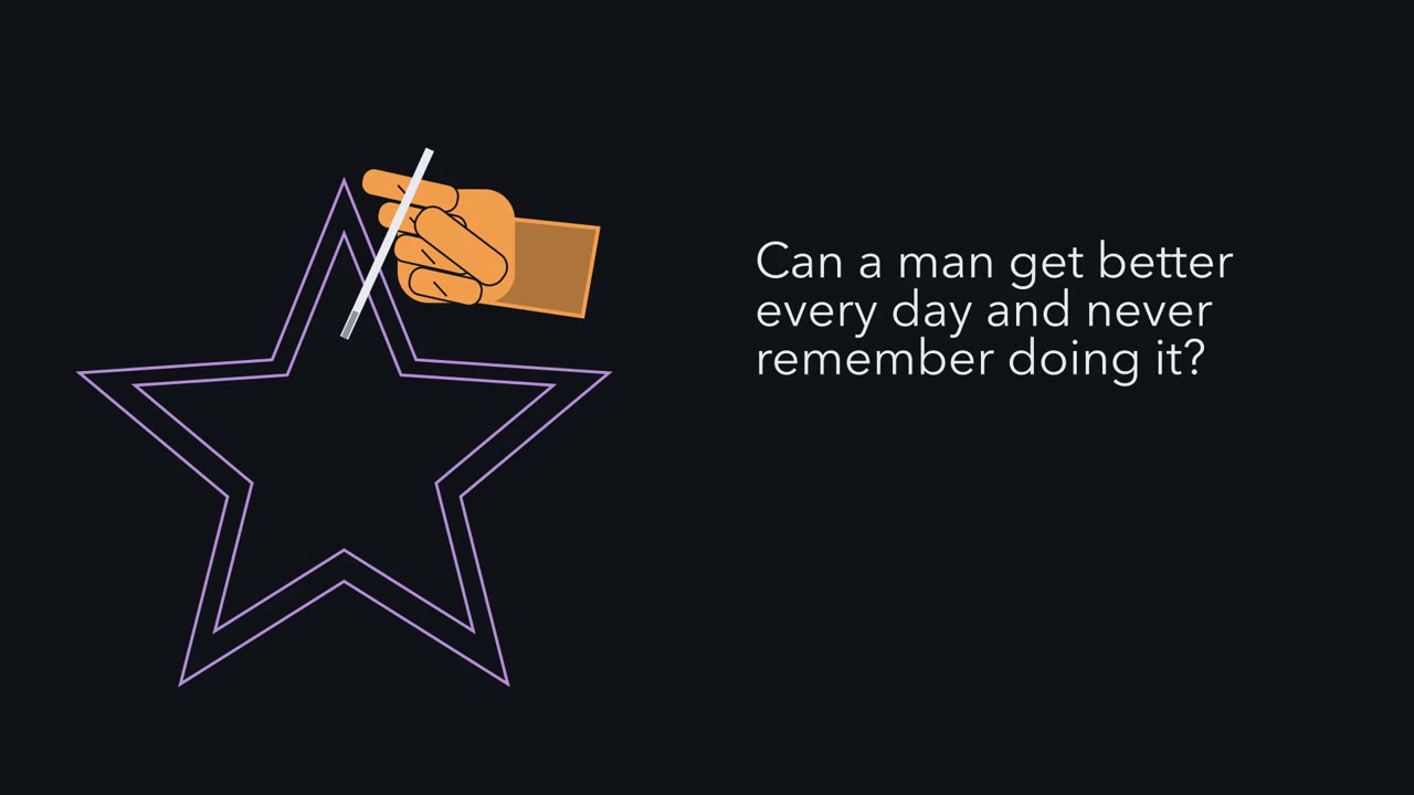
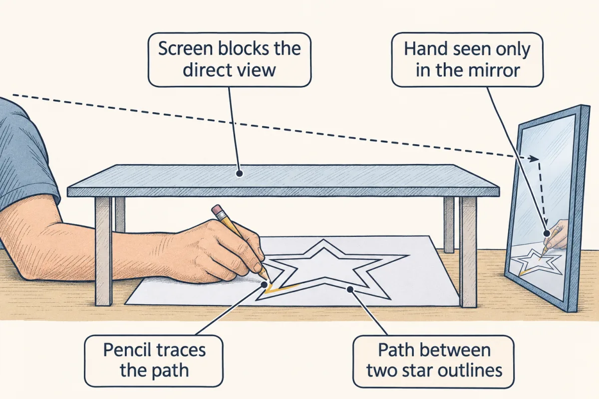
Can a man get better at something every day, and never remember doing it? One patient did. In 1953, a surgeon removed tissue from the inner part of both temporal lobes of a young man. Afterwards, the patient known as H.M. forgot new events within minutes, as soon as his attention moved on. Brenda Milner gave him a mirror-drawing task. He traced between two outlines of a star, seeing his hand only in a mirror. Across three days of practice, he improved, much as other people do. Yet he did not remember having done the task before. His drawing improved, so something kept a record of the practice. Why could he not tell anyone about it?
The record showed up only in how well he drew. It was not something he could recall and report. So there were two ways of remembering, and the operation badly damaged only one. Memory you can bring to mind and state is declarative. It covers events, and facts about the world. H.M. could almost never form new declarative memories. Memory expressed in performance is nondeclarative: skills, habits, priming and simple conditioning. He kept his skill learning. Working memory holds a few items for seconds, like a phone number while you dial. His was normal. Now a worry. Perhaps recall tests are simply harder, and any brain damage hurts hard tasks first.
What second result would rule out the idea that the failed task was just harder? A patient with damage somewhere else, who fails the task H.M. passed, and passes the task he failed. Here is the one idea of this lesson. Memory systems are told apart by crossed failures. Damage to one structure impairs task A but spares task B. Damage elsewhere does the reverse. Then weakening one shared capacity cannot explain both. The tasks rely, at least partly, on separable processes. That crossing is a double dissociation. It shows each structure is needed for its task in that experiment, not that the memory is stored there. Needed, not stored. That is the key to reading every lesion study.
From the film
- H.M. improved at mirror drawing over three days without remembering the task, so memory can show in performance without being recalled.
- Memory you can bring to mind and state is declarative, memory expressed in performance is nondeclarative, and working memory holds a few items for seconds.
- Memory systems are told apart by crossed failures, a double dissociation, which shows a structure is needed for its task, not that the memory is stored there.
In 1953 a surgeon removed tissue from the inner part of both temporal lobes of a young man with severe epilepsy. Afterwards he forgot new events within minutes, as soon as his attention moved on, yet he could still learn a new hand skill.
That patient, known in the literature as H.M., made a strange question answerable: is memory one ability or several? This lesson works through the evidence that it is several, and through the limits of that evidence, because nearly all of it comes from damaged brains.
One patient, two ways of remembering
Brenda Milner, a psychologist working in Montreal, tested him. In one task he traced a path between the two outlines of a star while seeing his hand and the paper only in a mirror, so every movement looked reversed. Across three days of practice H.M. improved, as people normally do, yet he did not remember having done the task before.
Predict. Something in H.M.’s brain kept a record of the practice, because his drawing improved. Why could he not tell Milner about it?
Check your prediction
The record showed up only in how well he drew, not in anything he could recall and report. The operation had removed tissue he needed to form new memories he could bring to mind, but not what he needed to improve with practice. So there are at least two ways of remembering, and the operation severely impaired only one of them.
Those two ways, and a third, have names.
- Declarative memory is memory you can bring to conscious recollection and state. It comes in two kinds. Episodic memory is memory for events placed in a time and a place: what happened at breakfast. Semantic memory is general knowledge of the world: that a spider has eight legs.
- Nondeclarative memory is expressed in performance rather than recollection. It includes skills and habits (mirror drawing), priming (being faster or more accurate with something you met recently, such as reading a word you saw a minute earlier) and simple classical conditioning (a tone that overlaps a puff of air to the eye comes to trigger a blink; Going deeper says why the overlap matters).
- Working memory is the temporary, active holding and manipulating of a few items, such as a phone number you repeat while you dial it.
Predict. Read H.M. a short list of digits and ask him to repeat it straight back. Then turn his attention to something else and ask again a few minutes later. What do you expect each time?
Check your prediction
Straight back, he did normally. His digit span, the number of digits he could repeat back immediately, was normal, and he could hold information as long as he kept rehearsing it. Once his attention moved on, new material was not retained, so the second request should fail. Working memory worked, but almost nothing held in it became a lasting declarative memory.
One way to keep the three apart: declarative memory is a diary you can read aloud, nondeclarative memory is the skill a job leaves in your hands, and working memory is a small notepad that is soon cleared once you stop rehearsing it or your attention moves on. Nondeclarative does not mean motor: it includes learning to predict outcomes, such as the weather from cards, and conditioned autonomic responses, such as changes in heart rate or sweating (the Bechara study below measured skin conductance).
The tissue H.M. lost lies in the medial temporal lobe, the inner (medial) part of each temporal lobe, which holds the hippocampus, the amygdala and the surrounding entorhinal, perirhinal and parahippocampal cortex. His operation was bilateral, on both sides. The nervous system map places it.
William Scoville and Brenda Milner described H.M. and other patients from Scoville’s operations in 1957 in the Journal of Neurology, Neurosurgery and Psychiatry. H.M.’s main loss was anterograde amnesia: he could almost never form lasting new declarative memories. He also lost some memories from before the operation, a retrograde amnesia, whose reach has been estimated differently over the years.
What he kept matters as much. His language and perception were retained, and formal intelligence testing did not show a general decline. Over many years he also acquired, slowly, a few new facts. So “he could learn nothing new” is wrong for skills and, slowly, for facts.
His anterograde amnesia shows that some part of the medial temporal lobe is needed to form new declarative memories. His factual knowledge of early life was largely spared, so it does not seem to be needed in the same way to recall facts learned long before. Neither result shows where memories are stored, and how the dependence changes with time is the subject of consolidation. (Going deeper has the caveats from his late-life testing.)
Why one patient is not enough
One ability failing after damage while another survives, as in H.M., is a dissociation, and with one patient or one group it is a single dissociation. It has a weak point. Perhaps the declarative tests were simply harder, and any brain damage hurts hard tasks first.
Predict. What second result would rule out “the failed task was just harder”?
Check your prediction
A second patient or group, with damage somewhere else, who fails the task H.M. passed and passes the task he failed. If recall were simply harder, nobody who passes it should fail the easier drawing task.
The one idea to carry through this lesson. Memory systems are told apart by crossed failures. If damage to one structure impairs task A but spares task B, and damage elsewhere does the reverse, that crossing is a double dissociation. One shared capacity weakened by different amounts cannot produce it in individual patients, and in groups it argues against that account, so the two tasks rely, at least in part, on separable processes. It shows each structure is needed for its task in that preparation (that species, lesion and task), not that the memory is stored there.
Why can weakening one capacity not produce a crossing? Suppose damage only turns down a single capacity, by different amounts in different patients, and performance on every task rises with that capacity. If patient A scores below patient B on task 1, A has less of the capacity, so A cannot score above B on task 2. The order of the patients stays the same on every task, however hard each task is (ties are possible near floor or ceiling). A crossing breaks that order.
The rule has a scope. It covers long-term memory evidence from lesions (areas of damaged tissue) and from temporary silencing with drugs, one task in one preparation at a time, and the argument is exact only for single patients with noise-free scores. Group averages are weaker evidence: a group mixing mild and severe cases can cross another group’s average with one capacity, if the tasks respond to it differently. Give one group capacities of 0 and 1 and the other 0.6 and 0.6; a task scored as the capacity averages 0.5 against 0.6, and one scored as its square 0.5 against 0.36. Reliable measurement does not remove that possibility, so individual scores and the spread within each group matter too. A crossing of group means supports separable processes without proving them, still less separate anatomical boxes (Going deeper has a network model that crosses without them).
The key experiment: lines that cross
Read the transcript of “Lines That Cross”
Film transcript: Lines That Cross
Can two lines that cross show that memory has more than one system? Watch one experiment. In 1996, Knowlton, Mangels and Squire gave people a weather-prediction task. Cue cards appeared, and the person guessed one of two outcomes. Each card predicted it only with some probability. No single trial is decisive, so the task is learned gradually, from feedback. Afterwards, a questionnaire asked about the training sessions themselves. Three groups took part: controls, amnesic patients, and patients with Parkinson's disease, whose striatum loses dopamine input. Over the first fifty trials, controls and amnesic patients learned the classification together.
The Parkinson's patients failed to learn it. Take the simplest difficulty model: one common capacity. Suppose the questionnaire is simply the harder task. The Parkinson's patients failed the easier one. How should they do on the questionnaire?
On that model, they should fail it too. Instead, they remembered the training. The amnesic patients had severely impaired memory for the training. Yet they learned the classification. So no single order of difficulty fits both groups. Each group does far worse on the task the other does well, and the lines cross. These are group averages, so the crossing argues against the simplest difficulty account rather than ruling out every single-capacity one. Now, a trap. Some answers are right by guessing. In this illustrative design, guessing alone gets half right on the two-choice task. On a four-choice questionnaire, it gets a quarter.
So measure each score up from that baseline, as a share of the controls' gain above it. An invented bar that looks like half of normal shrinks to about a quarter. The correction changes how big a loss looks. As long as controls beat chance, it cannot change which group is higher. So the crossing stays. Parkinson's disease damages more than the striatum. So this crossing supports separable learning processes, without pinning each to one structure. Now a second crossing, in three patients. A signal was paired with an aversive stimulus. Did a sweat response appear? Could the patient report the pairing? A patient with damage to both amygdalae could report the pairing, but showed no conditioned response.
A patient with damage to both hippocampi showed the reverse. Predict the third patient, with damage to both structures. That patient learned neither. Two crossings. In these studies, new memory that could be reported and memory shown in performance depended on different structures.
From the film
- In the weather prediction study, amnesic patients learned the classification but remembered the training poorly, while Parkinson's patients showed the reverse.
- These are group averages, so the crossing argues against the simplest difficulty account rather than ruling out every single-capacity one.
- Scores are measured above the chance baseline; the correction changes how big a loss looks, not which group is higher.
- In the conditioning studies, memory that could be reported and memory shown in performance depended on different structures.
Barbara Knowlton, Jennifer Mangels and Larry Squire (Science, 1996) asked whether nondeclarative learning extends past motor skills such as H.M.’s, and whether it depends on a particular structure. They used a weather-prediction task. On each trial a participant saw a combination of cue cards, guessed one of two outcomes (rain or sunshine) and was then told the answer. Each card predicted the outcome only with some probability. Because no single trial is decisive, the task is hard to solve by remembering episodes; it is learned gradually from the feedback across many trials. Afterwards a questionnaire asked about the training sessions themselves.
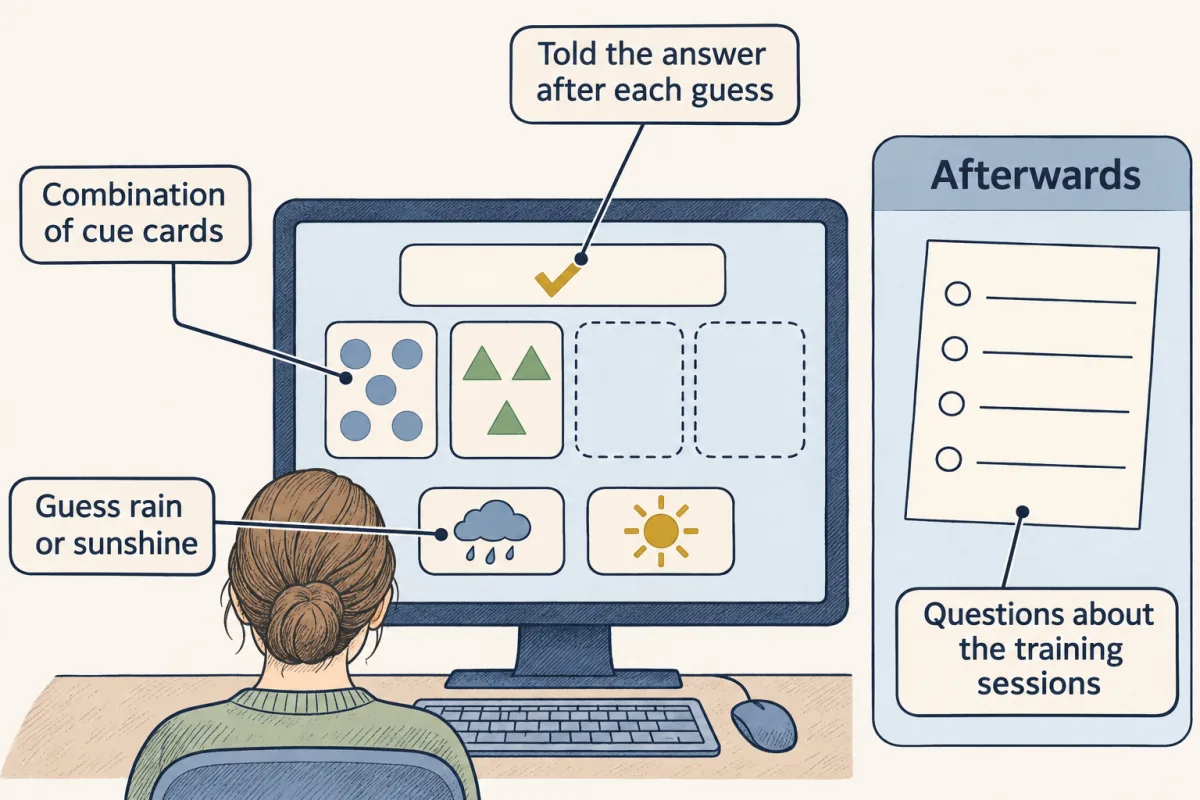
Three groups took part: controls, amnesic patients, and nondemented patients with Parkinson’s disease, in whom dopamine input to the striatum, a deep forebrain structure in the basal ganglia, is depleted (see the basal ganglia).
Picture the results as two panels, a sketch of the pattern rather than the published figure.
- Left panel: classification. Percent correct runs up the side and trials along the bottom, over the first 50 trials, with a dashed line at chance, 50%. The controls’ and the amnesic patients’ curves rise together. The Parkinson’s curve stays low, near chance.
- Right panel: the questionnaire. One bar per group. Now the order flips. The Parkinson’s patients’ bar is high, and the amnesic patients’ bar is low.
So amnesic patients learned the classification but had severely impaired memory for the training sessions, and the Parkinson’s patients showed the reverse. Could one task simply be harder? Suppose the questionnaire were the harder task, as the worry about H.M. assumed. Then the Parkinson’s patients, who failed the easier classification, should have failed the questionnaire too. They did not. Suppose instead the classification were harder. Then the amnesic patients should not have learned it while failing the questionnaire. No single order of difficulty fits both groups, so the lines cross. The authors read this as the neostriatum supporting the learning of nonmotor dispositions as well as motor behaviour.
Two caveats, both in Going deeper: the picture changed with longer training, and later studies questioned parts of the result.
Reading a crossing from numbers
Here is a worked problem with invented numbers in the design of the Knowlton study, not its data.
Suppose a two-choice classification task (chance 50%) and a four-choice questionnaire about the training (chance 25%). Controls score 70% and 80%. Amnesic patients score 68% and 40%. Parkinson’s patients score 52% and 78%.
Step 1: read the order of the groups on each task. On classification the amnesic group is higher (68 against 52). On the questionnaire the Parkinson’s group is higher (78 against 40). The order flips between tasks, so the lines cross. In single patients that would rule out the one-capacity account; in group averages it argues against it. You need no correction to see the crossing.
Step 2: size each loss above chance, as a share of the controls’ gain.
relative score = (patient score − chance) / (control score − chance)
Amnesic classification: (68 − 50) / (70 − 50) = 18 / 20 = 90%. Amnesic questionnaire: (40 − 25) / (80 − 25) = 15 / 55 = 27%. Parkinson’s classification: (52 − 50) / 20 = 2 / 20 = 10%. Parkinson’s questionnaire: (78 − 25) / 55 = 53 / 55 = 96%.
Each group is near normal on one task and far below it on the other. That is the double dissociation. As long as controls score above chance, the correction subtracts the same chance level from every group on a task and divides by the same positive control gain, so it cannot change which group is higher. What it changes is how big each loss looks, and so where a score falls against a fixed cut-off, such as half of normal. Real studies judge impairment against the spread of control scores, with statistical tests, not a single cut-off.
Step 3: notice the raw-score trap. The amnesic questionnaire score, 40% against 80%, looks like half of normal but is 27% above chance, about a quarter, because a quarter of answers are right by guessing. The Parkinson’s classification score, 52% against 70%, looks like about three quarters of normal but is 10%, because half are right by guessing. Raw scores fool the eye about size, not about whether the lines cross.
Step 4: state the limits. These are group averages, Parkinson’s disease damages more than the striatum, and amnesic patients’ lesions differ from one another. So the crossing supports two separable learning processes without proving them or pinning each to one structure.
In the simulation, switch between raw scores and scores above chance. Watch how the bars change size while the order on each task stays the same, and when a group crosses the sketch’s cut-offs for failing and passing.
- Controls (fixed: 70% and 80%)
- Amnesic
- Parkinson’s
- ▲ hollow dot: above the plot, drawn at 140%; the label gives the true value
| Group | Classification: raw → above-chance share | Questionnaire: raw → above-chance share |
|---|---|---|
| Controls | ||
| Amnesic | ||
| Parkinson’s |
In this sketch: invented numbers, controls fixed at 70% and 80%, chance known exactly (50% and 25%); the picture is a sketch of the logic, not data.
Score each task above chance: relative score = (patient − chance) / (control − chance). Parkinson’s disease damages more than the striatum, and amnesic patients’ lesions differ from one another, so a double dissociation does not by itself pin each process to one structure.
A second crossing, in three patients
Bechara and colleagues (Science, 1995) paired a visual or auditory stimulus with an aversive one and measured two things in each patient. Did the stimulus come to evoke a conditioned autonomic sweat (skin-conductance) response? And could the patient say which stimulus had been paired with which?
Two patients gave opposite results.
| Patient | Conditioned sweat response | Could report the pairing |
|---|---|---|
| Both amygdalae damaged (selectively) | No | Yes |
| Both hippocampi damaged | Yes | No |
Predict. A third patient had damage to both the amygdalae and the hippocampi. What did that patient learn?
Check your prediction
Neither. With both structures damaged, the patient acquired no conditioned response and could not report the pairing.
Here the crossing is between single patients, on one autonomic measure, so it is suggestive rather than decisive, and it does not show that the amygdala stores all emotional memory. Emotion and stress takes up the fear side.
Put the two crossings together. In these studies, new memory that could be recalled and reported was lost with medial temporal damage (and diencephalic damage, in the mixed amnesic group), while memory shown only in performance was lost with damage elsewhere, and which structure depended on the task. That is the pattern these studies found, not a law: H.M. could still report facts from early life, and reportability alone does not identify a memory system.
Needed is not stored
Read the transcript of “Needed Is Not Stored”
Film transcript: Needed Is Not Stored
If damage to a structure wipes out a memory, was the memory stored there? In rabbits, a tone is paired with a puff of air to the eye. After training, the tone alone closes the eyelid before the puff. The puff itself always triggers a reflex blink. In 1982, Thompson's laboratory removed the lateral cerebellum on the same side as the trained eye. The learned response to the tone was abolished.
Was the eyelid simply paralysed? If so, what would happen to the reflex blink to the puff? It would fail too. But the reflex blink survived, so the eyelid could still move. the other eye could still be conditioned. So the rabbits could still hear the tone, and still learn. With these controls, the lost response is good evidence that the cerebellum is needed for it. But needed for what? A lost response cannot tell a lost store from a cut output route. A later experiment silenced the output route with a drug, only during training. No learned response appeared. Once the drug wore off, it was there. The rabbits had learned. H.M.'s brain raises a second limit: which structure was needed?
After his death in 2008, it was cut into thousands of thin sections and rebuilt in three dimensions. The operation had taken the front of the hippocampus, most of the amygdala, and nearly all the entorhinal cortex. But the back of the hippocampus remained on both sides, with neurons still present.
The entorhinal cortex is the main cortical route into the hippocampus. What remained may have been largely cut off. The authors offered that as a model. Living cells in a section do not prove they were connected or working. So his amnesia followed a medial temporal lesion on both sides. His case alone cannot assign it to one structure. Crossed failures show that the damaged tissue is needed for a task. They do not tell you where memories live.
From the film
- In rabbit eyeblink conditioning, removing the lateral cerebellum abolished the learned response while the reflex blink survived, so the cerebellum is needed for it.
- A lost response cannot tell a lost store from a cut output route; silencing the output route during training still left the rabbits having learned.
- H.M.'s amnesia followed a medial temporal lesion on both sides, and his case alone cannot assign it to one structure.
A lost ability has two limits. It shows a structure is needed, not that the memory lives there, and when a lesion is impure it may not say which structure was needed. The eyeblink shows the first limit and H.M.’s brain the second.
The eyeblink: a single dissociation with strong controls
In well-trained rabbits, a tone paired with a puff of air to the eye comes to trigger eyelid closure before the puff arrives: a conditioned response. The puff itself always produces a blink, the unconditioned reflex. David McCormick, Gregory Clark, David Lavond and Richard Thompson (PNAS, 1982) removed the lateral cerebellum ipsilateral to (on the same side as) the trained eye. The learned response was abolished.
Predict. Suppose the lesion had simply paralysed the eyelid. What would have happened to the reflex blink to the puff?
Check your prediction
It would have failed too. It was spared, so the animals could still close the eye; what had gone was the learned response to the tone. The study had a second control: the eye on the other side could still be conditioned after the lesion, so the rabbits could still hear the tone and still learn.
With no second lesion showing the reverse effect, this stays a single dissociation. Its controls rule out paralysis, deafness and a general loss of learning, but not that the learned response is simply more fragile than the reflex. The authors suggested that at least part of the plasticity for the learned response might reside in the cerebellum. They could go no further, because a lost response alone cannot separate a lost store from a cut output route.
Krupa, Thompson and Thompson (1993) narrowed this by silencing regions with a drug only during training. With the red nucleus, on the output route, silenced, no learned response appeared, yet once the drug wore off the rabbits showed it: they had learned. With the cerebellar region around the interpositus nucleus silenced, they had not learned. A structure can be needed to express a memory without being where it is learned. The cerebellum gives the circuit.
H.M.’s own brain: which structure was needed?
The 1957 paper compared operations of different extent, and a lasting loss of recent memory went with resections (surgical removals) that took in the anterior hippocampus and the hippocampal gyrus. But every resection that reached the hippocampus also took the amygdala. Removals limited to the uncus and amygdala did not appear to impair memory, which argued against the amygdala alone, but the series could not show whether hippocampal damage alone was enough, or whether the amygdala added to the loss.
The postmortem study of H.M.’s brain, published in 2014 by Jacopo Annese and colleagues, measured what had actually been removed. His name, Henry Molaison, was published after his death in 2008. The resection took the anterior hippocampus, most of the amygdala and nearly all the entorhinal cortex. About 2 cm³ of posterior hippocampus remained on each side, and Nissl staining showed neuronal cell bodies in fields CA1 to CA4 and the subiculum. “Both hippocampi were removed” is therefore false.
The entorhinal cortex is the main cortical route into the hippocampus, so the remaining hippocampus may have been largely cut off. The authors proposed that this disconnection contributed more to H.M.’s amnesia than the loss of anterior hippocampus, but they offered it as a model. Living cell bodies in a section do not show the cells were connected or working. The safe statement is that H.M.’s amnesia followed a bilateral medial temporal lesion, and that his case alone cannot assign it to one structure.
Systems that interact
The lesion evidence supports this map, one task in one preparation per row.
| Task | Structure needed | Preparation | Kind of evidence |
|---|---|---|---|
| Recalling new facts and events, such as memory for the training sessions | Medial temporal lobe | H.M.; amnesic patients | Single dissociation in H.M.; double when set against the Parkinson’s group and against the amygdala patient |
| Probabilistic classification (weather prediction) | Striatum, implicated through Parkinson’s disease | Patient groups | Double dissociation across groups |
| Eyeblink conditioning | Lateral cerebellum, on the trained side | Rabbits | Single dissociation with reflex and other-eye controls |
| Conditioned autonomic (skin-conductance) response | Amygdala | Single patients | Double dissociation in single cases |
The map is a useful specialization, not a set of sealed boxes. In male rats trained on a spatial and a cued-response maze task, Ferbinteanu (2020) found that the hippocampus supported both tasks when they were learned close in time, and the dorsolateral striatum supported both when they were learned in the same environment. Which system does the work depended on the training history.
Memories also change their dependence over time, which consolidation covers. The cellular change underneath all of these is synaptic plasticity, and the prefrontal side of working memory is in attention and decisions. Whether new neurons in the adult human dentate gyrus contribute is debated; see adult neurogenesis.
Design problem
After a cardiac arrest a patient has memory problems, and MRI shows damage limited to field CA1 of the hippocampus on both sides. Design an assessment that decides whether the deficit is declarative only.
Saved in this browser only.
One way to do it
Test against matched controls, scoring above chance where tasks have chance levels.
First, separate working from long-term declarative memory: digit span (immediate) against delayed recall and recognition of a story or word list after 30 minutes. A selective declarative deficit predicts normal span and poor delayed recall.
Second, probe retrograde memory with items from recent years and from long ago, to see whether older memories are spared.
Third, test nondeclarative memory: mirror drawing across several sessions and a priming task. A declarative-only deficit predicts normal learning on both, with poor memory for the training sessions themselves. Probabilistic classification is a less pure probe: in the 1996 study amnesic patients matched controls only over the first 50 trials, and hippocampal patients have been impaired on some versions (Going deeper). Read it alongside the other two, not alone.
Fourth, state the limits. A case of this kind has been reported before: patient R.B., described by Zola-Morgan, Squire and Amaral in 1986, had a marked anterograde amnesia and, at autopsy, a bilateral lesion limited to field CA1 with only minor damage elsewhere. MRI can miss damage elsewhere, so the conclusion stays provisional without postmortem histology. And one patient shows that CA1 damage can be enough for a declarative deficit; it does not show that CA1 is the declarative memory system.
Going deeper
H.M.’s lesion in numbers
The 2014 reconstruction measured the resection as extending 54.5 mm on the left and 44.0 mm on the right along the study’s reference axis. Of the entorhinal cortex, about 0.03 cm³ remained on the left and 0.11 cm³ on the right. About 2.02 cm³ (left) and 1.96 cm³ (right) of posterior hippocampus remained.
H.M.’s older memories
The sparing of his early-life factual knowledge is one reason the hippocampus is often described as needed to form declarative memories and, for a time, to retrieve them. Testing late in his life found his recollection of personal episodes poor across all remote periods, which some read as evidence that detailed episodic recall always needs the hippocampus. By then, though, scans showed new damage, including cortical thinning, abnormal white matter and small infarcts, so those late tests are hard to interpret.
Episodic and semantic learning
Whether episodic and semantic learning depend equally on the hippocampus is debated. Three people with hippocampal damage from early childhood had severe amnesia for daily events yet reached low-average to average language, literacy and factual knowledge (Vargha-Khadem and colleagues, 1997).
When conditioning needs the hippocampus
Simple classical conditioning, as defined in this lesson, is the kind in which the signal overlaps the event it predicts. In trace conditioning a gap separates them, and that version needs the hippocampus and, in people, awareness of the pairing.
Who the amnesic patients were, and what came after 50 trials
In the Knowlton study the 12 amnesic patients were a mixed group: seven had medial temporal lobe damage and five had diencephalic damage. With longer training the amnesic patients did less well on the classification, and the Parkinson’s patients improved somewhat.
The weather-task crossing, revisited
Later studies questioned parts of the 1996 result. Hopkins, Myers, Shohamy, Grossman and Gluck (2004) found an impairment on the weather task in amnesic patients with hippocampal damage. Shohamy and colleagues (2004) found that the Parkinson’s impairment depended on how the task was trained: it appeared when the task was learned from feedback, not when it was learned by observation. The flagship crossing holds in its own design, but which processes the task calls on depends on the patients and the training.
A crossing without separate boxes
The main-text argument rules out the simple one-capacity account for single patients and argues against it for group averages. It does not rule out every account with a single system. Plaut (1995) showed that damaging a single connectionist network, with no separate modules, can produce a double dissociation. That is why the guiding idea concludes “separable processes” rather than “separate systems”.
Store or route in the eyeblink circuit
The 1982 ablations were large and took in the deep cerebellar nuclei. In the 1993 experiment the regions were silenced with infusions of muscimol, a drug that activates GABA-A receptors for the inhibitory transmitter GABA. The result narrows where the learning happens to the cerebellum, without identifying the synapses that hold the trace.
An engram timetable in mice
An engram cell is a neuron in the ensemble that holds a memory trace. In these experiments, cells active during learning were tagged, and reactivating the tagged ensemble with light could drive recall. In mice, Kitamura and colleagues (2017) found that prefrontal engram cells for a contextual fear memory were formed during initial learning but were at first silent: natural recall cues did not reactivate them, though light could drive recall through them. They matured over about two weeks. By then, tagged hippocampal (dentate gyrus) engram cells were no longer preferentially reactivated by natural cues, yet light could still drive recall through them. Do not stretch that mouse timetable to human autobiographical memory.
Your notes
Select any text in the lesson to highlight it or add a note to it. Your highlights and notes are saved in this browser and collected in your notebook.
This browser is not letting the site save anything (a private window or strict privacy settings), so highlights and notes will be lost when you leave the page.
How we measure it
Lesion-behaviour comparison
You compare people or animals with damage to different structures on the same tests, and ask which abilities fail and which survive. A selective loss with other abilities intact shows a structure is needed for that ability in that preparation; it does not by itself show the memory is stored there.
Probabilistic classification with a memory questionnaire
Participants learn to predict an outcome from cues that are only probabilistically related to it, and are later asked about the training sessions. Scoring both measures in the same people lets you separate gradual habit-like learning from memory for the episode.
Eyeblink (nictitating membrane) conditioning
A tone is paired with a puff of air to the eye until the tone alone triggers closure. Because the same eyelid also gives a reflex blink to the puff, you can test whether a manipulation removes the learned response while sparing the movement itself.
Postmortem histology and 3D reconstruction
The fixed brain is cut into serial sections, imaged and stained (for example with a Nissl stain for cell bodies), and the lesion is rebuilt in three dimensions. It measures what was removed and what remains more finely than MRI, but cannot by itself show whether the remaining tissue was connected or working.
Practice questions on this lesson (3) → MCAT-style, original, each with an explanation
Where this lesson fits
Before thisReview first
- Descending Control and the Basal GangliaThis course · Lesson 16
The striatum and its dopamine input matter here, because Parkinson's patients fail the weather-prediction task.
- The CerebellumThis course · Lesson 17
You need the cerebellum's circuit from there to follow the eyeblink conditioning evidence.
- The Map: How the Nervous System Is OrganizedThis course · Lesson 1
It places the medial temporal lobe, hippocampus and striatum that the lesion evidence refers to.
After thisStudy next
- Plasticity, Networks and ConsolidationThis course · Lesson 23
It takes up how memory's dependence on the hippocampus changes over time, which this lesson only mentions.
- Attention, Executive Control and DecisionsThis course · Lesson 25
It covers the prefrontal side of working memory, which this lesson only defines.
- Designing a Causal Circuit ExperimentThis course · Lesson 28
It extends the needed-versus-stored limit into designing experiments that show what a circuit does.
Elsewhere on the siteFilms, simulations and method films in other courses
- Extracellular single-unit and nerve recordingMethod film
The cerebellum eyeblink section asks whether a store or only an output route was lost, which recording from single cells can test.
Suggested by an AI model from the lessons themselves, then checked. How we use AI
Finish line
Test yourself
8 questions
Practice and MCAT-style, with explained answers.
Next · Lesson 23
Plasticity, Networks and Consolidation
Shine light on cells tagged during learning in a mouse's hippocampus and it freezes as if afraid. Has anyone found where the memory is stored?
Continue →