Neuroscience · Unit 1: Neurons and their signals
Neurons and Glia at Work
Rewritten on : new text, figures, flashcards and film. Your notes and highlights stay, but the text around them may have moved.
Let potassium pile up outside neurons during seizure-like firing and E_K can move by more than 30 mV. Who cleans it up?
Pause when the reading reaches a film, and offer to play it. Saved on this device.
Watch the film now, or keep listening
Builds on:The Map: How the Nervous System Is OrganizedWhy these?
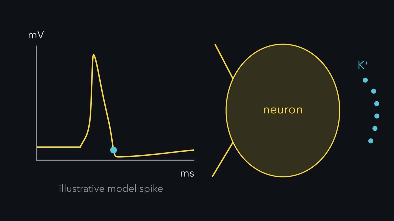 ▶ Watch Small Space, Big Change · 2:50
Read the transcript of “Small Space, Big Change”
Film transcript: Small Space, Big Change
Every spike ends with potassium leaving the neuron. Where does all that potassium go? Into the gaps between brain cells. Those gaps are narrow. Together they are only about a fifth of the tissue. Inside a neuron, potassium is high. Outside, it is low. A burst of firing lets some potassium out. Which concentration changes more, inside or outside? And which change matters more for the voltage?
In proportion, the outside, by a wide margin. Take a simple model: the space outside is a fifth of the tissue, and the firing neurons fill a third of the space inside. Then the same potassium changes the outside concentration about one and a third times as much as the inside one. the outside starts low. One extra mM raises it by a third. Losing 0.75 mM inside changes it by about half a percent. Potassium's equilibrium potential, E_K, depends on the ratio of outside to inside. In this model, the outside change moves it about 60 times as much as the inside change. In cat cortex, strong repetitive stimulation raised outside potassium to a ceiling of about 10 mM.
At that ceiling, E_K sits about 32 mV more positive than at rest. Does the neuron's resting potential move that far too? Less far, but the same way. In a simple model, it moves about half as far. A resting neuron is mostly, but not only, permeable to potassium. Sodium pulls on its voltage too. A modest rise brings the neuron closer to threshold, so it fires more easily. More firing releases more potassium. That loop can feed a seizure.
A large, sustained rise can do the opposite. Sodium channels inactivate, and the neuron can stop firing. So here is the rule. Small space outside, big change outside. A little released potassium is a big change outside, in proportion, and a tiny one inside. So in models like this one, E_K's shift comes mostly from outside.
So a neuron's resting potential depends on cells that keep clearing that space.
From the film
- The space outside brain cells is only about a fifth of the tissue, so in a simple model a little released potassium changes the concentration outside much more, in proportion, than inside.
- In cat cortex, strong repetitive stimulation raised outside potassium to a ceiling of about 10 mM, which in the lesson's example moves E_K about 32 mV.
- A modest rise in outside potassium can make a neuron fire more easily, which can feed a seizure, so its resting potential depends on cells that keep clearing that space.
When a small patch of brain fires very hard, as in a seizure, potassium leaking from its neurons can pile up outside them fast enough to shift EK, the voltage potassium pulls the cell toward, by tens of millivolts. Ordinary firing adds less, but it adds up. Something has to put it back.
Astrocytes, a kind of glial cell, do a large share of that work. How these cells were discovered and built belongs to Developmental Neuroscience (the neuron doctrine, origins of glia); here you meet them at work in the mature brain.
The puzzle: a small space outside
In a typical neuron, each action potential ends as potassium flows out and sodium current declines; the action potential shows why. Where does that potassium go? Into the space between cells, and that space is small. In the adult brain it is about a fifth of the tissue volume, a web of gaps tens of nanometres wide. The other four fifths is inside cells: one part outside for every four parts inside. A neuron’s resting voltage depends largely on EK, and EK depends on the ratio of potassium outside to inside.
Predict. Firing neurons lose some potassium into that space. Which concentration changes more, the one inside or the one outside? And which change matters more for the cell’s voltage?
Check your prediction
In millimolar, it depends on how much cell volume is exchanging potassium; in proportion, the outside, by a wide margin. Take an illustrative model with fixed, well-mixed volumes: the space outside is a fifth of the tissue, and the firing neurons fill a third of the four fifths inside. The inside-to-outside volume ratio is (0.8/3)/0.2 = 1.33, so for each 1 mM added outside the firing neurons lose about 0.75 mM. The bigger factor is the starting point, about 3 mM outside against about 140 mM inside. Adding 1 mM outside raises it by a third; losing 0.75 mM inside changes it by about half a percent. Since EK depends on the ratio, the outside contribution to its shift is about (140/3) × 1.33 = 62 times the inside one. In many ordinary firing conditions the outside dominates like this, but the actual ratio depends on which volumes take part (Going deeper).
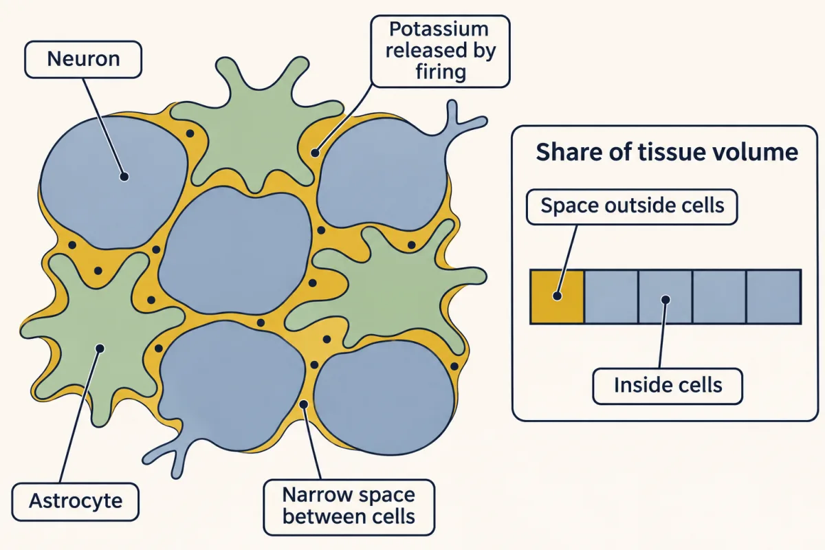
The one idea to carry through this lesson. Small space outside, big change outside. The space between brain cells is small and holds little potassium, so activity can change extracellular potassium strongly, and the shift in EK usually comes mostly from the outside; how much depends on the volumes taking part. A neuron’s resting potential therefore depends on cells that keep clearing that space.
The rule covers the potassium and glutamate that activity releases into grey matter. It explains why astrocytes buffer, pump and transport, and why deleting astrocytic GLT-1 caused seizures in mice. It does not explain myelin, metabolic support or microglia, other jobs glia do for the axon and the tissue, and it says nothing about whether astrocytes also send signals of their own, which is still argued.
A map of the neuron
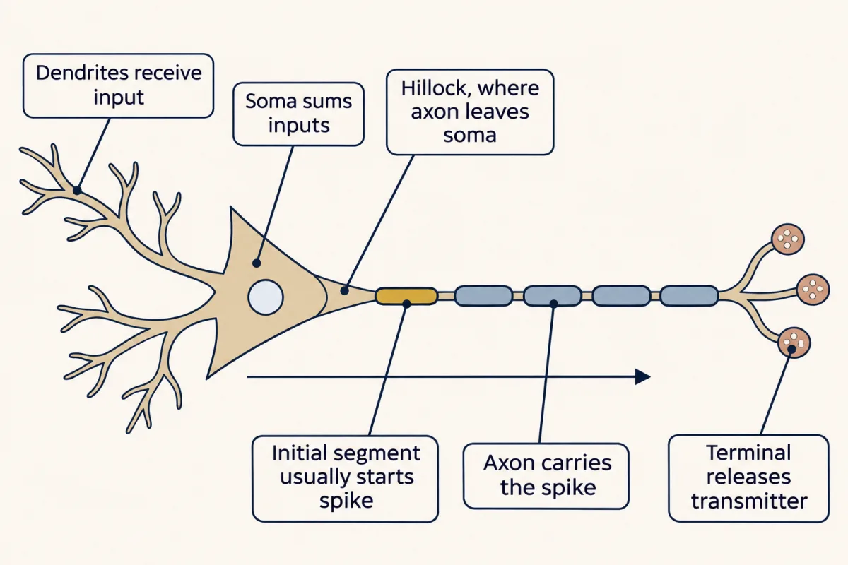
Before the glia, the neuron they look after. Its five compartments each mainly do one thing to the signal, and some leave something in the space outside. Read the last column: what a compartment leaves tells you which glial cell to look for.
| Compartment | What it does to the signal | What it leaves outside | Who looks after it |
|---|---|---|---|
| Dendrites | receive input as graded potentials | not followed here | — |
| Soma | sums inputs; holds the nucleus | not followed here | — |
| Axon initial segment | usually starts the spike | potassium, as each spike ends | astrocytes (as for the axon) |
| Axon | carries the spike, regenerating it | potassium, after every spike | astrocytes; myelinating cells insulate and fuel it |
| Terminal | turns the spike into transmitter release | glutamate, at excitatory synapses | astrocytes, through GLT-1 |
| The tissue around them all | — | — | microglia, which survey it |
Dendrites receive input. A synapse onto a dendrite produces a postsynaptic potential, a local change in membrane voltage whose size depends on how much transmitter arrived. It is graded: bigger input, bigger potential, with no threshold. An excitatory one is an excitatory postsynaptic potential (EPSP), a depolarization that moves the cell toward firing. In a passive dendrite it shrinks as it spreads, the way a ripple fades from where a stone fell. The next lesson, on the resting membrane and passive signals, puts numbers on how far it travels.
The soma, or cell body, holds the nucleus and most of the machinery that makes proteins. It is also a place where inputs meet. Spatial summation adds potentials arriving at different synapses at about the same time; temporal summation adds potentials arriving at one synapse in quick succession. Dendrites also do some of this adding and transform inputs before they arrive.
The axon initial segment (AIS) is the first stretch of axon. Packed with voltage-gated sodium channels a little way out from the large cell body, it is where the action potential usually starts (density is only part of the reason; see Going deeper).
The axon carries the spike away, regenerating it as it goes (in myelinated axons, only at the gaps in the myelin; see below). It also carries cargo both ways on microtubule tracks, described in molecular motors.
The terminal turns the spike back into chemistry. Depolarization opens voltage-gated calcium channels, and calcium entry triggers fusion of transmitter-filled vesicles with the membrane. At fast synapses this release can follow the spike in less than a millisecond, though the delay is not the same at every synapse. Synaptic transmission takes this apart.
The picture is the canonical one, drawn from cortical and hippocampal pyramidal neurons and cerebellar Purkinje cells, and it has exceptions: some mammalian neurons fire spikes in their dendrites, and the trigger zone is not always where textbooks put it.
Where the spike starts
Many textbooks say the action potential begins at the axon hillock, the cone where the axon leaves the soma. A somatic recording cannot test this, since a spike starting tens of micrometres away arrives almost at once. So Lucy Palmer and Greg Stuart imaged the axon at several points simultaneously with a voltage-sensitive dye. They worked in layer 5 pyramidal neurons in acute slices of rat somatosensory cortex, from rats usually three to five weeks old.
Picture the record: one voltage trace for each point along the axon, stacked on a shared time axis. The traces look much alike, but one begins slightly before the rest. That earliest signal appeared about 35 µm from the hillock, in the distal part of an initial segment about 40 µm long. Experiments targeting the first node suggested it is not involved in initiation. This is a measurement in one cell type, not a rule for every neuron.
How the AIS is assembled is covered in neuronal polarity; what it does, in the action potential.
How far EK moves
Back to the potassium. An ion’s equilibrium potential is the voltage at which its concentration gradient and the electrical force on it balance. For potassium at 37 °C the Nernst equation, derived in membrane transport, gives
EK = 61.5 mV × log10([K+]o / [K+]i)
Read it as a rule of thumb: every tenfold change in the ratio moves EK by about 61.5 mV.
Worked problem. Take [K+]i = 140 mM, hold it steady, and treat potassium on its own. The values are illustrative.
At rest, [K+]o = 3 mM: EK = 61.5 × log10(3/140) = −102.6 mV.
After intense firing. In cat cortex, even strong repetitive stimulation and seizure-like afterdischarges raised extracellular potassium only to a ceiling of about 10 mM, and ordinary activity raises it much less. At 10 mM, EK = 61.5 × log10(10/140) = −70.5 mV, about 32 mV more positive.
Check it with the rule of thumb. Going from 3 to 10 mM is a little more than a threefold rise, which on the log scale is about half a tenfold step (the square root of 10 is about 3.2). So EK should move by about half of 61.5 mV. It does.
- [K+]o at the cursor
- 12.0 mM
- EK at the cursor
- −65.6 mV
- EK at rest, 3 mM
- −102.6 mV
- EK at the peak
- −65.6 mV
- Shift at the peak
- +37.0 mV
- Excess [K+]o left at the cursor
- 100%
In this sketch (illustrative values from the lesson’s worked problem): potassium alone, [K+]i fixed at 140 mM, rest 3 mM, 37 °C, one exponential return to 3 mM with an illustrative τ (not a measured value).
Rule to keep: any fourfold rise in [K+]o shifts EK by 61.5 × log10(4) = 37.0 mV, whatever [K+]i is. The 1.85 s and 1.13 s of Tyurikova and colleagues are decays of an astrocyte current, not this τ.
The simulation starts at a fourfold rise, to 12 mM, an extreme case beyond the measured ceiling, and lets you vary how fast the excess clears. Its clearance time is illustrative, not measured.
Predict. When EK moves 32 mV, does the neuron’s resting potential move 32 mV, more, or less?
Check your prediction
Less, but in the same direction. A neuron at rest is mostly but not only permeable to potassium; sodium and other ions also pull on its voltage. In a simple model of the resting membrane (Going deeper), it moves about half as far as EK: about 16 mV here, and roughly 10 to 21 mV across plausible sodium permeabilities. The next lessons make this precise. At steady rest with no current injected, the total membrane current, pump current included, is zero. In a simplified model with fixed conductances and no other net current, the resting voltage is a conductance-weighted average of the ions’ equilibrium potentials; with two ions it lies closer to the one with the larger conductance.
What does the neuron feel? A modest rise brings it closer to threshold, so it fires more easily, and more firing releases more potassium: a loop that can feed a seizure. A large, sustained rise can do the opposite. Held depolarized, sodium channels inactivate, threshold moves up, and the neuron can stop firing altogether (see “threshold moves” in the action potential). Either way, something has to clear the space.
Astrocytes clear the potassium
Read the transcript of “Who Puts the Potassium Back”
Film transcript: Who Puts the Potassium Back
Something has to clear the potassium from the space outside neurons. Who does it, and how? In 1966, Stephen Kuffler, John Nicholls and Richard Orkand recorded from glial cells. They worked in the optic nerve of an amphibian, the mudpuppy Read the record first. The glial cell's voltage sits steady and very negative. Then the neighbouring axons fire a train of impulses. The glial cell slowly depolarizes. Longer and faster trains depolarize it more. When the axons stop, it recovers over seconds. What is the glial cell sensing?
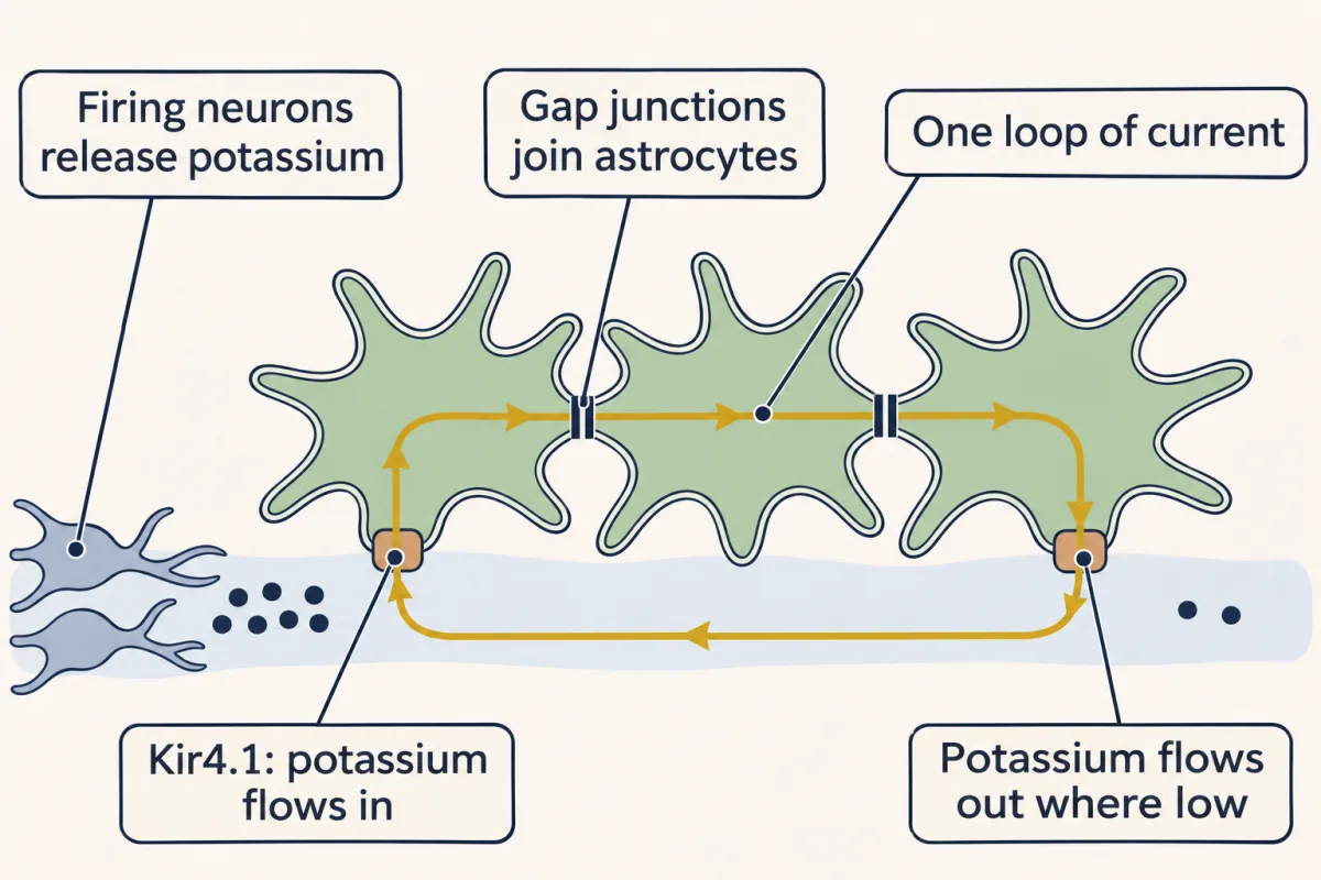
Potassium. Changing the potassium in the bath moved the glial voltage as a potassium electrode's would. So the glia were sensing the potassium the axons released as it built up. Astrocytes are joined by gap junctions, channels that link the insides of neighbouring cells. Their membranes are almost purely permeable to potassium. In this simplified, strongly coupled network, the astrocytes stay at nearly the same voltage. What differs from place to place is the local E_K, set by the local potassium. Now let one spot fire hard. There, the local E_K is more positive than the network's voltage. So potassium flows in. In this model it enters through the potassium channel Kir4.1.
That entry depolarizes the network slightly. Where potassium is still low, potassium now flows out. Entry and exit happen at the same time, as one loop of current. This is spatial buffering. It moves potassium from where it is high to where it is low. On balance, the potassium stays outside, only spread thinner. Putting it back inside cells is a separate job. Sodium-potassium pumps in astrocytes and neurons take potassium up. How much each mechanism contributes is still debated. Ken McCarthy's laboratory deleted Kir4.1 from glia in mice. In hippocampal slices, astrocytes lacking the channel took up potassium poorly. What else might fail when this potassium channel is gone?
Glutamate uptake. That does not mean the channel carries glutamate. Glutamate transport depends on the astrocyte's voltage. And Kir4.1 helps hold that voltage negative. One limit. The deletion began in development, and it also reached oligodendrocytes. So the result cannot be pinned on mature astrocytes alone. So astrocytes sense the potassium that firing releases. They spread it through their network, and pumps take it back inside cells.
From the film
- Glial cells in the mudpuppy optic nerve depolarized as neighbouring axons fired, because they were sensing the potassium the axons released.
- In a simplified model of spatial buffering, potassium enters astrocytes through Kir4.1 where it is high and leaves where it is low, as one loop of current through astrocytes joined by gap junctions.
- Spatial buffering spreads potassium thinner and leaves it outside, while sodium-potassium pumps in astrocytes and neurons put it back inside cells, and how much each contributes is still debated.
- In mouse hippocampal slices, astrocytes lacking Kir4.1 took up potassium poorly, though the deletion began in development and also reached oligodendrocytes.
The evidence: glia that read potassium
Early direct evidence that glia sense the potassium released by active axons came in 1966, when Stephen Kuffler, John Nicholls and Richard Orkand recorded from glial cells in the optic nerve of an amphibian, the mudpuppy Necturus.
Picture the record: a glial cell’s membrane potential, steady and very negative, drawn under a train of impulses in the neighbouring axons. During the train the glial cell slowly depolarizes, more for longer and faster trains, and it recovers over seconds once the axons stop.
Changing the potassium in the bath moved the glial voltage as a potassium electrode’s would, so the glia were sensing the potassium the axons released as it built up outside them. That record led to the idea of spatial buffering.
The mechanism: a current loop through the astrocyte network
Astrocytes are joined to one another by gap junctions, channels that link the insides of neighbouring cells, and their membranes are almost purely permeable to potassium. In a simplified, strongly coupled network the astrocytes therefore sit at nearly the same voltage, near a conductance-weighted average of the local EK values (real networks can have voltage gradients and pump or transporter currents). What differs from place to place is the local EK. Now let one spot fire hard.
- Where potassium is high, the local EK is more positive than the astrocyte’s voltage, so potassium flows in, in this model through the potassium channel Kir4.1. (Kir4.1 is inwardly rectifying by name, but it rectifies only weakly compared with other Kir channels, so it can also carry the outward limb in step 3.)
- That entry depolarizes the connected network slightly.
- Where potassium is still low, the network’s voltage is now more positive than the local EK, so potassium flows out.
Entry and exit happen at the same time, as one loop of current, so potassium can leave far away without waiting for the ions that entered to travel there. This is spatial buffering: it moves potassium from where it is high to where it is low. On balance it leaves the potassium outside, only spread thinner, where it does less harm. Pumps instead put it back inside cells: sodium-potassium ATPases in astrocytes and in neurons take potassium up. How much each mechanism contributes is still debated. In rat hippocampal slices, Kir4.1 buffered potassium while it was rising, and blocking the pumps slowed clearance after stimulation ended.
Predict. Ken McCarthy’s laboratory deleted Kir4.1 from glia in mice. What should happen to astrocytes’ potassium uptake? Is there any reason their glutamate uptake might change too?
Check your prediction
In hippocampal slices from mice 15 to 20 days old, CA1 astrocytes without Kir4.1 took up potassium poorly, and glutamate poorly too (Djukic and colleagues, 2007). That does not mean Kir4.1 carries glutamate. Glutamate transport is driven by ion gradients and carries net positive charge into the cell, so it depends on the astrocyte’s voltage, which Kir4.1 helps hold negative. Two limits: the deletion began in development and also reached oligodendrocytes, so the result cannot be pinned on mature astrocytes alone.
Glutamate: the same rule, more sharply
Glutamate, the main fast excitatory transmitter, must be cleared from outside synapses too, and the lesson’s rule applies even more sharply: transporters keep glutamate outside cells very low between releases, so each release makes a large, brief rise near the synapse. The transporter GLT-1 (also called EAAT2) is mostly astrocytic, with a smaller pool in excitatory terminals.
Predict. Petr and colleagues deleted GLT-1 in mice, from astrocytes in one line and from neurons in another. Which deletion should matter more for the animal? And which should look bigger in an uptake assay in synaptosomes, the pinched-off nerve terminals made from brain tissue?
Check your prediction
For the animal, the astrocytic deletion. Made with an inducible Cre (a gene deletion switched on by a drug), it removed about 80 percent of GLT-1 protein, and the mice had seizures and early death. In the assay, the neuronal one: deleting GLT-1 from neurons cut uptake into crude forebrain synaptosomes by about 40 percent. The two fit once you see what the assay samples: made from terminals, it over-samples the small neuronal pool of GLT-1. Most GLT-1 is astrocytic, and the seizures point to that pool for clearance in the intact brain.
The axon’s other partners: myelin, fuel and microglia
Myelin is a membrane wrapped many times around an axon. It gives the axon a high-resistance, low-capacitance sheath, so current spreads farther between the gaps called nodes of Ranvier. One myelinated stretch between two nodes is an internode. The spike is regenerated at each node, and current flows through the axon from one node to the next: excitation skips the internode, but current does not. In the peripheral nervous system a myelinating Schwann cell makes exactly one internode. In the CNS one oligodendrocyte can make several, often on different axons, with no fixed count. Some Schwann cells never myelinate: Remak cells hold small unmyelinated axons. How myelin is made is in myelination.
Myelin is not frozen in adults. Hughes and colleagues imaged adult mouse somatosensory cortex for weeks and saw newly generated oligodendrocytes add new sheaths, while in mice 10 to 14 months old only about 1 percent of existing sheaths changed length over seven weeks.
Oligodendrocytes also feed axons. Lee and colleagues knocked down the lactate transporter monocarboxylate transporter 1 (MCT1) in oligodendrocytes of the optic nerve, and the axons were damaged; in slice culture, added lactate prevented injury when MCT1 was blocked and glucose withheld. The fuel may differ by region: in mouse corpus callosum slices, Meyer and colleagues found that oligodendrocytes kept axons working mainly by delivering glucose, and lactate rescued them less well. The fuller story is in glia as metabolic partners.
Microglia are the brain’s resident immune cells. Using two-photon imaging in healthy adult mouse neocortex, Nimmerjahn, Kirchhoff and Helmchen saw them in their presumed resting state with cell bodies in place but processes that extended and retracted continually, surveying the tissue. Most such imaging has been done in anesthetized mice; in awake mice, Stowell and colleagues found less process surveillance, because noradrenaline acts on β2-adrenergic receptors on microglia. Their role in removing synapses during development is told in microglia and complement pruning.
What is still argued: gliotransmission
Read the transcript of “Settled and Argued”
Film transcript: Settled and Argued
Astrocytes clear potassium and glutamate. Do they also send fast signals of their own? Start with what is settled. Glutamate follows the same rule as potassium, even more sharply. Between releases, transporters keep outside glutamate very low. So each release makes a large, brief rise near the synapse. Most of the glutamate transporter GLT-1 sits in astrocytes, with a smaller pool in terminals. Deleting GLT-1 from astrocytes in mice caused seizures and early death. Now the open question. Do astrocytes also release glutamate that acts quickly on synapses? This is called gliotransmission. Astrocytes show calcium signals. In principle, a calcium rise could trigger release, as it does in a terminal.
One test deleted a receptor that releases stored calcium, the IP3 receptor type 2. Hippocampal transmission did not change. Does that prove astrocytes never release glutamate onto synapses? No. In adult mice, astrocytes lacking that receptor still showed calcium fluctuations, mostly in their processes. So the null result cannot exclude effects driven by the calcium that remains. A later study found a subgroup of adult mouse hippocampal astrocytes with synaptic-like release machinery. Deleting a vesicular glutamate transporter from astrocytes suppressed the glutamate hotspots linked to that subgroup. That is positive evidence, but in one subgroup and one species.
Neither study refutes the other. They used different manipulations and asked different questions. So keep two lists. That astrocytes clear potassium and glutamate is settled. Fast signalling by astrocytes is still argued. Your design problem is to test it in one group of adult mouse astrocytes.
From the film
- That astrocytes clear potassium and glutamate is settled: GLT-1 is mostly astrocytic, and deleting it from astrocytes in mice caused seizures and early death.
- Whether astrocytes also release glutamate that acts quickly on synapses, called gliotransmission, is still argued.
- A null result from deleting the IP3 receptor type 2 cannot exclude effects of the calcium that remains, and the positive evidence so far is for one subgroup of adult mouse hippocampal astrocytes.
Gliotransmission is the proposal that astrocytes release transmitters, such as glutamate, that act quickly on synapses. Astrocytes show calcium signals (see calcium signalling), and a calcium rise could in principle trigger vesicle release, as it does in a terminal.
The evidence conflicts. Agulhon and colleagues raised or disrupted astrocyte calcium signalling in mouse hippocampus, including by deleting the inositol trisphosphate (IP3) receptor type 2, which releases calcium from internal stores, and found no change in excitatory transmission or plasticity.
Predict. Does that null result show that astrocytes do not release transmitter that acts on synapses? What would you want to check first?
Check your prediction
Not on its own. A null result can speak only to what the manipulation actually removed, under the conditions tested, so check whether the deletion really removed astrocyte calcium. Srinivasan and colleagues later showed that adult mouse astrocytes lacking that receptor still have calcium fluctuations, mostly in their processes, in slices and in vivo. So the null result does not exclude effects driven by the remaining calcium.
On the other side, de Ceglia, Volterra and colleagues found a specialized subgroup of adult mouse hippocampal astrocytes expressing synaptic-like release machinery, including VGLUT1, and showed that deleting VGLUT1 from astrocytes suppressed the glutamate hotspots linked to that subgroup. That is positive evidence, restricted to one subgroup, in one species. Neither study refutes the other: they used different manipulations and asked different questions.
Design problem
Do adult hippocampal astrocytes release glutamate that changes synaptic transmission? Starting from the IP3R2 null result and the VGLUT1 subgroup above, design an experiment in mice that could show whether release from those astrocytes changes transmission, and say what your result could and could not establish. Plan three things: what you remove and when, what you compare against, and what readout would change.
Saved in this browser only.
One way to do it
Manipulation. Delete VGLUT1 from astrocytes only in adults, with a tamoxifen-inducible Cre driven by an astrocyte promoter. This avoids a developmental confound and targets the release machinery rather than one calcium pathway.
Check the deletion. Confirm that VGLUT1 is lost in astrocytes and that neurons and oligodendrocytes did not recombine. Many excitatory neurons in hippocampus and cortex are full of VGLUT1, so even sparse neuronal recombination could confound the interpretation.
Measure release and its effect. A glutamate sensor reports glutamate from any source, so first image it with neuronal firing and synaptic release blocked, to see the astrocyte signal on its own. Then, with transmission intact, record from CA1 pyramidal neurons in slices: evoked EPSPs, spontaneous events and, if predicted, plasticity. Compare with littermates given tamoxifen but lacking the floxed allele.
Test sufficiency. In separate animals, drive only astrocytes (for example with a light-gated or chemically gated receptor expressed in them) and ask whether a glutamate signal and a synaptic change appear, and whether they vanish after VGLUT1 deletion. Check that the deletion did not change glutamate uptake or ion handling, which could explain a loss of signal without any vesicular release.
State the scope. A positive result would show that VGLUT1-dependent release from this astrocyte subgroup can change transmission in adult mouse hippocampus under the conditions tested. It would not show that all astrocytes do this, in other regions or species, or that it matters for behaviour. A negative result would hold only for the readouts and activity patterns tested.
Going deeper
Why the distal initial segment
Sodium-channel density is only part of the usual explanation for why the spike starts in the distal part of the initial segment. Reviews also point to the sodium-channel subtype concentrated there, which opens at less depolarized voltages, and to the segment’s electrical distance from the soma’s large membrane.
Watching saltatory conduction
In the myelinated axons of mouse layer 5 pyramidal neurons, voltage imaging by Popovic and colleagues (2011) showed the spike propagating saltatorily, from node to node, toward the axon’s branches.
How much the outside wins
For small changes near rest, with [K+]o = 3 mM, [K+]i = 140 mM and 61.5 mV per tenfold change, each extra millimolar outside moves EK by about 8.9 mV and each millimolar lost inside by about 0.19 mV: a ratio of 140/3, about 47. (These are slopes at rest; a full step from 3 to 4 mM moves EK by 7.7 mV.) Multiply by the participating inside-to-outside volume ratio to get the outside’s lead, as in the opening prediction. A separate, conditional example: if all the intracellular volume, four fifths of the tissue, exchanged potassium with the fifth outside, the volume ratio would be (1 − 0.2)/0.2 = 4 and the outside would lead by roughly 190-fold, or about 140- to 260-fold if the space is anywhere from 0.25 to 0.15 of the tissue. Within that fixed model, with the space at 0.20, 190 is an upper value. In the same shared-pool model the outside’s millimolar change is the larger one only if the participating cells fill more than a quarter of the inside volume, and its contribution to EK is the larger one unless they fill less than about 0.5 percent of it.
Any fourfold rise
If [K+]i stays constant, a fourfold rise in [K+]o shifts EK by 61.5 × log10(4) = 37.0 mV, whatever that fixed [K+]i is: from 3 to 12 mM, for example, EK goes from −102.6 to −65.6 mV. That is the simulation’s starting case.
How far the resting potential follows
The body’s figures use the Goldman-Hodgkin-Katz equation with [K+]i held at 140 mM and [Na+]o at 145 mM, chloride and the pump ignored, and a fixed sodium-to-potassium permeability ratio. For a rise from 3 to 10 mM, ratios of 0.02 to 0.08 give the 21-to-10 mV range, two-thirds to a third of EK’s shift; 0.04 gives 15.6 mV. Other choices give other fractions.
When the inside starts to matter
In very thin axons during long, high-frequency trains, potassium lost from the inside and sodium gained stop being negligible. Then the outside no longer automatically dominates, and you have to compare the actual changes on each side.
A clearance time constant
Suppose astrocytes remove the excess potassium with a single-exponential time constant of 2 s (an illustrative value, not a measured one). The excess falls to 10 percent when e−t/2 s = 0.1, so t = 2 s × ln(10) = 2 × 2.303 = 4.6 s.
Read a real number correctly
In mouse hippocampal slices, Tyurikova and colleagues (2025) measured the decay of the potassium current recorded in astrocytes after a train of five stimuli at 50 Hz: 1.85 s in control astrocytes and 1.13 s in astrocytes overexpressing Kir4.1. The decay is (1.85 − 1.13)/1.85 = 38.9 percent shorter. That is a property of a current, not a time constant for extracellular potassium, so it cannot stand in for the simulation’s clearance time or the calculation above.
Your notes
Select any text in the lesson to highlight it or add a note to it. Your highlights and notes are saved in this browser and collected in your notebook.
This browser is not letting the site save anything (a private window or strict privacy settings), so highlights and notes will be lost when you leave the page.
How we measure it
Voltage-sensitive dye imaging
A dye in the membrane changes its fluorescence with voltage, so a camera can follow the membrane potential at many points along a dendrite or axon at once. Comparing when the signal appears at each point shows where an action potential starts.
In vivo two-photon time-lapse imaging
A laser that excites fluorescence only at its focus images labelled cells below the brain surface of a living animal. Imaging the same cells repeatedly shows how processes move over minutes, or how myelin sheaths are added and change over weeks.
Conditional (cell-type-specific) gene deletion
A recombinase such as Cre, expressed from a promoter active in one cell type, removes a floxed gene (one flanked by loxP sites that Cre cuts out) only in those cells, sometimes only after a drug is given. The method is only as specific as the recombination: you must check which cells actually lost the gene, and when.
Whole-cell recording and genetically encoded sensors
A patch pipette on an astrocyte records the currents that flow when nearby axons fire, and blockers separate the potassium and transporter components. Fluorescent sensors such as GCaMP report calcium inside a cell, and sensors of the iGluSnFR family report glutamate outside it.
Practice questions on this lesson (3) → MCAT-style, original, each with an explanation
Where this lesson fits
Before thisReview first
- The Map: How the Nervous System Is OrganizedThis course · Lesson 1
It introduces the neurons and glia and the barrier around brain tissue that this lesson then follows in action.
After thisStudy next
- The Resting Membrane and Passive SignalsThis course · Lesson 3
It uses E_K and potassium leak to explain the resting voltage, and how passive signals spread along dendrites.
- The Action Potential and ConductionThis course · Lesson 4
It explains how spikes begin and how potassium leaves the axon, the source of the potassium that glia clear here.
- Synaptic Transmission and Quantal ReleaseThis course · Lesson 5
It follows glutamate release at the terminal, the transmitter that astrocyte GLT-1 clears afterwards.
Elsewhere on the siteFilms, simulations and method films in other courses
- Two Pushes, One HillFilm · Cell Biology
It shows how concentration difference and voltage together set which way potassium flows.
- Cut the AxonFilm · Developmental Neuroscience
It shows how a neuron sets up one axon and many dendrites, the compartments mapped here.
- Why Wrap, and How MuchFilm · Developmental Neuroscience
It shows how glial wrapping makes the signal jump between nodes of Ranvier.
- When the Body Pushes BackFilm · Biology
It shows how opposing responses hold internal conditions in range, as astrocytes do for outside potassium.
Suggested by an AI model from the lessons themselves, then checked. How we use AI
Finish line
Test yourself
9 questions
Practice and MCAT-style, with explained answers.
Next · Lesson 3
The Resting Membrane and Passive Signals
In a resting neuron potassium leaks out and sodium leaks in all the time. Why does the voltage hold still?
Continue →