Developmental Neuroscience · Unit 9: Experience refines the wiring
Microglia and Complement Prune Synapses
Rewritten on : new text, figures, flashcards and film. Your notes and highlights stay, but the text around them may have moved.
Why can a developing neuron make the first protein of an immune cascade that marks bacteria for destruction?
Pause when the reading reaches a film, and offer to play it. Saved on this device.
Watch the film now, or keep listening
Builds on:Activity Before Experience: Spontaneous Retinal WavesOcular Dominance and the Critical PeriodToo Many Neurons: Naturally Occurring Cell DeathWhy these?
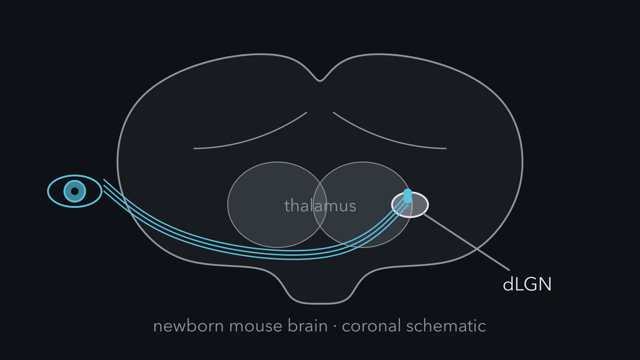 ▶ Watch Which Inputs Lose? · 2:22
Read the transcript of “Which Inputs Lose?”
Film transcript: Which Inputs Lose?
In a newborn mouse brain, some connections will be lost. Which ones, and what carries them away? Start in the visual thalamus, the relay where axons from the eye end. In a newborn mouse, inputs from the two eyes overlap there. Over roughly the first week and a half, the two eyes' inputs sort into separate territories. Each side ends up mostly the territory of the opposite eye. The eye on the same side keeps a small patch. One relay neuron starts with many retinal inputs, and ends with only a few. Inputs that lose are removed. So, which inputs lose? And what carries them away? The sorting is driven partly by spontaneous waves of activity. They sweep the young retina before the eyes open.
Predict. If activity biases the outcome, which inputs should lose more often: the busier ones, or the quieter ones? In this circuit, experiments that change one eye's activity point to the quieter ones. Later, an experiment that silences one eye tests whether microglia follow.
For the second question, the obvious suspects are microglia, the brain's resident immune cells and its main phagocytes. Here is the one idea to carry through. Relative activity biases which retinal inputs lose, and microglia take relatively more from the quieter eye. Complement helps them eat part of that.
The rule has a scope. This is the mouse visual thalamus, in the first week and a half after birth. Complement is needed for normal sorting. These studies did not show that it carries the activity information. And part means part. Some eating goes on without complement's receptor. Astrocytes eat synapses too.
So keep the two findings side by side. How activity and complement connect is still open.
From the film
- In the young mouse visual thalamus, the two eyes' inputs start out overlapped and sort into separate territories over roughly the first week and a half.
- Relative activity biases which retinal inputs lose, and microglia take relatively more material from the quieter eye.
- Complement helps microglia take up part of that material, and the evidence does not show that it carries the activity information.
A developing retinal neuron can make C1q, the first protein of an immune cascade that marks bacteria for destruction.
Complement is a set of proteins best known from the blood. They bind microbes, dead cells and clumps of antibody, and they coat them so that phagocytes, the cells that eat other cells, will swallow them. Finding complement in a young neuron looked like a category mistake. It turned out to be a clue to one way a developing brain removes connections it has made too many of. This lesson follows that clue from a gene list to the inside of a microglial lysosome.
Two eyes, one relay: which inputs lose?
The best-studied case is the dorsal lateral geniculate nucleus (dLGN), the visual relay in the thalamus where retinal axons end. Its relay neurons receive the retinal input and pass it on to the visual cortex. Ages here are written in postnatal days: P5 means the fifth day after birth.
In a newborn mouse, inputs from the two eyes overlap in the dLGN, and many relay neurons receive inputs from both. Over the first postnatal week and a half, mostly between about P3 and P10, they sort into separate territories, a process called eye-specific segregation. Each dLGN ends up mostly the territory of the opposite eye, with a small patch for the eye on the same side. A single relay neuron also goes from many retinal inputs to a few, a refinement that continues for weeks after the territories have sorted.
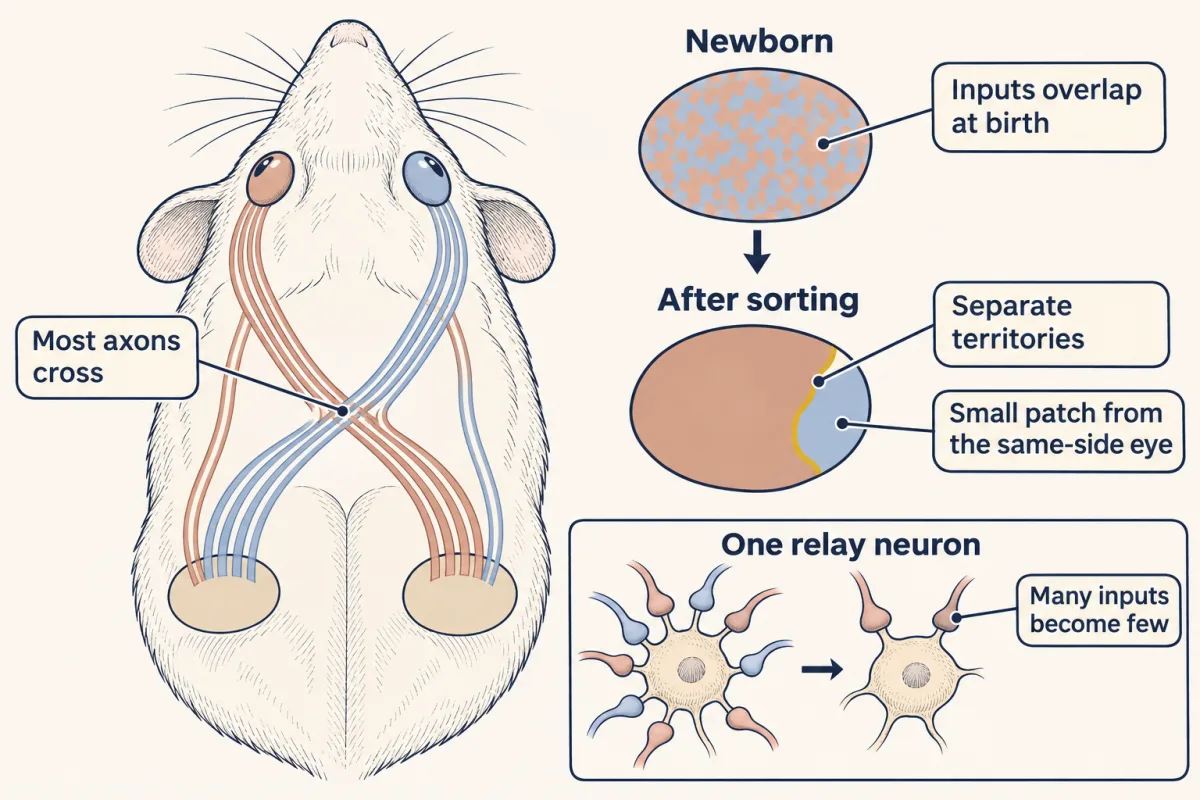
The sorting is driven partly by spontaneous retinal waves, bursts of activity that sweep the young retina before the eyes open (see the lesson on spontaneous retinal waves). Inputs that lose are removed. Two questions run through this lesson: which inputs lose, and what carries them away? For the first, eye-manipulation and retinal-wave experiments point to the less active inputs losing (Penn et al., 1998; Stellwagen and Shatz, 2002); this lesson tests whether microglia follow the same bias. The obvious suspects for the second are microglia, the brain’s resident immune cells and its main phagocytes.
The one idea to carry through this lesson. Relative activity biases which retinal inputs lose, and microglia take up relatively more material from the less active eye; complement helps them take up part of it.
Scope. Mouse dLGN, mainly during eye-specific segregation (about P3 to P10), with some readouts extending to about a month. The limits come with the evidence.
A tag from the immune system
Read the transcript of “A Tag and a Reader”
Film transcript: A Tag and a Reader
How does an immune protein mark something to be eaten? And does the young brain use the same trick? In the blood, complement marks targets for eating cells. Three steps matter here. Step one. C1q binds. It is shaped like a bunch of six tulips, and each head can grip a surface. Step two. Enzymes in the C1q complex cut two more proteins. Their pieces build an enzyme that cuts C3. C3 splits in two. The large piece, C3b, can fix itself to the nearby surface. Step three. C3b is trimmed to iC3b. Complement receptor 3, on an eating cell, binds it, which promotes engulfment. Now to neurons. Beth Stevens, in Ben Barres's laboratory, grew purified retinal ganglion cells with astrocytes in a dish.
The neurons raised their C1q. In the young mouse visual thalamus, C1q and C3 lay at developing synapses. One caution. Neurons can make C1q, but most brain C1q comes from microglia. Which cell supplies it at these synapses is not settled. Predict. If complement helps remove losing inputs, what should a mouse without C1q look like?
The two eyes' territories failed to refine normally. They still overlapped more than in normal littermates. Single relay neurons agreed. Most knockout cells still received several inputs, where normal cells had few. Mice lacking C3 showed a similar overlap.
What this shows: complement is needed for normal refinement in this circuit. What it does not show: which synapse got a tag, what read it, or which cell carried the input away. So the knockouts point to a tag. The next step is to catch the eater in the act.
From the film
- Complement marks a target for eating in three steps: C1q binds a surface, C3 is cut and its large piece fixes to that surface, and complement receptor 3 on an eating cell binds the tag.
- In mice lacking C1q or C3, the two eyes' territories failed to refine normally and still overlapped more than in normal littermates.
- These knockouts show that complement is needed for normal refinement in this circuit, but not which synapse got a tag, what read it, or which cell carried the input away.
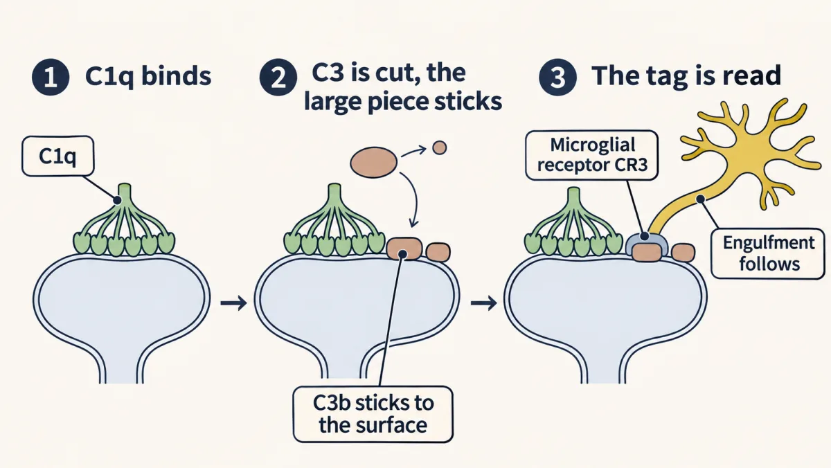
The classical complement pathway has three steps that matter here.
- C1q binds. C1q is shaped like a bunch of six tulips, each head able to grip a surface.
- C3 is cut, and the large piece sticks. Binding activates a protease in C1q’s complex, C1r, which activates a second, C1s. C1s cuts C4 and C2, and the pieces assemble into an enzyme, a C3 convertase, that cuts C3 into a small fragment, C3a, and a large one, C3b. Cutting exposes a reactive bond in C3b that can fix it to a nearby surface.
- The tag is read. C3b is trimmed to iC3b. A phagocyte carrying complement receptor 3 (CR3) binds iC3b, which promotes engulfment of what it coats. CR3 is an integrin, the CD11b and CD18 pair, of the adhesion-receptor family taught in the lesson on integrins and Rho GTPases.
Beth Stevens, working in Ben Barres’s laboratory at Stanford, found that purified rat retinal ganglion cells, the neurons that carry the eye’s output to the brain, raised their expression of C1q when grown with astrocytes. That finding sent the team looking at synapses in the brain. Later work found that microglia make most of the C1q in the mouse brain, so which cell supplies the C1q at dLGN synapses is not settled.
In the young mouse, Stevens and her colleagues found C1q in the retina and dLGN early in postnatal life; neuronal C1q is normally turned down in the adult. In the dLGN, C1q and C3 lay at developing synapses. C1q often sat at small synaptic contacts, which the team took to be immature or disappearing ones. That was a proposal: fixed images could not show which contacts later disappeared.
The test was genetic. In a whole-body knockout mouse, both copies of one gene are disrupted in every cell. To estimate how many retinal inputs one relay neuron has, stimulate the optic tract in a brain slice harder and harder while recording the cell’s current: each new step in the current marks one more retinal axon recruited. Weak inputs can be missed, so this is an estimate, not a full count.
Predict. If complement helps remove losing inputs, what should the dLGN of a mouse without C1q look like, in its eye territories and in single relay neurons?
Check your prediction
In mice lacking C1q, the two eyes’ territories failed to refine normally and still overlapped more than in normal littermates. In recordings, most normal relay cells had pruned down to few inputs, while most C1q-knockout cells still received several. Mice lacking C3 showed a similar overlap. The authors proposed that unwanted synapses are tagged by complement for elimination.
Notice what the knockouts did not show: which synapse got a tag, what read it, or which cell carried the input away.
Caught in the act: microglia take the quieter eye
Read the transcript of “Caught in the Act”
Film transcript: Caught in the Act
If microglia eat retinal inputs, can we catch them doing it? And do they choose what they eat? In 2012, Dorothy Schafer, in the Stevens laboratory, set out to look. Each eye got its own colored tracer at postnatal day four. A day later, they looked in the visual thalamus. A speck beside a microglial cell does not show it was eaten. So only label inside the cell counts. Retinal material sat inside microglial lysosomes, the cell's digestive compartments. A dye that glows only in acid agreed. The swallowed material was mainly presynaptic, from retinal terminals.
Eating had a clock. It was high at postnatal day five, and much lower by day nine. Eating also used a tag and a reader. Without complement receptor 3, the reader, or C3, the tag's source, microglia took up about half as much. Those mice later had more overlap between the eye territories, and extra retinal synapses. But half the eating remained. Complement is one route of several. Now the key question. Silence one eye with tetrodotoxin, which blocks sodium channels. Predict. If microglia remove less active inputs, whose material should they hold more of?
More of the silenced eye's material, relative to how much of that eye's input is there. Boost one eye with forskolin instead, and they take relatively more of the untreated eye's. The two drugs push in opposite directions. A drug that simply damaged axons would raise eating of its own eye both times.
Why relative to what is there? Near one microglial cell, one eye's input can far outnumber the other's. Say 90 units from one eye, and 10 from the other. An eater with no preference takes a tenth of each, so it holds 9 to 1. Now silence the minor eye, and let the eater take twice the fraction of it: two tenths instead of one tenth. It holds 2 units, still fewer than 9. So a fair test compares each eye with its own input, measured where the microglia are sampled. Now the limits. This compares whole eyes, not single synapses. It does not show how activity becomes a mark. And later imaging elsewhere suggests microglia often take bites, not whole synapses.
Here is the idea again. Relative activity biases how much material microglia take from each eye. Complement helps them take part of it. How the two connect is still open.
From the film
- Microglia in the young mouse visual thalamus held retinal material inside their lysosomes, and this uptake was high at postnatal day five and much lower by day nine.
- Without complement receptor 3 or C3, microglia took up about half as much, so complement is one route of several.
- With one eye silenced, microglia held relatively more of that eye's material, and with one eye boosted, relatively more of the untreated eye's, so the comparison must be made against how much of each eye's input is there.
- This compares whole eyes, and how activity and complement connect is still open.
In 2012 Dorothy Schafer, then a postdoctoral fellow in the Stevens laboratory, went after the missing step with Barres and others. Seeing a fluorescent speck beside a microglial cell does not show that the cell ate it, so engulfment has to be measured inside the cell:
engulfment index = (volume of retinal label inside a microglial cell) ÷ (volume of that microglial cell) × 100
Dividing by the cell’s volume means a large cell does not seem to eat more merely because it is large. The team injected red and blue tracers, dyes that retinal axons carry from the eye to their terminals, into the two eyes of mice at P4 and looked in the dLGN a day later, in mice whose microglia were marked green. They rebuilt each microglial cell in three dimensions and kept only the label inside it. Retinal material sat inside microglial lysosomes, the cell’s digestive compartments. A dye that glows only in acid, and a genetic label of retinal neurons, gave the same result. Electron microscopy and staining for VGlut2, a protein of retinal terminals, showed that the engulfed material was mainly presynaptic.
When, and through what. Engulfment had a clock. It was high at P5, when segregation is under way, and much lower by P9. It also had a receptor. In mice lacking complement receptor 3 (CR3), the microglial reader, or lacking C3 and so the tags cut from it (C3b, iC3b), microglia took up roughly half as much retinal material as normal microglia. Those mice later had more overlap between the eye territories, and more retinal synapses about a month after birth.
Predict. Without CR3, microglia ate about half as much. Should a CR3 knockout keep every input it would normally have lost, or only some of them?
Check your prediction
Only some, as it turned out. Residual uptake shows that microglia still engulf material by routes other than CR3, and astrocytes eat dLGN synapses too, but a halved P5 index cannot by itself predict how much refinement is lost. The index is a snapshot of cargo at P5, not a count of inputs removed, and these experiments do not say whether eating starts the removal or clears material that neurons shed. The wiring answered: refinement fell short rather than failing, and the excess of structural synapses was modest, of the order of 1.3-fold (about 30 percent more) by array tomography, an imaging method that counts synapses in ultrathin serial sections.
Now the question that matters most: does the eater choose?
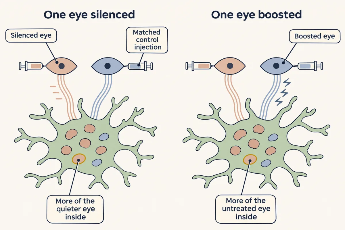
Predict. Two drugs change one eye’s activity. Tetrodotoxin (TTX) blocks sodium channels and silences neurons. Forskolin raises cAMP, a messenger inside the cell, and increases their activity. At P4 you treat one eye of each mouse and give the other eye a matched control injection; each eye gets its own tracer. At P5 you measure how much of each eye’s material dLGN microglia hold. If microglia preferentially remove less active inputs, what should each drug do? And before you compare the two eyes, what must you correct for?
Check your prediction
With one eye silenced, microglia took relatively more material from that eye. With one eye boosted, they took relatively more from the untreated eye. The two drugs push in opposite directions, which is stronger evidence than either alone: a drug that simply damaged axons would raise engulfment of its own eye both times.
The correction is for how much of each eye’s input is there, around the microglia you sample. Take an invented example. Suppose a cell’s neighbourhood holds 90 units of labelled input from one eye and 10 from the other. An eater with no preference that takes a tenth of everything holds 9 units and 1 unit. Now silence the minor eye and let the eater take a fifth of its input: it holds 2 units against 9. The silenced eye is still the smaller raw share, yet per unit of its own input it was eaten twice as much (2 ÷ 10 = 0.2, against 9 ÷ 90 = 0.1).
So compare each eye with its own input, matched or measured where the microglia are sampled. A whole-nucleus territory map cannot give those local numbers. Dividing by microglial volume, as the index does, is needed too, but it does not fix this.
So the bias is between whole eyes: in this circuit, microglia engulf relatively more of the less active eye’s material.
Design problem
You want to test whether the activity bias needs complement. Using the TTX experiment above, design the comparison that would answer it, name the result that would mean “yes,” and list the controls.
Saved in this browser only.
One way to do it
Repeat one-eye TTX in normal mice and in CR3 (or C3) knockouts. In each mouse, measure the bias: the silenced eye’s eaten fraction against the control eye’s, each taken relative to that eye’s locally available labelled input. Then ask whether the bias differs by genotype (in statistical terms, a genotype-by-treatment interaction).
- If the bias is lost in the knockout, the deleted protein is needed for the bias to show.
- If the bias survives, the bias can be expressed without that protein.
Neither result says which molecule encodes relative activity. A knockout could lose the bias because it cannot eat tagged targets, not because the tag carries the activity information. A surviving bias could ride on another receptor, and losing CR3 leaves the complement tags in place. Rescue experiments, or deleting the gene only in microglia and only at this age, would sharpen the answer.
Controls:
- Give the other eye a matched saline or vehicle injection, so both eyes are handled the same.
- Measure each dLGN separately, because the silenced eye is the main input to the opposite nucleus and the minor one on its own side.
- Treat the left eye in some mice and the right in others, so a left-right difference cannot pose as an activity effect.
- Check that TTX silences the eye equally well in both genotypes, and that it does not kill retinal ganglion cells, since dying cells would be eaten for a different reason.
- Correct for each eye’s local input within each genotype, because the knockouts keep more overlap.
- Check that enough uptake remains in the knockouts to measure a bias.
- Normalize to microglial volume, and score the images blind to genotype and treatment.
Find me, eat me
Any pruning claim raises two questions: how does the eater get there, and what marks the target? “Find me” signals draw the eater in; “eat me” signals mark the target. Complement is on the eat-me side; none of the dLGN experiments above tested a find-me signal.
Find-me signals act at a distance. ATP released from damaged cells draws microglial processes toward the damage. Fractalkine, also called CX3CL1, is a membrane protein on neurons that can be cut free; its receptor, CX3CR1, is on microglia. In 2011 Rosa Paolicelli, in Cornelius Gross’s laboratory, reported that mice lacking CX3CR1 had fewer microglia and more dendritic spines in the developing hippocampus, with signs of immature connections.
Predict. The extra spines in the CX3CR1 knockouts were gone by about P40. Does that mean pruning failed, or something else?
Check your prediction
Something else. The excess was transient, so the result is best read as delayed pruning in that region, not its absence. Later studies in other circuits have given mixed answers about whether CX3CR1 is required. In mouse barrel cortex, the part of the cortex that maps the whiskers, its loss blocked engulfment without blocking recruitment, so there the find-me label does not hold: the receptor was needed for eating, not for arrival.
Eat-me signals sit on the target: complement tags, and phosphatidylserine (PS), a membrane lipid normally kept on the inner, cytoplasmic face of the membrane (see the lesson on membranes). A dying cell moves it to the outer face, where phagocytes recognize it. In 2020 a study from Stevens’s and Michela Matteoli’s groups, jointly led by Nicole Scott-Hewitt and Fabio Perrucci, injected a PS-binding probe into the brain ventricles of P4 mice. At P5, PS turned up on the presynaptic side of a small fraction of dLGN synapses, a fraction that fell between P5 and P10 (in the hippocampus the signal peaked later), and mice lacking caspase-3, an enzyme of apoptosis (programmed cell death), showed no significant difference in the signal. So a synapse can display a death signal locally while its neuron lives. A synapse may then carry two kinds of eat-me mark, a lipid and a protein, possibly read by different receptors. C1q can bind PS, so the two tags may act in series, with the lipid recruiting the protein, or in parallel; which is open.
Two other forms of removal belong elsewhere. Axons that prune themselves using caspases and p75 are in the lesson on axons that cut themselves back, and the loss of extra inputs at the neuromuscular junction is in the lesson on synapse elimination.
When pruning goes wrong: links, not causes
If pruning shapes circuits, too much or too little might matter for disease. Changes in synapse number have been proposed to contribute to autism and to schizophrenia. These are hypotheses, supported by associations, and the disorders themselves are covered in the lesson on disorders of the synapse.
The strongest human evidence linking complement to a disorder is genetic. In 2016, Aswin Sekar, Steven McCarroll and colleagues, including the Stevens laboratory, studied C4, the next protein in the cascade, cut by C1q’s enzymes. Humans have two C4 genes, C4A and C4B, in varying copy numbers. Variants predicted to raise C4A expression in the brain were associated with a higher risk of schizophrenia. In mice, loss of C4 reduced pruning in the dLGN.
Predict. Put the human and mouse results together. Which way do they suggest pruning might be changed in schizophrenia, and how strong is the case?
Check your prediction
Together they suggest that more complement activity might mean more pruning, which fits the long-standing observation of fewer dendritic spines in some schizophrenia brains. The case is a hypothesis, not a cause. The human result is an association, the common C4 variants each change risk only modestly, and the mouse result comes from the visual thalamus, not the human cortex.
Putting it together
| Experiment | Animal and age | Observed result | What it supports | What it does not establish |
|---|---|---|---|---|
| Look for C1q and C3 | mouse retina and dLGN, early postnatal | at developing synapses | a candidate tag in the right place | which contacts later disappear; which cell supplies the C1q |
| C1q knockout | mouse dLGN | eye territories overlap more; relay cells keep several inputs | complement is needed for normal refinement | which synapse was tagged, what read it, which cell removed it |
| C3 knockout | mouse dLGN | similar overlap | the same | the same |
| Label each eye, look inside microglia | mouse, tracers at P4, looked at P5 | retinal, mainly presynaptic material in microglial lysosomes | microglia engulf retinal inputs | that they are the only route |
| Engulfment over time | mouse, P5 and P9 | high at P5, much lower by P9 | uptake is high while sorting is under way | when it starts or what sets the decline |
| CR3 or C3 knockout | mouse, P5 and about a month | about half the uptake; later more overlap and more synapses | complement is one route of eating | what carries the other half |
| TTX or forskolin in one eye | mouse, P4 to P5 | relatively more of the less active eye’s material eaten | relative activity biases eating, eye by eye | how activity becomes a mark; choice at single synapses |
| CX3CR1 knockout | mouse hippocampus, postnatal | fewer microglia; extra spines, gone by about P40 | delayed pruning in that region | a role in the dLGN |
| C4 variants; C4 knockout | humans; mouse dLGN | higher C4A linked to schizophrenia risk; less pruning without C4 | a hypothesis worth testing | that pruning causes the disorder |
Within this scope the experiments support a narrower statement than the guiding idea: relative activity biases how much of each eye’s retinal material microglia engulf, and complement helps microglia engulf about half of it. That this engulfment accounts for which inputs lose remains the working idea, not a result. Its limits, gathered in one place:
- A bias, eye by eye. The activity evidence compares whole eyes; no choice was seen at single identified synapses.
- Needed, not shown to carry the information. Without C1q, C3 or complement receptor 3 (CR3) refinement falls short, so they are needed in the sense of the lesson on the neurotrophic hypothesis: without one, the normal outcome is not reached. These studies did not show that complement carries the activity information or marks the quieter inputs.
- Part means part. About half the microglial eating remains without CR3, and astrocytes eat dLGN synapses too. Later imaging, in hippocampal slice cultures and in the living Xenopus retinotectal system, suggests that microglia often take partial bites of presynaptic membrane (trogocytosis) rather than whole synapses. Mice that lack microglia from birth still reach normal synapse and spine numbers in the hippocampus, so other cells can compensate there.
- One circuit. Microglia prune in other regions too, but which signals they use varies by circuit and age.
Going deeper
Who does the eating
The origin of microglia, outside the neural tube, belongs to the lesson on where glia come from. They carry many of the sensors of an immune cell, including Toll-like receptors that switch on NF-κB, which the lesson on innate immunity covers. Astrocytes, the star-shaped glia that wrap synapses, eat too: in the mouse visual thalamus they engulf synapses through the receptors MEGF10 and MERTK.
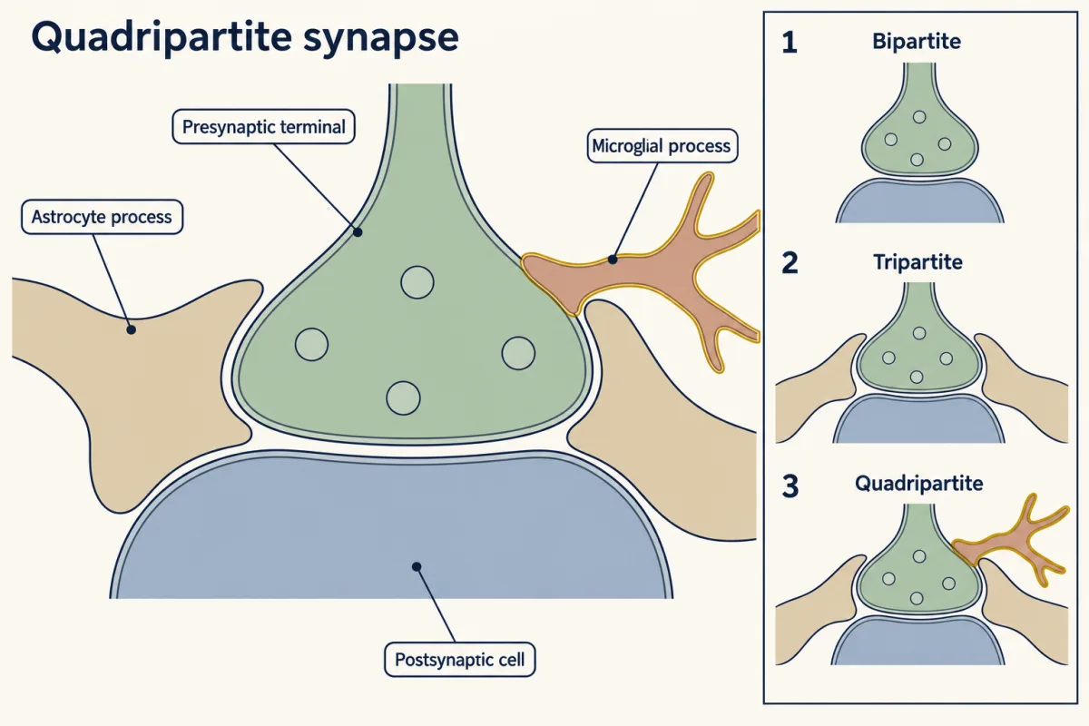
So a synapse is not only a meeting of two neurons. The classical picture is bipartite, a presynaptic terminal and a postsynaptic cell. Adding the astrocyte that enwraps it gives the tripartite synapse. Adding the microglial process that touches it and can remove it gives what can be called the “quadripartite” synapse.
Candidates for the mark
How relative activity becomes a molecular mark is the central open question. Candidate protective “don’t eat me” signals such as CD47 and its receptor SIRPα (shown to limit engulfment in the mouse dLGN), local regulators of complement, and activity-dependent exposure of phosphatidylserine have all been proposed. Which operate in a given circuit at a given age, and how far the dLGN rules generalise, remains to be shown.
Your notes
Select any text in the lesson to highlight it or add a note to it. Your highlights and notes are saved in this browser and collected in your notebook.
This browser is not letting the site save anything (a private window or strict privacy settings), so highlights and notes will be lost when you leave the page.
How we measure it
Eye-specific tracer injection
Two cholera toxin fragments carrying different fluorescent dyes are injected into the two eyes. Retinal axons carry them to the visual thalamus, where the overlap between the two colours measures how far eye-specific segregation has gone, and dye found inside microglia shows whose inputs they took.
Engulfment quantification by 3D reconstruction
Confocal image stacks of fluorescently labelled microglia are rebuilt in three dimensions, and labelled material outside the cell's volume is removed. The volume of internalized material is divided by the volume of the microglial cell, so a large cell does not appear to engulf more merely because it is large.
Knockout mice
In the whole-body knockout mice used here, both copies of one gene are disrupted in every cell. Comparing knockout and normal littermates for C1q, C3, complement receptor 3 or CX3CR1 asks whether that protein is needed for normal pruning, though not where or when it acts.
Whole-cell recording of relay neurons
In a brain slice, the optic tract is stimulated with increasing strength while the current in one thalamic relay neuron is recorded. Each new step in the response marks one more retinal axon recruited, which gives an estimate of how many inputs the cell still has.
Practice questions on this lesson (3) → MCAT-style, original, each with an explanation
Where this lesson fits
Before thisReview first
- Activity Before Experience: Spontaneous Retinal WavesThis course · Lesson 38
Retinal waves drive the activity differences between inputs that this lesson says microglia and complement act on.
- Ocular Dominance and the Critical PeriodThis course · Lesson 39
You need its idea that the more active eye's inputs win and the quieter eye's lose before asking what removes them.
- Too Many Neurons: Naturally Occurring Cell DeathThis course · Lesson 28
It sets up the idea that a nervous system makes too much and then removes some of it.
After thisStudy next
- Disorders of the Synapse: Built, Then KeptThis course · Lesson 49
It takes up the proposed links between altered synapse number and autism or schizophrenia that this lesson only introduces.
- Sensitive Periods in Behavior: Song, Language and SightThis course · Lesson 42
It uses the idea that experience-dependent removal of connections closes off circuit change as development goes on.
Elsewhere on the siteFilms, simulations and method films in other courses
- Cell-free extracts and reconstitutionMethod film
It shows how a cell process is rebuilt in a tube to test which parts are needed, as for the C1q, C3 and CR3 tagging steps.
- A Cell Keeps Its Answer ReadyFilm · Cell Signaling
It shows how innate immune cells recognize targets they have never met, the system whose tagging proteins neurons borrow here.
Suggested by an AI model from the lessons themselves, then checked. How we use AI
Finish line
Test yourself
8 questions
Practice and MCAT-style, with explained answers.
Next · Lesson 42
Sensitive Periods in Behavior: Song, Language and Sight
A young songbird that never hears an adult sing grows up singing a simple, abnormal song. Why can't it catch up later?
Continue →