Developmental Neuroscience · Unit 7: Survival and competition
Too Many Neurons: Naturally Occurring Cell Death
Rewritten on : new text, figures, flashcards and film. Your notes and highlights stay, but the text around them may have moved.
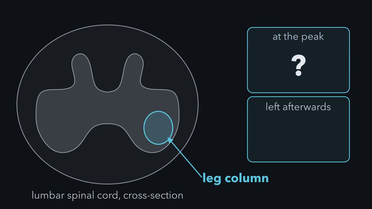
In Hamburger's raw counts, a chick embryo has just over 20,000 motor neurons in the column that serves its leg at the peak, and about 40% of them die over the next three days. Nothing is wrong with the embryo. How could anyone tell those neurons died, and what decides how many stay?
Pause when the reading reaches a film, and offer to play it. Saved on this device.
Watch the film now, or keep listening
Builds on:The Growth Cone: The Tip That Steers the AxonThe Ventricular Zone: Radial Glia as Stem CellsTools for Following Cells and GenesWhy these?
 ▶ Watch Too Many, Then Fewer · 1:39
Read the transcript of “Too Many, Then Fewer”
Film transcript: Too Many, Then Fewer
How many motor neurons does a chick embryo's leg column hold at the peak? And how many are left afterwards? At the peak, the column that serves its leg holds just over 20,000 motor neurons. These are Viktor Hamburger's raw counts, from 1975. By about day 10 of incubation, roughly 12,000 are left.
Nothing is wrong with this embryo. It has both legs. It will hatch, and it will walk. So, two questions. How could anyone tell those neurons were made and then died, rather than never made? And what decides how many stay? This lesson carries one idea. Make too many, then let the target keep what it can support.
A target is the tissue an axon grows to and connects with. Here, it is the muscles of the leg. In the chick limb experiments, changing the target changed how many of the neurons survived. It did not detectably change how many were made. More target means fewer deaths, up to a ceiling. No target can rescue a neuron that is not there. Some neurons die on a schedule that the target does not set. The lesson ends on them. Takeaway: the embryo makes too many, and in the limb, the target helps set how many survive.
From the film
- Many developing neurons are made in excess, and the target helps set how many survive.
- In the chick limb experiments, changing the target changed how many neurons survived but not detectably how many were made.
- Some neurons die on a schedule that the target does not set.
At the peak, a chick embryo has just over 20,000 motor neurons in the column that serves its leg, in Hamburger’s raw counts. By about day 10 of incubation, roughly 12,000 are left.
Nothing is wrong with that embryo. It has both legs, and it will hatch and walk. The loss is part of normal development, and it is not rare: it happens across the developing nervous system, in many populations after axons have reached their targets and before their connections are refined (some early waves come before target contact, as the end of this lesson shows). This lesson asks two questions about it. How could anyone tell that those neurons were made and then died, rather than never made? And what decides how many stay?
The one idea to carry through this lesson. Make too many, then let the target keep what it can support. In the chick limb experiments that follow, changing the target changed how many of the neurons already present stayed alive, with no detected change in how many were present before the loss.
Too small, or lost?
Read the transcript of “Count Across Time”
Film transcript: Count Across Time
A count comes out low. Were the missing neurons never made, or made and then lost? In 1934, Viktor Hamburger removed one wing bud from chick embryos about three days old. Days later, the operated side had fewer motor neurons than the intact side, its control. Hamburger read this as the limb setting how many neurons the cord makes. That reading fits the count. So do others. Think of a bakery whose shelf ends the day half empty. It might have baked fewer, sent some away, or baked some as rolls. Or it baked a full batch and threw half away. The shelf looks the same every time.
A low neuron count is that shelf. A count made only at the end cannot tell the histories apart. Pause, and predict. What would you count, and when, to tell never made from made and then lost? Count at closely spaced stages, on both sides. Count healthy neurons, and count dying ones. A dying neuron often has a small, dark, condensed nucleus. It is called pyknotic. Never made: the operated side never catches up, and no extra dying cells appear. But dying cells are cleared fast. So count often, and birthdate the cells, before trusting a missing hump. Made, then lost: the sides start equal. Then dying cells appear, and the count falls behind.
In wartime Italy, Rita Levi-Montalcini and Giuseppe Levi studied staged sections like these, in a laboratory at home. They found neurons that had already formed, and were now degenerating. The missing neurons had been made. In 1949, her joint paper with Hamburger, on the sensory neurons of chick spinal ganglia, supported that reading. The target's main job was not to call neurons into existence. It was to keep them alive. In 1975, Hamburger counted the death itself, in normal embryos with all their limbs. The leg's motor neurons peaked near day 6. Then about two in five of them died, most within three days.
In 1976, Margaret Hollyday and Hamburger grafted an extra leg beside the normal one. Predict. If more target meant more births, where would the grafted side's count be at day 6? At day 6, before the normal loss, the grafted side had no more motor neurons than the control. Later, in embryos whose grafts moved vigorously, it had more. About a tenth to over a quarter more.
More target did not mean more births. It meant fewer deaths. Takeaway: a count tells you a history only if you also counted along the way.
From the film
- A low count at the end cannot tell neurons that were never made from neurons that were made and then lost, so you must count across time.
- In Hamburger's raw counts, about two in five leg motor neurons died after the peak near day six.
- An extra leg did not add motor neurons before the normal loss; more target meant fewer deaths, not more births.
A few words first. A target is the tissue an axon grows to and connects with, here the muscles of a limb. A limb motor neuron has its cell body in the spinal cord and sends its axon out to a muscle. The motor neurons for one limb sit in a column along the side of the cord, the lateral motor column; the column for the leg lies in the lumbosacral segments, in the lower back.
In 1934 Viktor Hamburger removed one wing bud, the small swelling on the flank that becomes the wing, from chick embryos about three days old. He was building on earlier limb-removal work by Marian Shorey. The other side of the same spinal cord served as the control. Days later the operated side had fewer motor neurons. Hamburger read the deficit as the limb setting how many neurons the cord makes. In his model the limb sends a message back along the nerves that reach it, and the message acts on cell division or differentiation.
That reading fits the count. So do others. Think of a bakery that ends the day with half its usual loaves on the shelf. It might have baked half as many. It might have baked a full batch and sent some to another shop, or some might have come out as rolls and not been counted as bread. Or it might have baked a full batch and thrown half away. The shelf looks the same every time.
A low neuron count is that shelf. Too few made, too few arriving where you count, too few becoming the type you count, or made and then lost: a count made only at the end cannot tell them apart.
Predict. You can fix embryos at any stage after the operation. What would you count, and when, to tell “never made” from “made and then lost”? Sketch both histories as the operated side’s count against the intact side’s, over time.
Check your prediction
Operate on a large batch of embryos at the same stage, then fix some of them at closely spaced stages afterwards, every half day through the period when motor neurons form and connect. In each embryo, count two things on both sides, in the same spinal segments: healthy neurons and dying ones. A dying neuron can often be spotted in a section. Its chromatin collapses into a small, dark, condensed nucleus, a pyknotic nucleus.
- Never made: the operated side never catches up with the intact side, and no extra dying cells appear.
- Made, then lost: the two sides start equal. Then dying cells appear on the operated side, and its count falls behind.
Two cautions. Dying cells are cleared fast, so a missing burst of them means something only if you sampled often enough to catch it. And a pause in production can look like a loss. To guard against it, mark the cells as they are born; a neuron’s birth date is the time of the last division of the precursor that made it. Give a pulse of tritiated thymidine at a known stage, so that cells which stop dividing soon after stay heavily labelled (birthdating, under Techniques). Then confirm that labelled motor neurons were made in normal numbers on both sides.
In wartime Italy, Rita Levi-Montalcini and Giuseppe Levi carried out a staged histological version of that design. Italy’s racial laws of 1938 had barred Levi-Montalcini, who was Jewish, from her university post in Turin, so she built a small laboratory at home, first in Turin and then in the Piedmont countryside, and worked there with Levi, her former teacher. They removed limb buds from chick embryos and studied sections at a series of stages afterwards, using a silver stain that makes neurons and their fibres stand out; birthdating came later. In the parts of the spinal cord and ganglia that had lost their limb, they found neurons that had already formed and differentiated, and were now degenerating. The missing neurons had been made. They published the series in 1942, 1943 and 1944.
That contradicted Hamburger’s reading. He read those papers and invited her to St. Louis to settle it. She arrived in 1947. Their 1949 paper turned to sensory neurons. It followed chick spinal ganglia, the clusters of sensory neurons beside the cord, through division, differentiation and degeneration, in normal embryos and after limb removal or grafting. Many neurons formed and sent out fibres before the deficit appeared; dying cells, and their removal by macrophages, cells that engulf debris, became conspicuous later. Later studies of cell division and birthdating after limb-bud removal agreed. The target’s main job was not to call neurons into existence. It was to keep them alive.
A count tells you a history only if you also counted along the way.
Counting the death itself
In 1975 Hamburger came back to the question in normal embryos with all their limbs, and counted the death itself. In the lumbar lateral motor column, the count peaked at just over 20,000 at days 5.5 to 6.5 of incubation. It settled at a plateau of about 12,000 after day 9.5. Most of the loss came between days 6.5 and 9.5.
That is a loss of about 8,000 neurons, or about 40% of the peak. Divide by the peak, the number that was there to lose. Dividing the same 8,000 by the plateau gives about 67%, which answers a different question.
These are raw counts: a nucleus cut across two neighbouring sections can be counted twice. Hamburger also gave corrected figures, a peak of over 17,000 and a plateau of about 10,300, and the share lost is still about 40%. So treat the absolute numbers as approximate; the fraction lost is the robust result.
If removing target raised the death, would adding target lower it?
More target
In 1976 Margaret Hollyday and Hamburger grafted an extra leg onto chick embryos, beside the normal one. That gave the motor neurons on that side an extra limb to innervate, though only some segments reached it. How much more support it gave them is unknown: the grafts were innervated by varying combinations of nerves from the thoracic (chest-level) and upper lumbar cord.
Predict. Compare the grafted side’s motor-neuron count with the control side’s at day 6, before the normal loss, and again later, after it. What would you see if more target meant more births? What would you see if it meant fewer deaths?
Check your prediction
- More births, other processes unchanged: the grafted side is already higher at day 6.
- Fewer deaths, equal cohorts at the start: the two sides are equal at day 6, and the grafted side pulls ahead only during the death period.
The result was the second. At day 6, before the normal loss, the grafted side had no more motor neurons than the control. Later, in embryos whose grafts moved vigorously, it had more: about 11% to 28% more in their counts, roughly a tenth to over a quarter.
So the extra leg did not detectably add neurons by day 6; the later difference fits fewer deaths among the neurons already present. The graft went in around the time these neurons were being born, so the experiment says little about whether a target can influence production earlier.
A survival effect predicts no difference before the death period and a difference after it. Other histories, such as delayed differentiation or later loss of a marker, could mimic that pattern, so fewer dying cells and tracing would argue further.
The target, then, supplies something limited that the neurons innervating it need to stay alive. Hollyday and Hamburger left open whether that something was synaptic space or a trophic agent, a substance that sustains neurons, which is why this lesson says “support”. The search for a trophic agent had already produced nerve growth factor, a story told in that lesson. The tests of whether NGF, a target-derived survival signal, is the limiting factor, for sensory and sympathetic neurons rather than motor neurons, are in the next lesson.
A dial with a ceiling
Read the transcript of “A Dial With a Ceiling”
Film transcript: A Dial With a Ceiling
The extra leg enlarged the target. Suppose it doubled the support. Could the survivors double too? Twice 12,000 is 24,000. But only about 20,000 were present at the peak. If no new neurons join afterwards, no target can rescue more than that. That is the ceiling. And the real rise was well below it. In embryos with vigorously moving grafts, survivors rose by a tenth to over a quarter.
So the target turns a dial. More target, fewer deaths, up to the ceiling. Why did it turn so little? The experiment cannot tell. The graft may be only partly innervated. Activity at the muscle also matters. In chick embryos, paralysing the muscle with curare rescues many motor neurons that would otherwise die. Is every developmental death the target's doing? One test adds cells instead of target. Derek Southwell and colleagues transplanted precursors of cortical interneurons into host mice, across a 200-fold range of doses. Predict. Take a toy model, where the host can support only 1,000 transplanted cells. What fraction dies as the dose rises?
In the toy model, none die until the dose passes 1,000. Then the fraction climbs, to 95%. percent. The real result was different. About the same fraction died at every dose tested. the cells died at their own age, not the host's. They carried their own clock. Later work showed that, during this window, circuit activity can tune how many survive.
The chick has another case, in the neck-level, or cervical, cord. Around day 4 to 5, a wave of cervical motor neurons dies, before the later death that follows target contact. A bigger target did not change this early wave. Nor did paralysis, or the trophic factors tested. Drugs that block RNA or protein synthesis reduced it. So it needs new RNA and protein, an active program. Moved to another level of the cord, the cervical tube still lost its neurons on schedule.
So, make too many, then let the target keep what it can support. That holds for many peripheral neurons, and for motor neurons at their muscles. Takeaway: in the limb, the target helps set survival, like a dial with a ceiling. Count across time, and you can see it.
From the film
- The target turns a dial: more target, fewer deaths, up to a ceiling set by the neurons present.
- In mice, about the same fraction of transplanted interneuron precursors died at every dose tested, and they died at their own age.
- An early wave of chick cervical motor neuron death is not controlled by the target and needs new RNA and protein.
Predict. Suppose the extra leg had doubled the support available to the column. If the number of survivors simply copied the support, how many motor neurons would survive? Could that many survive?
Check your prediction
A copy would double the survivors, from about 12,000 to about 24,000. That cannot happen. Only about 20,000 were present at the peak. If no neurons join the column afterwards, and the graft left the day-6 count unchanged, rescuing every one still cannot give 24,000; it would raise survivors by about two-thirds at most.
The table sets that ceiling beside what was seen. The last row applies the 1976 percentages to the 1975 counts, which come from different experiments, so treat it as an illustration of scale, not a measurement.
| Case | Survivors | Change from normal |
|---|---|---|
| Normal embryo | about 12,000 | — |
| Survivors copy a doubled support (hypothetical) | about 24,000 | +100%, more than were present |
| Ceiling: every neuron present at the peak survives | about 20,000 | about +67% |
| Extra leg, illustrative scale | about 13,300 to 15,400 | +11% to +28% |
So the target turns a dial: more target, fewer deaths, up to a ceiling set by the neurons present. Why the dial turned so little here, the experiment cannot tell. It shows a partial response, not the shape of the response in general. The graft may be only partly innervated, or survival may also depend on activity at the muscle, as the curare experiments below show.
How a developing neuron dies
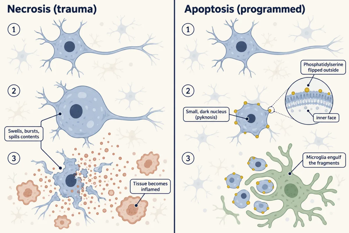
The dying neurons are not injured. Necrosis is typically death from injury: the cell swells, bursts and spills its contents, and the tissue becomes inflamed. Most developing neurons die by apoptosis instead, a programmed death run by the cell’s own machinery of Bcl-2 family proteins and caspases (see that lesson).
The chromatin condenses into the small, dark pyknotic nucleus, and the cell shrinks and breaks into fragments. It also flips phosphatidylserine, a lipid normally kept on the inner face of the membrane, to the outside. Phagocytes read that lipid as an “eat me” signal and engulf the corpse: microglia, the resident phagocytes, in the brain and spinal cord, and macrophages and other phagocytes elsewhere, as in the spinal ganglia of the 1949 paper. The clean-up is fast, which is one reason the death went unnoticed for so long: at any one moment only a few dying cells are visible. How the neuron’s death machinery is triggered is the subject of a later lesson.
How much, and where
In the populations that have been counted, between about a fifth and four-fifths of the neurons die, depending on the population. Each number belongs to a system. In the chick lumbar lateral motor column it is about 40% of the peak. In the rat retina, about half of the ganglion cells, the neurons whose axons form the optic nerve, are eliminated. Frog limb motor neurons are also lost in large numbers in the tadpole, and in mouse cortical interneurons about 40% die after birth.
You will often hear that “half of all neurons die.” That rounds a range into a rule. The “roughly half” comes from Ronald Oppenheim’s 1991 review in the Annual Review of Neuroscience, which gathered direct counts in many vertebrate populations, each made inside a defined developmental window. Within those windows, around half was a common value, but some populations lost far less and some far more. The review concerned development, not adult loss.
Why make neurons just to kill them?
Predict. Give two jobs that overproduction followed by death could do for a developing nervous system, then read on.
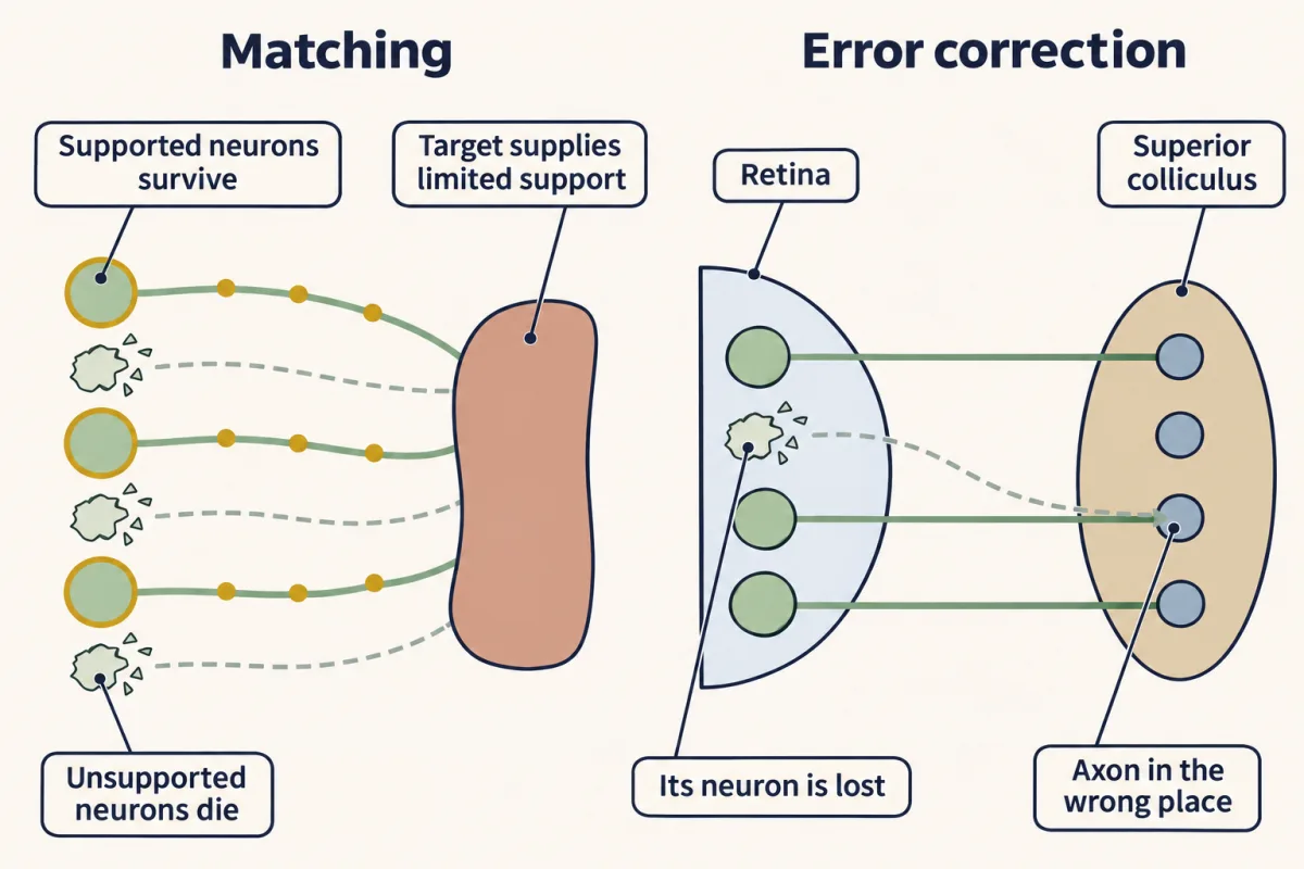
Matching. The number of neurons a target needs is hard to specify in advance. Limbs, muscles and organs vary in size between individuals and change as the embryo grows. Overproduction followed by target-limited survival, the bakery’s strategy, settles the number without a plan; the extra limb showed the dial turning.
Activity at the muscle also matters. In chick embryos, paralysing the muscle with curare, which blocks transmission from nerve to muscle, rescues many motor neurons that would otherwise die, as Oppenheim’s experiments with R. H. Pittman showed. That shows survival depends on what happens at the muscle, not that death picks out wrong connections.
Error correction. Axons sometimes grow to the wrong place or make weak connections. If survival depends on support received at the right target, neurons that went wrong can lose out. The clearest case is in the newborn rat: retinal ganglion cells whose axons reach the wrong part of the superior colliculus, the midbrain target where retinal axons form a map, are removed preferentially during the normal period of ganglion-cell death. Death is not the only fix, though. Many wiring errors are corrected by pruning axon branches or synapses, and a neuron can sometimes survive on a wrong connection. How the competition plays out is the subject of a later lesson.
Where the target account stops
If the target set all death, every wave of it would answer to the target. Two studies show that some does not.
The first is an early wave of motor-neuron death in the chick cervical, or neck-level, cord, described in 1996 by Hiroyuki Yaginuma, Ronald Oppenheim and colleagues. The wave comes around days 4 to 5 of incubation, before the later death that follows target contact. Enlarging the peripheral target, paralysing the embryo with curare and supplying muscle extracts or a long list of trophic factors all failed to change the early wave, though they helped the later one. Drugs that block RNA or protein synthesis reduced it, so it needs new RNA and protein, an active program. Cervical neural tube transplanted to other segmental levels kept its early death, so the schedule was carried within that tube. Whether it was carried by each doomed neuron or by its neighbours, the grafts could not say.
The second study turned the extra-leg logic around. Instead of adding target, it added cells. In 2012 Derek Southwell, Arturo Alvarez-Buylla and colleagues transplanted precursors of cortical interneurons, the local inhibitory neurons of the cortex, into the cortex of host mice, across a 200-fold range in the number transplanted.
Predict. Take a toy model. Suppose, hypothetically, that the host could support exactly 1,000 transplanted cells, that any cell within that limit survives, and that nothing else kills them. What fraction of the transplanted cells would die at each of these illustrative doses?
| Cells transplanted (illustrative) | 100 | 1,000 | 5,000 | 20,000 |
|---|---|---|---|---|
| Fraction dying, if a fixed host supply sets survival | ? | ? | ? | ? |
Check your prediction
| Cells transplanted (illustrative) | 100 | 1,000 | 5,000 | 20,000 |
|---|---|---|---|---|
| Fraction dying, if a fixed host supply sets survival | 0% | 0% | 80% | 95% |
Survivors are the smaller of the dose and 1,000. Nothing dies until the dose passes the limit; then the dying fraction climbs steeply.
The real result did not look like that. About the same fraction died at every dose tested. And, as a separate observation, the transplanted cells died when they reached the age at which the host’s own interneurons die, not when the host reached that age.
The constant fraction argued against a fixed host supply; the timing showed that the transplanted cells carried their own clock. These are separate results: timing by itself does not tell you what fraction dies. The study could not tell whether the clock was set in each cell or through competition among the interneurons.
Later work added a second layer. In 2018 Fong Kuan Wong, Oscar Marín and colleagues showed that the activity of pyramidal cells, the excitatory neurons of the cortex, during a postnatal window regulates how many interneurons survive, acting through phosphatase and tensin homolog (PTEN), an enzyme that brakes a survival-signalling pathway inside the interneurons. The interneurons carry their own clock, but the local circuit helps set how many survive.
So target-dependent survival explains much developmental neuron death, especially in peripheral populations and in motor neurons after they reach their muscles. It is one source of control among several, and some populations die on their own clocks.
Putting it together
Each history of a low count predicts a different set of observations. Counting across time, tracing the cells, and changing the target or the number of cells help narrow down which one happened.
| History | Count before the death period | Dying (pyknotic) cells | Count at the end | What the further tests show |
|---|---|---|---|---|
| Fewer made | low | not raised | low | birthdating shows fewer born; if the target set production, more target would raise the count before the death period (not seen with the extra leg) |
| Made, then moved or became another type | normal in a lineage-labelled count | not raised | low where you count | a permanent lineage label finds the cells elsewhere, or carrying another marker |
| Made, then lost; target sets survival | normal | raised during the window | low | more target: equal before the window, fewer deaths during it, up to the number present |
| Made, then lost on the cells’ own clock | normal | raised at a time set by the cells’ age | low | timing follows the cells’ age, not the target or host; test the dying fraction separately, since activity can still tune it |
The chick lumbar motor neurons fit the third row. The early cervical wave and the cortical interneurons fit the fourth: the cervical wave ignored the tested target changes and kept its schedule in transplanted neural tube, and the transplanted interneurons died at the donor cells’ age, not the host’s.
Design problem
Suppose a population in the mouse hindbrain loses 50% of its neurons in the first postnatal week. Design experiments that tell whether its target sets the number or the cells die on their own schedule.
Saved in this browser only.
One way to do it
Walk the table’s rows.
- Confirm the loss is death. Count healthy and pyknotic neurons at closely spaced ages through the week, and birthdate the neurons so that a pause in production cannot pass for a loss. Normal numbers early, then a burst of dying cells and a falling count, means made and then lost. Mark the cells with a permanent lineage label, so that neurons that moved or changed type are not mistaken for deaths.
- Change the target. Reduce or enlarge the target, and count across the same ages. If the target sets survival, the counts match before the death period and diverge during it: more death with less target, fewer deaths with more, up to the number present. If neither the timing nor the number follows the target manipulations you tried, those manipulations are not setting them.
- Change the number of cells, and their age. Transplant the precursors into hosts at several doses, from donors younger or older than the host. A fixed host supply makes the dying fraction climb once the dose passes what the host can support. Death timed by the donor cells’ age, not the host’s, points to a schedule carried by the cells. Read the timing and the fraction separately.
- Keep the two layers apart. A cell-carried schedule does not rule out tuning by the surrounding circuit, as in the cortical interneurons. Test activity separately.
Going deeper
Error correction in the rat retina
In the newborn rat, blocking retinal ganglion-cell impulse activity with tetrodotoxin, a sodium-channel blocker, stops the preferential loss of cells with mistargeted axons, though about the same total number still die.
Death without the usual caspases
Apoptosis is not the only route. In mice lacking caspase-3 or caspase-9, about the same number of developing neurons still die, more slowly and without the apoptotic look. In some genetic backgrounds, caspase-3 mutant mice have relatively normal brain morphology.
Your notes
Select any text in the lesson to highlight it or add a note to it. Your highlights and notes are saved in this browser and collected in your notebook.
This browser is not letting the site save anything (a private window or strict privacy settings), so highlights and notes will be lost when you leave the page.
How we measure it
Unilateral limb-bud extirpation
You remove a wing or leg bud from one side of a young chick embryo and let it develop. The intact side is the control in the same animal, so a difference between the two sides of the spinal cord measures what the missing target did.
Serial-stage cell counts with pyknotic profiles
You section embryos fixed at closely spaced stages, count healthy neurons in a defined region, correct for section thickness, and count pyknotic nuclei (condensed, dark chromatin) as dying cells. A falling count with a burst of pyknosis means cells were made and lost; a count that is low from the start, with no burst of pyknosis, points to fewer made, provided sampling began early enough to catch any loss.
Normal staging of the chick embryo
Hamburger and Howard Hamilton's 1951 series of morphological stages lets embryos be compared by form rather than by hours of incubation. Without it, counts from embryos of the same age but different rates of growth could not be lined up.
Tritiated-thymidine birthdating
A pulse of radioactive thymidine is built into the DNA of cells that are copying their DNA to divide at that moment, and cells that stop dividing soon after stay heavily labelled. It shows whether neurons are still being made while others die, so a falling count can be read correctly.
Practice questions on this lesson (3) → MCAT-style, original, each with an explanation
Where this lesson fits
Before thisReview first
- The Growth Cone: The Tip That Steers the AxonThis course · Lesson 3
Axons reaching their targets, whose growth you follow there, is the starting point for the later loss of neurons.
- The Ventricular Zone: Radial Glia as Stem CellsThis course · Lesson 13
You need to know how neurons are first produced to see why a count at the peak is the supply that survivors cannot exceed.
- Tools for Following Cells and GenesThis course · Lesson 4
Birthdating and lineage labels from there are how you tell neurons never made from neurons made and lost.
After thisStudy next
- The Neurotrophic Hypothesis TestedThis course · Lesson 29
It tests whether a target-derived protein, NGF, is the limiting support that decides which neurons survive.
- The Death Machinery in NeuronsThis course · Lesson 31
It explains how the apoptotic program of Bcl-2 proteins and caspases is triggered in these neurons.
- Competition: How Neurons Win a TargetThis course · Lesson 32
It shows which neurons win when the target's limited support cannot keep all of them alive.
Elsewhere on the siteFilms, simulations and method films in other courses
- Sealed or TornFilm · Cell Biology
It shows apoptosis taking a cell apart with the plasma membrane intact, the death mode behind most of this loss.
- Cell death assaysMethod film
It shows how apoptotic cells are told from necrotic and live ones, which is how dying neurons are identified in counts.
- Reading annexin V and PISimulation · Cell Biology
It lets you practise reading annexin V and PI to separate dying from dead cells.
Suggested by an AI model from the lessons themselves, then checked. How we use AI
Finish line
Test yourself
9 questions
Practice and MCAT-style, with explained answers.
Next · Lesson 29
The Neurotrophic Hypothesis Tested
Neutralize one protein in a newborn mouse and its sympathetic ganglia waste away. Neurons compete for a limiting amount of target-derived factor, and the amount limits how many live. Does NGF decide how many neurons there are?
Continue →← Lesson 27 · Choosing a Partner: Layers, Self-Avoidance and Smell