Cell Biology · Skim version
Cell Death and Clearance
Read the full lesson → Skim the whole course →
The guiding idea
In a developing worm, the same 131 cells die on schedule, yet in healthy tissue you rarely see a corpse. How does a cell die without leaving a mess?
Death is a normal, genetically controlled part of building a body: in a worm, the same 131 cells die on schedule, yet in healthy tissue dead cells are cleared so fast that corpses are rarely seen.
From the film: Sealed or Torn
- In apoptosis, the cell's own caspases take it apart while its plasma membrane is still intact, at least at first.
- While still sealed, an apoptotic cell can send find-me signals and expose phosphatidylserine, an eat-me flag, on its outer face.
- In healthy tissue, a phagocyte usually eats the cell before it leaks, so nothing spills.
- In a dish with no phagocyte, an apoptotic cell often leaks later: secondary necrosis.
- In necrotic deaths the membrane generally opens early, and spilled alarm signals can cause inflammation.
From the film: A Look Is Not a Cause
- Some deaths that tear the membrane are genetic programs, not accidents.
- In the mammalian model, necroptosis needs RIPK3 and MLKL; pyroptosis needs pore-forming gasdermins; ferroptosis is driven by iron-dependent lipid damage.
- A death's look names an endpoint, not a cause; to find the cause, block the machinery and see whether the death changes.
- Annexin V reads phosphatidylserine it can reach from outside, and propidium iodide reads a leaky membrane.
- Mouse macrophages lacking NINJ1 died but released little LDH, so LDH reports the final tear, not the death.
-
The worm shows the cell kills itself
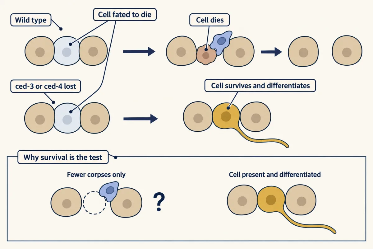
In worms lacking ced-3 or ced-4, cells normally fated to die survived, and some differentiated. A missing corpse could mean fast clearance; a living, differentiated cell shows the death did not happen. AI-generated illustration -
Clearance: separating each step from the death
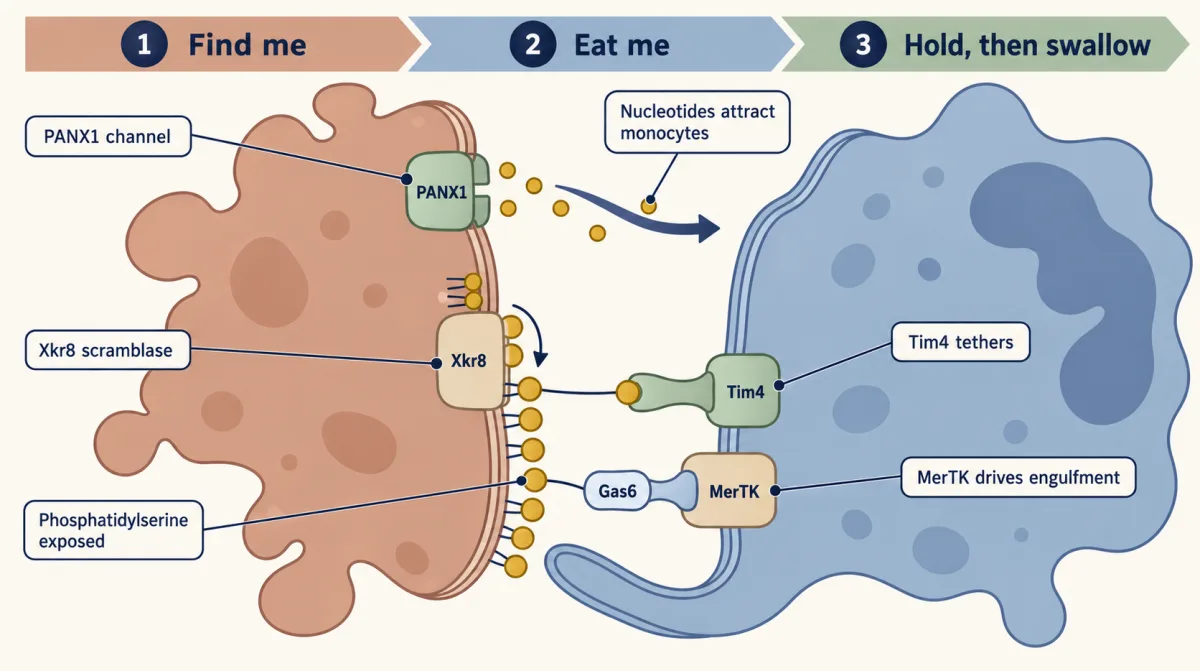
Clearance in steps: nucleotides released through PANX1 call a phagocyte, Xkr8 exposes phosphatidylserine, and Tim4 tethers while MerTK drives engulfment (shown in resident mouse peritoneal macrophages). AI-generated illustration -
Necrotic deaths can be programs too
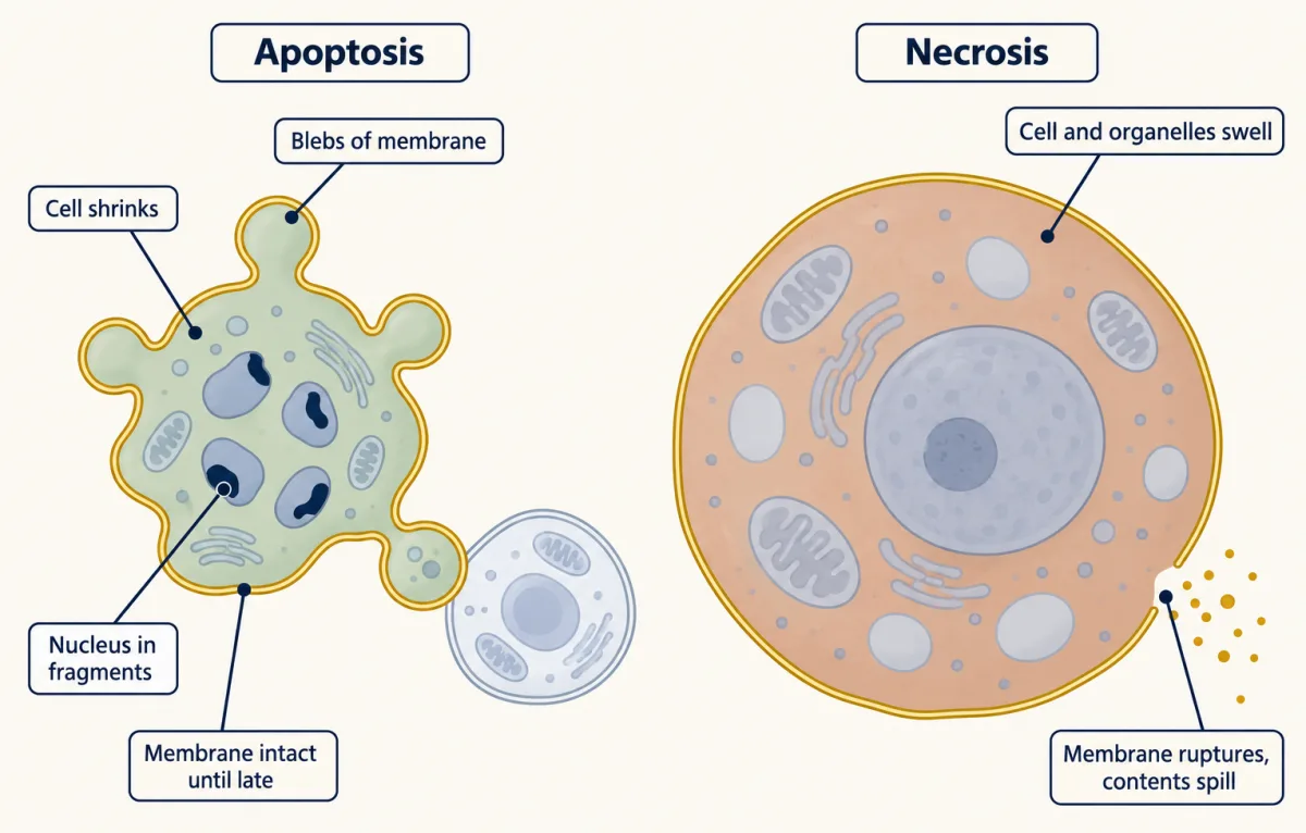
Apoptosis before the membrane fails (left) and necrosis (right) as they look under the microscope. These looks name an endpoint, not a cause: a cell can die with necrotic looks through dedicated machinery. AI-generated illustration -
Reading the flag and the leak with two dyes
-
Going deeper
Where this lesson fits
Before thisReview first
- Membrane Organization and TopologyThis course · Lesson 4
You need the two-leaflet membrane and its lipid asymmetry from there to follow how phosphatidylserine moves to the outer face.
- Vesicle Budding, Targeting and FusionThis course · Lesson 11
Engulfment and phagosome fusion with lysosomes build on how membranes bud, target and fuse.
After thisStudy next
- Stem Cells and Tissue RenewalThis course · Lesson 24
Tissue renewal balances cell loss against replacement, so the steady-state clearance idea applies directly.
- Cell Identity, Cell State and DiseaseThis course · Lesson 25
Disease states often involve altered death and survival, using the programmed-death machinery learned here.
- Designing a Cell Biology ExperimentThis course · Lesson 26
Combining ambiguous markers over time and then perturbing is the design reasoning practised there.
Suggested by an AI model from the lessons themselves, then checked. How we use AI