Cell Signaling · Unit 6: Channels, the nucleus, and life or death
Stress Signals: p38 and JNK, p53, Unfolded Proteins and Oxidants
Your cells build p53 all day and destroy it almost as fast. Why make a guardian only to shred it?
Pause when the reading reaches a film, and offer to play it. Saved on this device.
Watch the film now, or keep listening
Builds on:Ubiquitin and Regulated Proteolysis as SignalsRas to MAPK: The Kinase CascadeCircuits: Amplification, Feedback, Adaptation and SwitchesWhy these?
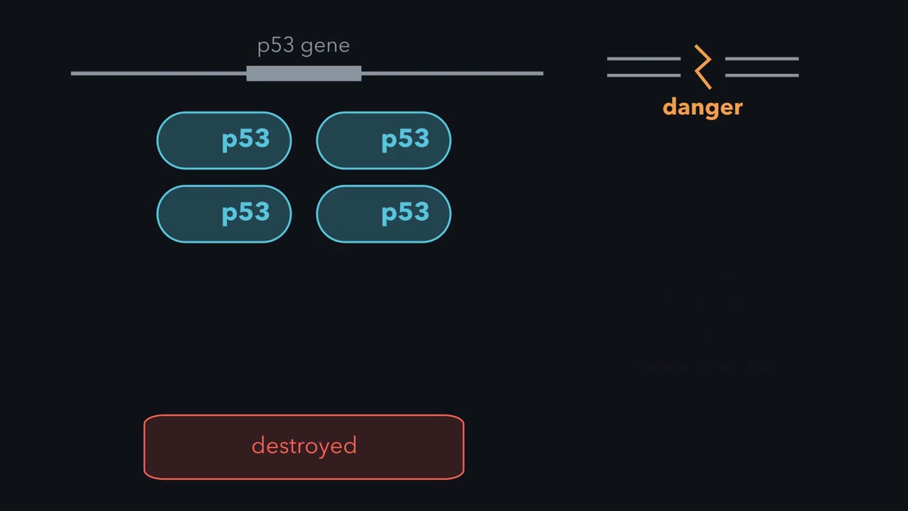 ▶ Watch Made All Day, Held Back Until Needed · 2:39
Read the transcript of “Made All Day, Held Back Until Needed”
Film transcript: Made All Day, Held Back Until Needed
Your cells make a protein called p53 all day long. Half of any new batch is destroyed within about 20 minutes. So why build a guardian, only to destroy it? Because a cell in danger cannot wait for a gene to be read and a protein to be built. If the guardian is already there, and something holds it back, the cell only has to let go. p53 is a transcription factor, a protein that binds DNA and switches genes on. Its restraint is a protein called MDM2. MDM2 grips a short piece at the front end of p53. That grip covers the part that switches genes on. It also tags p53 with ubiquitin, a mark that sends it to the proteasome to be destroyed. Now DNA breaks across both strands.
That switches on a kinase called ATM. ATM and the kinases beneath it add phosphates to MDM2. They also add phosphates to the stretch of p53 that MDM2 grips. The grip loosens, and destruction slows sharply. So p53 piles up without any new gene being read. Free of its restraint, it switches on the gene for p21. That protein blocks the kinases that drive cell division. It switches on genes for DNA repair as well. If the damage is severe, it also switches on genes that kill the cell. Chemical threats use the same design, with a responder called Nrf2. Nrf2 switches on genes for protective enzymes, and the cell makes it constantly. Keap1 grips it as part of a ubiquitin ligase that tags it for destruction.
Then a reactive chemical hits Keap1. On the current view, tagging stops, so new Nrf2 goes to the nucleus. Put the two side by side. In each, a responder is made all the time. Something holds it back, or destroys it. Stress removes the restraint, and the responder is already there. So keep this rule: a stress responder is made all the time and held back, until stress removes the restraint.
Your cells make a protein called p53 all day long, and half of any newly made batch is destroyed within about twenty minutes. Why build a guardian only to shred it? Because a cell in danger cannot wait for a gene to be read and a protein to be built. If the responder already exists and something is holding it back, the cell only has to let go.
The idea: the responder is made all the time and held back
You have met this design before: the α subunit of HIF is made constantly and destroyed while oxygen is present (the lesson on ubiquitin), β-catenin is destroyed until Wnt arrives, and NF-κB waits in the cytoplasm until IκB is destroyed. Think of a car at a red light with the engine running and a foot on the brake. Lifting the foot is faster than starting the engine.
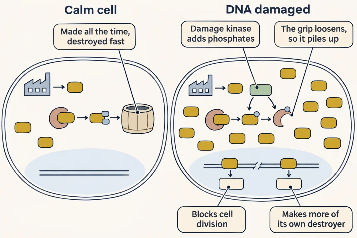
p53 is a transcription factor, a protein that binds DNA and switches genes on. Its restraint is MDM2, an E3 ubiquitin ligase. MDM2 grips a short helix at the front end of p53. The grip covers the part of p53 that activates genes, and MDM2 tags p53 with ubiquitin, which sends it to the proteasome.
One of the genes p53 switches on is the gene for MDM2. p53 orders its own destroyer: negative feedback, as in the lesson on signaling circuits.
DNA damage breaks the pair apart. A break across both strands switches on a kinase called ATM. ATM and the kinases beneath it add phosphates to the stretch of p53 that MDM2 grips, and to MDM2 itself. The grip loosens and destruction slows sharply, so p53 piles up without any new gene being read. It then switches on the gene for p21, a protein that blocks the kinases driving cell division, along with genes for DNA repair. If the damage is severe it also switches on genes that kill the cell, the subject of the next lesson.
A cell without working p53 can keep dividing with broken chromosomes, which is why TP53, the gene for p53, is the most frequently mutated gene in human cancer. Most of those mutations fall in the domain that touches DNA. Some tumors, sarcomas among them, keep p53 intact and carry extra copies of the MDM2 gene instead, so the brake stays pressed down hard.
Pulses, not a dial: the timing of a signal carries information
Grind up millions of irradiated cells, measure p53 on a blot, and you see it rise and then fade, at most with a damped wave or two. The natural reading is a dial: more damage, more p53.
Galit Lahav tested that reading by watching one cell at a time. Working with Uri Alon at the Weizmann Institute, she built human cells in which p53 carried a cyan fluorescent tag and MDM2 a yellow one, irradiated them, and filmed them for hours. In single cells p53 rose and fell in separate pulses. The average height and width of a pulse did not depend on the dose. What rose with the dose was the average number of pulses, and genetically identical cells in one dish gave none, one, two or more.
Why had blots hidden this? The cells pulse out of step, so the peaks of some fill the troughs of others. Average a thousand strobe lights flashing at different moments and you record a steady glow.
The pattern depends on the stress. Pulses of fixed size belong to gamma rays, which break both DNA strands. After ultraviolet light, p53 rises in a single pulse whose height and length grow with the dose. And in cancer cells given chemotherapy, whether a cell dies depends on how high p53 climbs and how soon, so the level of p53 carries information too.
Does the shape of the signal matter? In 2012 Lahav’s laboratory used timed doses of nutlin-3, a small molecule that blocks MDM2, to hold p53 steady at about the height of one natural pulse. In the cell line they studied, irradiated cells left to pulse tended to recover and divide again. Cells held steady more often entered senescence, a permanent exit from division. The protein and its peak level were the same. Holding p53 up also meant more p53 in total, but when the group compared cells at equal total p53, sustained p53 still switched on senescence genes more strongly. The shape of the signal over time mattered.
Design problem
After a dose of radiation, the amount of p53 in a single cell rises and falls in pulses of about the same size. More damage gives more pulses, not bigger ones. Design a circuit of two proteins that gives pulses and not a steady rise. Then say how a cell could use the number of pulses to choose between recovering and leaving the cell cycle for good.
Saved in this browser only.
One way to do it
Use an activator A and a destroyer D. A switches on the gene for D, and D marks A for destruction. That is negative feedback, and enough delay, with a steep enough response, turns it from a thermostat into a pulse generator. Making D takes time, so A keeps climbing and overshoots. When D arrives in bulk it knocks A down. With A gone no new D is made, the old D decays, and A can climb again.
To let damage set the number of pulses, make the damage signal a gate: while breaks remain, a damage kinase keeps loosening D’s grip so each cycle can start. The loop sets the height of a pulse and the damage sets how many there are.
To count, give A two kinds of target gene. Arrest and repair genes make short-lived products that rise and fall with A. A gene for permanent arrest makes a long-lived product, a little with each pulse. A few pulses leave it below a threshold. Many pulses, which mean the damage is still there, push it over.
The stress kinases: the three-tier plan again
In the lesson on Ras and mitogen-activated protein kinase (MAPK) you followed three kinases in a row: Raf, MAPK/ERK kinase (MEK), extracellular signal-regulated kinase (ERK). Stress pathways have co-opted this plan, ending in p38 and c-Jun N-terminal kinase (JNK). Above p38 sit the MAP2Ks MKK3 and MKK6, above JNK sit MKK4 and MKK7, and above those sit many MAP3Ks, each wired to a different kind of trouble. In the standard route, p38 and JNK, like ERK, are fully switched on only when a MAP2K adds two phosphates, to a threonine and a tyrosine one residue apart.
What starts them? Ultraviolet light, heat, a sudden change in salt concentration, oxidants, bacterial products, and the inflammatory signals tumor necrosis factor (TNF) and interleukin-1. One MAP3K, ASK1, repeats this lesson’s design: it is held off by the small protein thioredoxin until an oxidant makes thioredoxin let go.
p38 was reached from several directions in 1994. At Scripps, Jiahuai Han in Richard Ulevitch’s laboratory cloned a protein that gained phosphate on tyrosine when macrophages met lipopolysaccharide, a piece of bacterial outer wall. It also gained phosphate when cells were put in a solution with too much solute, and its sequence resembled HOG1, the kinase yeast use to survive high salt. At SmithKline Beecham, John C. Lee’s group used labeled versions of their own anti-inflammatory compounds as bait and recovered the same kinase. Two more groups reported it that year as a kinase switched on by stress and by interleukin-1.
Remember two jobs of p38. In immune cells it raises the output of TNF and interleukin-1, partly through a kinase beneath it called MK2. MK2 also phosphorylates Hsp27, a small chaperone that protects a stressed cell’s proteins.
JNK is named for its substrate: c-Jun N-terminal kinase. It was found in the early 1990s as a kinase that ultraviolet light and other stresses switch on. It adds phosphates to serines 63 and 73 of c-Jun, the AP-1 partner from the previous lesson, making c-Jun a stronger activator of genes. When stress persists, JNK also pushes a cell toward death, a link the next lesson takes up. In neurons a MAP3K called DLK feeds JNK, and in 2011 Ghosh and colleagues reported that cultured sensory neurons from mouse embryos lacking DLK resist both the axon degeneration and the cell death that follow loss of nerve growth factor.
A sensor that works by being attacked: Keap1 and Nrf2
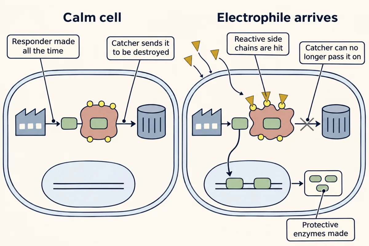
Some threats are chemicals: oxidants, and electrophiles, molecules short of electrons that react with DNA and protein. The responder is Nrf2, a transcription factor that switches on genes for protective enzymes, including those that join an electrophile to glutathione so it can be excreted. Nrf2 is made constantly. A protein called Keap1 catches it. Keap1 is the part of a ubiquitin ligase that grips Nrf2, so the ligase tags Nrf2 for destruction, and in a calm cell Nrf2 lasts well under an hour.
How does Keap1 learn that an electrophile has arrived? It gets hit by one. A cysteine side chain ends in a sulfur atom that gives up electrons easily, and mouse Keap1 has 25 cysteines among its 624 amino acids, a few of them unusually reactive. An electrophile such as sulforaphane, a compound from broccoli, reacts with them. The current view is that the altered Keap1 can no longer pass Nrf2 on for destruction, so newly made Nrf2 goes to the nucleus instead.
Unlike a receptor that fits its ligand in a shaped pocket, Keap1 reads a chemical property, hunger for electrons, so one protein can report on a large family of unrelated compounds. Which cysteine does the sensing depends on the compound. In mice whose Keap1 has serine in place of cysteine 151, sulforaphane does much less to Nrf2, while some other inducers work as before.
Paul Talalay’s laboratory in Baltimore isolated sulforaphane in 1992, and Ken Itoh and Masayuki Yamamoto in Tsukuba reported Keap1 in 1999. Together the groups then showed that inducers react with Keap1’s cysteines. One medicine, dimethyl fumarate for relapsing multiple sclerosis, is an electrophile that turns on Nrf2, though how much of its benefit comes that way is debated.
A sensor inside the folding compartment: repair first, then death
Read the transcript of “A Cut That Shifts the Frame”
Film transcript: A Cut That Shifts the Frame
Proteins headed for the cell surface fold inside the endoplasmic reticulum. If unfolded chains arrive faster than they can be folded, the cell needs to know. The unfolded protein response does this with three sensors. One is IRE1, which spans the membrane. When unfolded protein builds up, IRE1 molecules cluster. Clustering switches on an RNA-cutting activity in their cytoplasmic ends. In its best-known job, IRE1 cuts a segment out of one messenger RNA. In animals, that message is XBP1. A ligase then joins the ends. Only the spliced message yields an active transcription factor. Look at the piece that is removed. In XBP1 it is 26 nucleotides. That is not a multiple of three.
So the reading frame shifts, and the protein gains a new tail. That factor orders more chaperones. Chaperones help proteins fold, so folding capacity rises. This is the repair branch. Keep this order. IRE1 clusters. The message is cut and joined. The frame shifts. An active factor appears.
Proteins headed for the cell surface or for export fold inside the endoplasmic reticulum (ER), helped by chaperones such as BiP. If unfolded chains arrive faster than they can be folded, the cell needs to know. The unfolded protein response does this with three sensors, each spanning the ER membrane with one end inside. The leading accounts: BiP sits on the sensors and is pulled away when unfolded chains need it, and unfolded chains also bind IRE1 directly.
IRE1 was tied to this response twice in 1993, by Kazutoshi Mori in Dallas and by Peter Walter’s laboratory in San Francisco, in yeast screens for mutants that could not report a folding backlog. It looked like an ordinary kinase, and both groups first expected it to pass the signal on by phosphorylation. When unfolded protein builds up, IRE1 molecules cluster, and clustering switches on an RNA-cutting activity in their cytoplasmic ends. In its best-known job, IRE1 cuts a segment out of one messenger RNA, HAC1 in yeast and XBP1 in animals, and a ligase joins the ends. Only the spliced message yields an active transcription factor, which orders more chaperones. In mammalian XBP1 the piece removed is 26 nucleotides, not a multiple of three, so the reading frame shifts and the protein gains a new tail.
PERK is a kinase that phosphorylates eIF2α, a factor ribosomes need to begin translation, so protein synthesis slows and fewer new chains enter the ER. ATF6 works like Notch: under stress it moves to the Golgi, proteases cut it, and the freed piece enters the nucleus to switch on chaperone genes.
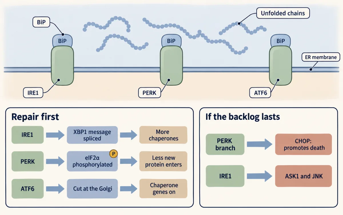
All three first repair, by lowering the load and raising folding capacity. If the backlog lasts, the same sensors change their message. PERK’s branch switches on CHOP, a transcription factor that promotes death, and IRE1 recruits an adaptor that feeds ASK1 and c-Jun N-terminal kinase (JNK). As with p53, timing carries part of the information: a brief stress mainly calls for repair, while a long or severe one suggests repair is failing. How strong the stress is and the state of the cell matter too. Heavy secretors depend on this especially: children born without working PERK develop diabetes within months of birth, and mice lacking PERK develop diabetes with too few insulin-producing cells in the pancreas.
Your notes
Select any text in the lesson to highlight it or add a note to it. Your highlights and notes are saved in this browser and collected in your notebook.
This browser is not letting the site save anything (a private window or strict privacy settings), so highlights and notes will be lost when you leave the page.
How we measure it
Live single-cell fluorescence imaging
Fuse the protein you care about to a fluorescent protein, keep the cells alive on a microscope, and photograph the same cells again and again for hours. Brightness tracks the amount of protein in each cell, so you see timing that an average over millions of cells hides.
Reporter gene screen in yeast
Join the control sequence of a stress-responsive gene to a gene whose product gives a color, mutate the yeast at random, and look for colonies that fail to change color under stress. Each such colony carries a mutation in a gene the pathway needs. Screens of this kind tied IRE1 to the unfolded protein response.
Photoaffinity labeling
Make a version of a drug that carries a radioactive atom and a group that light converts into a reactive one. Let it bind, flash it with light so that it bonds covalently to the protein it is sitting in, and purify the labeled protein. This names the target of a compound whose effect is known but whose partner is not.
Cysteine substitution
Replace one cysteine at a time with serine or alanine, which cannot react in the same way, and test whether the protein still senses its trigger. If the response to one compound is lost and the response to another survives, that cysteine is the site the first compound needs.
Method films · the methods library
- Engineered mutants: site-directed, dominant-negative and constitutively active 4:25
- Forward genetic screens 4:07
- Gel electrophoresis, SDS-PAGE and western blot 4:01
- Gene cloning: vectors, libraries and screening 4:04
- Gene targeting: knockout and knock-in mice 4:13
- Live-cell time-lapse imaging 4:01
- Mathematical and computational models 4:00
- Pharmacological blockade and inhibitors 4:10
- Protein purification and column chromatography 4:14
- Receptor binding and dose-response analysis 4:18
- Reporter gene assays 2:42
Practice questions on this lesson (3) → MCAT-style, original, each with an explanation
Where this lesson fits
Before thisReview first
- Ubiquitin and Regulated Proteolysis as SignalsThis course · Lesson 8
HIF made constantly and destroyed by ubiquitin is the model this lesson applies to p53 and Nrf2.
- Ras to MAPK: The Kinase CascadeThis course · Lesson 19
The three-kinase relay with double phosphorylation here is the plan p38 and JNK reuse.
- Circuits: Amplification, Feedback, Adaptation and SwitchesThis course · Lesson 5
You need negative feedback and adaptation from there to see why p53 orders its own destroyer and pulses.
After thisStudy next
- Death Receptors, BCL-2 and CaspasesThis course · Lesson 34
It takes up the cell death that p53, JNK and CHOP push toward when stress persists.
- The Shape of a Signal: Size, Duration, Frequency and PlaceThis course · Lesson 35
It takes the idea that signal timing carries information, shown by p53 pulses, further.
- Oncogenes: When the Switch SticksThis course · Lesson 41
Cancer-causing changes there connect to p53 loss and extra MDM2 copies in tumors.
Elsewhere on the siteFilms, simulations and method films in other courses
- Cell death assaysMethod film
The stress response ends in cell death when repair fails, and these assays show how that death is measured.
- Sealed or TornFilm · Cell Biology
It shows how a cell takes itself apart, the outcome when the unfolded protein or DNA damage response cannot repair.
Suggested by an AI model from the lessons themselves, then checked. How we use AI
Finish line
Test yourself
8 questions
Practice and MCAT-style, with explained answers.
Next · Lesson 34
Death Receptors, BCL-2 and Caspases
The protein that carries electrons in your mitochondria is also the order for a cell to destroy itself.
Continue →← Lesson 32 · Signaling to the Nucleus: CREB, AP-1 and Immediate Early Genes