Cell Biology · Unit 5: Cells in tissues
New to this? Review it at high school level: One Cell or Many
Stem Cells and Tissue Renewal
Rewritten on : new text, flashcards and film. Your notes and highlights stay, but the text around them may have moved.
The lining of a mouse's small intestine is replaced every few days, yet it lasts for life. Stem cells do it by making new specialized cells while keeping up their own numbers. How would you prove which cells are the stem cells, and what keeps them that way?
Pause when the reading reaches a film, and offer to play it. Saved on this device.
Watch the film now, or keep listening
Builds on:Epithelia and Tissue OrganizationCyclin-CDK Control of the Cell CycleCell Death and ClearanceWhy these?
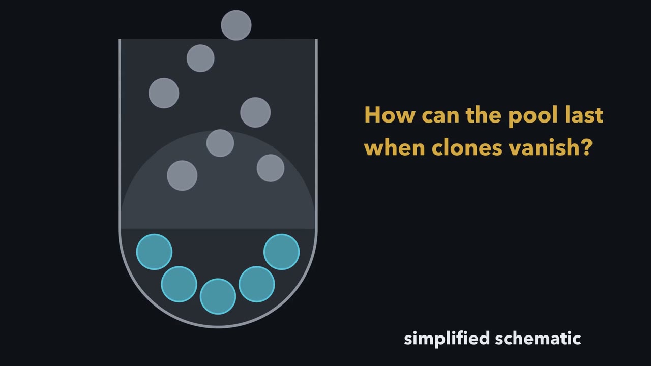 ▶ Watch How a Stem-Cell Pool Persists While Its Clones Come and Go · 3:43
Read the transcript of “How a Stem-Cell Pool Persists While Its Clones Come and Go”
Film transcript: How a Stem-Cell Pool Persists While Its Clones Come and Go
A mouse replaces its intestinal lining every few days. How can its stem-cell supply last when individual clones can vanish? Watch one crypt, the pocket where new lining cells begin. In this simplified picture, a handful of blue stem cells occupy the crypt base. The epithelium is the intestinal lining.
Cells need niche contact or signals to keep a base position. Stem cells have two jobs. First, they make specialized descendants that leave the base. Second, they maintain a stem-cell pool over time. Neighbours can fill positions as cells leave, so the pool may persist without preserving every cell. Shape or Lgr5 expression alone cannot prove those jobs.
Scientists give one expressing cell a permanent green lineage mark. Every daughter inherits green, even after it stops expressing Lgr5. The mark follows ancestry. Suppose the marked cell is only a short-lived precursor, a cell with limited output. What should happen to green after its descendants leave the lining?
A short-lived clone would fade away. Instead, observed green ribbons persisted for sixty days and contained every epithelial lineage. A lineage is a descendant family. That is multipotent output: several cell types within one tissue. But a long-lived ribbon does not mean its first green cell survives forever. The trace identifies a stem-cell population through descendant output over time. What descendants do, not a marker alone, supplies the evidence. Now give nearby stem cells permanent Confetti colours: green, yellow, red and cyan. In normally renewing mouse small intestine, Lgr5-high cells divide about once daily. Most divisions are symmetric.
Here that means both daughters retain stem positions, or both leave them. The clones appear to compete for niche-supported positions. Green expands when red leaves. Red disappears. Later, cyan pushes into green positions.
Colour shares change while the total pool stays constrained. Repeated replacements erase most colours and expand another until one clone fills the base. Different repeats produce different winners. The result is takeover by one clone, not survival of every starting clone. The number of supported positions stays roughly constrained while clone identities change. Balance belongs to the population, not every division. No cell is appointed the permanent winner. This pattern is neutral drift: chance competition among equivalent lineages. So follow descendants over time. During normal mouse-gut renewal, the pool can make specialized cells while most clones vanish and one may take over.
The population persists even as its membership changes. A stable stem-cell pool persists while most of its clones vanish.
From the film
- In the mouse gut, what descendants do over time—not a marker alone—identifies a stem-cell population.
- A permanent lineage mark follows descendants even after they stop expressing Lgr5.
- In the normally renewing mouse small intestine, Lgr5-high stem cells appear to compete for limited crypt-base positions.
- The stem-cell pool can stay balanced while most clones vanish and one may take over.
The first film followed an inherited mark and changing clone colours in one crypt; it did not judge what that evidence establishes, what limits the positions, who can refill them after injury or whether a specialized nucleus can be reset beyond its tissue. Throughout, the rule is to follow descendants over time and keep the experimental conditions attached to the conclusion.
What the lineage trace proves—and what it does not
A stem-cell population produces differentiated descendants and self-renews, meaning that a stem-cell pool persists over long periods. One division proves little because transit-amplifying cells, short-lived progenitors that divide a few times and then differentiate, also copy themselves briefly. Repeated growth and replating from one cultured cell or transplantation through successive hosts can test persistence.
The small-intestinal epithelium, its cellular lining, folds into finger-like villi above pocket-like crypts (see epithelia). Absorptive, mucus-secreting goblet and hormone-secreting enteroendocrine cells move up from crypts and are shed at villus tips. Paneth cells instead move down, live for weeks and secrete antibacterial proteins; slender crypt-base columnar cells sit between them.
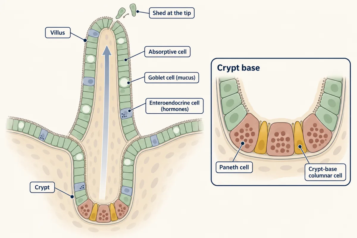
In 2007 Nick Barker, Hans Clevers and colleagues found that Lgr5, a gene activated by Wnt signalling (a local signal that supports stem cells, described in the niche section), is expressed in these cycling columnar cells in mouse small intestine and colon. Expression is a marker, not a fate test. In Lgr5-CreERT2 mice, one tamoxifen pulse permanently marks cells expressing Lgr5 at that moment, and every daughter inherits the mark (the construct is explained in Going deeper; more in tools for development). Label began at the crypt base and, over 60 days, filled ribbons containing every epithelial lineage.
That sustained, multilineage output supports the conclusion that the labelled population contains stem cells; it does not mean every initially marked cell survives. It shows multipotency, production of several cell types within one tissue, not pluripotency, the ability to make all lineages of the embryo proper. Stem cells need not be dormant, and their definition does not require one stem and one differentiated daughter from every division.
Put neutral drift to work
The first film’s changing colours suggest a population rule; a prediction makes that rule quantitative. If every division produced one stem cell and one transit-amplifying cell, pool size would be fixed by asymmetric division. In 2010 Hugo Snippert, Clevers and colleagues tested that idea, and a fixed hierarchy, with Confetti: a reporter that switches on one of four fluorescent proteins so neighbouring clones usually differ in colour. Mouse small-intestinal Lgr5-high cells divided about once a day, most divisions were symmetric, and over 1 to 6 months surviving clones grew while others vanished. The dynamics are consistent with daughters stochastically retaining a stem-cell position or entering the transit-amplifying pool.
Predict. A low tamoxifen dose labels 100 crypts, each starting with one marked stem cell. Suppose each crypt has an effective pool of five equivalent competitors—an illustrative number smaller than the number of Lgr5-high cells counted per crypt. After every labelled clone has vanished or filled its crypt, how many fully labelled crypts do you expect?
Check your prediction
Only one equivalent competitor eventually fills a crypt, so each of the five is equally likely to win. One starting clone therefore has probability 1/5, giving 100 × 1/5 = 20 fully labelled crypts; the other 80 labelled clones are lost.
The point of the arithmetic is population balance on average. The start has 100 × 1 = 100 labelled stem cells, and the expected end has 20 × 5 = 100. At an intermediate time with 50 surviving clones, their mean size is 100/50 = 2 cells: survivors grow because others disappear. This is not a promise about any one crypt.
That pattern is neutral drift, chance competition among equivalent lineages for limited stem-cell positions. The first film’s handful of positions was the simplest case; the fuller picture includes unequal short-term odds and a distinction between cells counted and cells effectively competing, developed in Going deeper.
labelled stem cell unlabelled stem cell clone lost clone survives, crypt mixed crypt fully labelled
| Of the 100 labelled crypts | Observed now | Expected |
|---|---|---|
| Clones surviving | ||
| Mean size of a surviving clone | ||
| Fully labelled crypts | ||
| Labelled stem cells in total |
Rule to keep: with N equivalent stem cells, a labelled clone takes over its crypt with probability 1/N, so 100 labelled crypts give about 100/N fully labelled ones, and labelled stem cells are conserved on average (100). In this sketch each event replaces one randomly chosen stem cell with a copy of a randomly chosen one, and the time axis is in events, not days. N here is an effective number for the model, which the lesson puts below the roughly 14 Lgr5-high cells counted per crypt (the slider runs up to 14). The numbers are round and illustrative, as in the lesson.
Every one of those drift calculations assumes a limited number of stem-cell positions. The second film picks up here: what limits those positions, and what keeps a cell in the stem-cell state?
Read the transcript of “What Keeps a Cell a Stem Cell?”
Film transcript: What Keeps a Cell a Stem Cell?
What keeps a cell in the stem-cell state? Chapter one showed a pool that lasts while its clones come and go. But the crypt base held only a few positions. What limits them? Watch the same crypt, now with its signals drawn in.
Start with the neighbourhood. A stem-cell niche is the local environment around stem cells. Its contact and signals bias whether a daughter keeps a stem-cell position or differentiates. In the mouse gut, Paneth cells in the lining and stromal cells beneath it both contribute. Two kinds of signal run in opposite directions.
Wnt is strongest at the crypt base and promotes self-renewal. R-spondin, EGF and Notch also support the base. BMP is strongest higher up, among differentiated cells, and it suppresses self-renewal. During cell turnover, a blue stem cell is displaced upward, away from the strongest Wnt. What do you expect it to do?
As cells move up, they become more likely to leave the stem-cell state. Less Wnt reaches them, and more BMP. The cell that remains at the base keeps its support. So the positions are limited because the support is local. Here is the key idea. In the mouse gut, being a stem cell looks less fixed. It looks more like a state that local signals support.
Chapter one's clones competed for places inside that support. A winner holds a place, not a permanent title. A dish tests how much surroundings matter. Single sorted Lgr5 cells grew into organoids with matrix and added factors. Organoids are crypt-villus structures grown in culture. They could be replated repeatedly. No stromal cells were present; the matrix and factors supplied a niche. If the state depends on support, a harder question follows. Could a cell that has already left the base come back? Injury tests that, when the usual stem cells are lost. Reprogramming pushes further, asking whether a specialized nucleus can be reset beyond its tissue.
So when you ask who is a stem cell, look at the place as well as the cell. Local niche signals help hold a cell in the stem-cell state. Whether a cell that left can return is the next test.
From the film
- A stem-cell niche is the local environment whose contact and signals bias whether a daughter stays a stem cell.
- In the mouse gut, Wnt is strongest at the crypt base and promotes self-renewal; BMP is strongest higher up and suppresses it.
- Cells that move up, away from the base signals, become more likely to leave the stem-cell state.
- Single Lgr5 cells grew into organoids when a matrix and added factors stood in for the niche.
- Injury asks whether cells can regain a tissue stem-cell state; reprogramming asks how far resetting can go.
The niche: what limits a stem-cell position
The second film showed the niche and its opposing Wnt and bone morphogenetic protein (BMP) signals (stromal cells are the connective-tissue cells beneath the epithelium); it did not show how to test which source of support matters. The experimental distinction is simple: adding support tests sufficiency, whereas removing it in the animal tests requirement. Loss followed by rescue is especially strong; no failure after removal can mean that another source compensated.
In 2009 Toshiro Sato and colleagues reported the second film’s organoid result: the matrix was Matrigel, a basement-membrane matrix, and the added factors were epidermal growth factor (EGF), R-spondin 1 and Noggin, a BMP inhibitor (see organoids).
Predict. In 2011 Sato and colleagues found that Paneth cells express Wnt3, EGF, TGF-alpha and the Notch ligand Dll4. Paneth co-culture greatly improved organoid formation, and a Wnt pulse could substitute for Paneth cells. What does that show, and what does it not show?
Check your prediction
Paneth cells can supply support under those culture conditions: a sufficiency result. It does not show that the living gut requires Paneth cells, because another source could substitute there. Requirement needs removal in the animal (see Wnt signalling and Notch signalling).
Design problem
Are Paneth cells indispensable in the living mouse gut? Design an in vivo test, state what “Paneth cells only” and “other sources can stand in” predict, and list controls.
Saved in this browser only.
One way to do it
Express the human diphtheria-toxin receptor from a Paneth-cell lysozyme gene, then give toxin to ablate Paneth cells while sparing other cells. Score Lgr5-positive cells, EdU incorporation (DNA synthesis in S phase; see cell-cycle control) and output from a separate Lgr5 tracing pulse over days to weeks.
Separately delete Porcn, which encodes the enzyme Porcupine that lipidates Wnt for secretion, either in the epithelium or in stromal FOXL1-positive telocytes, flat cells with long extensions beneath the epithelium. If Paneth cells are the only niche, their ablation should remove Lgr5 cells and stall renewal, while telocyte Porcn deletion should do nothing. If sources can substitute, stem cells may persist after ablation; telocyte deletion then tests whether that source is required.
Controls include toxin in mice without the receptor, receptor mice without toxin, staining to confirm Paneth-cell removal and tamoxifen-only controls for Cre lines. The published results favour distributed support: telocyte Porcn deletion reduced crypt Wnt signalling and proliferation within 24 hours in 2018, whereas Lgr5 cells survived Paneth-specific ablation in 2019.
After injury, other cells can refill the stem pool
The second film ended on whether a cell that left the stem-cell state can return. Injury is a distinct condition from the first film’s normal renewal, not an exception to that homeostatic result.
Predict. Secretory precursors are permanently labelled, and immediate staining finds no labelled Lgr5-positive cells. Lgr5-positive-cell killing is then confirmed. Weeks later, labelled Lgr5-positive cells and labelled epithelial ribbons appear. What is the narrowest conclusion?
Check your prediction
Labelled precursors regained a stem-cell state after injury: they became Lgr5-positive cells whose descendants rebuilt the epithelium. The result does not make them pluripotent or show that they are normal stem cells before injury.
In 2020 Murata and colleagues found that after lethal injury to Lgr5 cells in mouse gut, absorptive and secretory progenitors dedifferentiated, meaning that they regained a tissue stem-cell state. They switched on Ascl2 days before Lgr5, and restoration required ASCL2. Reviews describe related behaviour after other injuries, including irradiation, and from more than one precursor type. Cell fate is therefore less fixed than a one-way ladder suggests. Regaining gut stem activity is not becoming pluripotent; neural tissues pose related questions (see adult neurogenesis and injury and regeneration).
Resetting beyond one tissue: Gurdon to Yamanaka
Injury shows that committed gut progenitors can regain a tissue stem-cell state. Nuclear transfer and reprogramming use different tools to ask the second film’s broader question.
Potency is the range of cell types a cell can produce. A unipotent cell makes one type; a multipotent cell makes several types within one tissue; a pluripotent cell makes the embryo proper’s lineages; and a totipotent cell can make an entire conceptus, including placenta and other extra-embryonic tissues. The strongest test begins with one cell and measures its descendants.
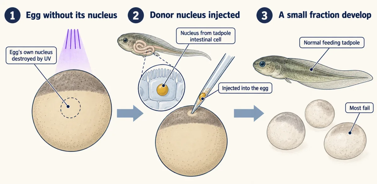
In 1962 John Gurdon transplanted nuclei from intestinal cells of feeding Xenopus tadpoles into eggs whose own nuclei had been destroyed by ultraviolet light. Most failed, but a small fraction became normal feeding tadpoles.
Predict. Does the large number of failed nuclear-transfer eggs show that specialized nuclei had discarded developmental genes?
Check your prediction
No. Each normal tadpole directed by a transplanted nucleus shows that at least that specialized nucleus retained the needed instructions and that egg cytoplasm could read them. Failures alone cannot identify what was missing, and a genetic nuclear marker ruled out a surviving egg nucleus.
In 2006 Kazutoshi Takahashi and Shinya Yamanaka used four transcription factors—Oct3/4, Sox2, Klf4 and c-Myc—to turn mouse fibroblasts into induced pluripotent stem (iPS) cells under embryonic-stem-cell culture conditions (see eukaryotic regulation). The cells formed tumours containing tissues from all three germ layers, the foundational layers of the embryo, and contributed to mouse embryos. In 2007 Yamanaka’s laboratory reprogrammed adult human skin fibroblasts with the same factors, and James Thomson’s laboratory reported human iPS cells made with a different set.
Predict. Mouse iPS cells contribute to tissues from all three embryonic germ layers. Which potency claim does that support: multipotent, pluripotent or totipotent?
Check your prediction
Pluripotent. The result spans the embryo proper’s lineages. It does not show that one cell can form an entire conceptus with extra-embryonic tissues, the test for totipotency.
At least some specialized nuclei therefore retain the instructions needed for development, and defined proteins can reset a specialized cell to pluripotency. Neither result shows that a specialized cell itself became totipotent, and passing a developmental test is separate from clinical safety.
Going deeper
The film chapters and main text use functional results first; this separate toolbox holds the historical assay, molecular mechanics and refinements needed to inspect those results closely.
The functional definition came before Lgr5
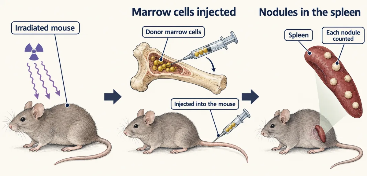
In 1961 James Till and Ernest McCulloch injected marrow cells into irradiated mice and found that spleen nodule counts rose with the number of cells injected. In 1963, with Andrew Becker, chromosome markers showed that each colony was a clone. One cell, its descendants and a count of lineages set the pattern; later work from the group found new colony-forming cells inside colonies, evidence of self-renewal.
How Cre lineage tracing works
Cre is a recombinase that cuts out DNA between two loxP sites pointing the same way. In CreERT2, fusion to a mutant estrogen-receptor domain keeps Cre mostly outside the nucleus until tamoxifen is given. Cre then removes a stop sequence from a Rosa26 reporter. Some Cre can leak into the nucleus without tamoxifen, so untreated mice are a necessary control.
The fuller neutral-drift picture
Live imaging by Ritsma and colleagues in 2014 found that cells at the crypt bottom survive better than cells at its edge, but movement between positions makes the months-long outcome look neutral. The effective competitor number can therefore differ from the roughly 14 Lgr5-high cells counted per mouse small-intestinal crypt. With N = 14, 100 starting clones give 100/14, about 7, fully labelled crypts in expectation; counting these outcomes can estimate the effective number.
What may replace Paneth-cell support
Sato and colleagues’ 2011 study reported that Paneth-cell removal coincided with Lgr5-cell loss. In the later cell-specific ablation experiment, enteroendocrine and tuft cells moved into the gaps and were proposed to supply Notch ligand. A review notes that too few of these cells touched every stem cell, so the substitute support remains unsettled.
Early and later iPS lines
The first mouse iPS clones, selected with an Fbx15 reporter, did not make adult chimaeras, animals containing cells from both the host embryo and introduced iPS cells. Nanog-selected clones in 2007 did, and one passed through the germline. Thomson’s human iPS cells used OCT4, SOX2, NANOG and LIN28. Reprogramming does not show that every iPS line matches embryonic stem cells in every test.
Your notes
Select any text in the lesson to highlight it or add a note to it. Your highlights and notes are saved in this browser and collected in your notebook.
This browser is not letting the site save anything (a private window or strict privacy settings), so highlights and notes will be lost when you leave the page.
How we measure it
Inducible Cre lineage tracing
A gene expressed in the cells of interest drives CreERT2, a Cre recombinase that enters the nucleus mainly after tamoxifen; a little leaks in without it, so untreated mice are a needed control. A pulse of tamoxifen lets Cre remove a stop sequence from a reporter gene, and the mark is inherited by every descendant. Looking weeks later shows what the labelled cells made.
Multicolour clonal analysis (Confetti)
A reporter that, after Cre acts, expresses one of four fluorescent proteins at random gives neighbouring clones different colours most of the time. Counting how many clones survive and how large they grow over time tests models of how stem cells compete.
Single-cell organoid culture and replating
One sorted cell is embedded in basement-membrane matrix with defined growth factors and grows into a crypt-villus organoid. Breaking the organoid up and replating it again and again tests sustained propagation under those stated conditions.
Spleen colony transplantation assay
Marrow cells are injected into irradiated mice, and the colonies that appear in the spleen are counted and analysed. Colony number per cell injected, colony clonality and the lineages in each colony measure blood-forming stem and progenitor cells.
Practice questions on this lesson (3) → MCAT-style, original, each with an explanation
Where this lesson fits
Before thisReview first
- Epithelia and Tissue OrganizationThis course · Lesson 23
The gut lining's villi, crypts and cell types are described there, and this lesson assumes that layout.
- Cyclin-CDK Control of the Cell CycleThis course · Lesson 17
Self-renewal and transit-amplifying divisions rest on how cells decide to divide or stop.
- Cell Death and ClearanceThis course · Lesson 21
Shedding and clearance of cells at villus tips is part of why the lining needs constant renewal.
After thisStudy next
- Cell Identity, Cell State and DiseaseThis course · Lesson 25
It uses stem-cell renewal and cell state to ask why mutated cells in healthy tissue stay or spread.
- Designing a Cell Biology ExperimentThis course · Lesson 26
It practices the same skill of designing tests that show requirement or sufficiency, as in the niche problem.
Suggested by an AI model from the lessons themselves, then checked. How we use AI
Finish line
Test yourself
8 questions
Practice and MCAT-style, with explained answers.
Next · Lesson 25
Cell Identity, Cell State and Disease
Much of the lining of an older person's healthy oesophagus can carry mutations found in cancer. Why is it still healthy?
Continue →