Cell Biology · Unit 3: Traffic and movement
Vesicle Budding, Targeting and Fusion
Rewritten on : new text, figures, flashcards and film. Your notes and highlights stay, but the text around them may have moved.
A yeast mutant keeps making secretory enzymes at 37 °C but cannot release them. Which step of delivery broke, and how could you tell?
Pause when the reading reaches a film, and offer to play it. Saved on this device.
Watch the film now, or keep listening
Builds on:Protein Sorting into CompartmentsMembrane Organization and TopologyExperiments That Distinguish MechanismsWhy these?
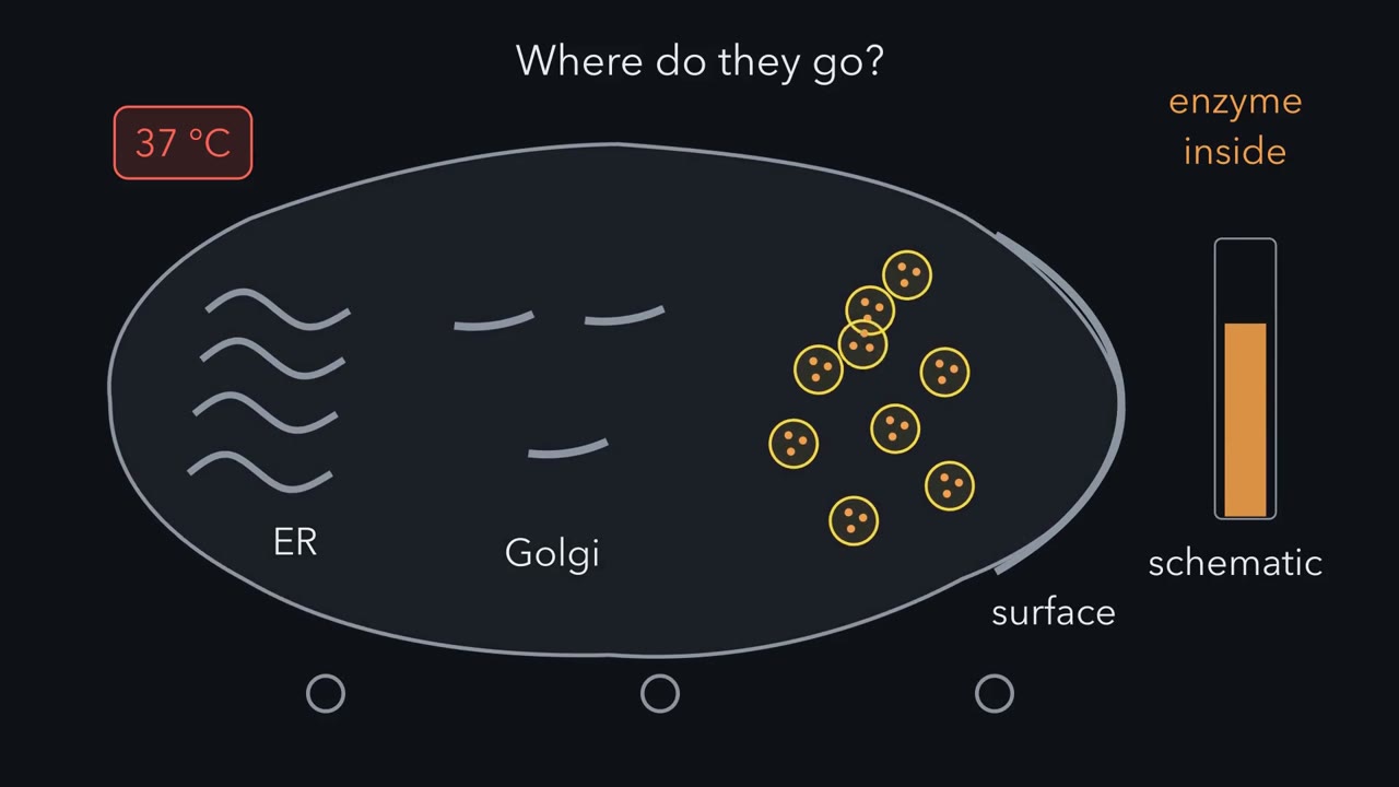 ▶ Watch Three Handoffs for a Vesicle · 4:58
Read the transcript of “Three Handoffs for a Vesicle”
Film transcript: Three Handoffs for a Vesicle
Where do the enzymes go? This yeast mutant keeps making them at 37 °C. But it releases little. Yeast secrete enzymes along a route: ER, then Golgi, then vesicles, then the cell surface. Small membrane spheres called vesicles carry the cargo. This mutant is called sec1. At 25 °C it grows and secretes. At 37 °C, its enzyme pool inside rises up to eightfold. The enzymes sit in membrane-bound vesicles that fail to fuse with the surface.
Back at 25 °C, with protein synthesis blocked, the cells secrete them. Every vesicle on that route has three jobs. Budding: pick up cargo and pinch off. Targeting: be caught by the right membrane. Fusion: merge with it. Back to sec1. For the last vesicle on the route, which job worked? And which could have failed?
Budding worked, because the vesicles exist. Targeting or fusion failed. A pile of vesicles alone cannot tell you which. Budding first. A coat of proteins assembles on a patch of membrane. Think of a cookie cutter with hands: it bends a bud and grabs the cargo. At the ER this coat is COPII. A switch called Sar1, holding GTP, pulls it on. When Sar1 hydrolyzes that GTP, the coat begins to let go. In a test tube, vesicles made with a GTP analogue that cannot be hydrolyzed still budded. But they kept their coat, and failed to target and fuse. Making a vesicle and delivering it are separate steps. Here is the one idea. A vesicle's trip is three handoffs, and each can fail on its own.
A coat buds it. A Rab and a tether catch it. SNAREs, guided by an SM protein, fuse it. Targeting next. A Rab is a small switch with a lipid tail. Holding GDP, it can be carried in the cytosol by GDI. At the right membrane, an exchange factor loads GTP, and the Rab holds on. Rab with GTP is one lit sign over a loading bay. It recruits tethers, which catch the vesicle. It is one sign among several. Fusion last. Two membranes resist merging, so SNARE proteins supply the pull. Four strands, called Qa, Qb, Qc and R, come from the two membranes. They zip into one rope, toward the membrane anchors. The zipping pulls the two bilayers together until they fuse.
The zip itself uses no ATP. In the main fusion steps studied, SNAREs also need an SM protein. It binds them and helps the right four zip together. Sec1 is the SM protein for the last step of secretion. That step is fusion of secretory vesicles with the plasma membrane. So at 37 °C, vesicles still bud and pile up. Back at 25 °C, Sec1 works again, and the stored vesicles fuse. But the pile alone never named fusion. A failed Rab, tether or SNARE for that step gives the same pile. Sec1's known job names it. On a linear route, cargo piles up in the last place it reached. Double mutants used that rule to put the steps in order. Test tubes add timing.
GDI, which pulls GDP-bound Rabs off membranes, blocks fusion when added at the start. What if it is added after vesicles dock? Its effect fades. Once vesicles had docked, fusion was no longer sensitive to GDI. A fast, specific inhibitor stops working once the state it targets is past. So each vesicle on this route relies on three separate handoffs: bud, catch, fuse. Any one can fail on its own. Where cargo stalls, and when an inhibitor stops mattering, help place the break.
From the film
- The sec1 mutant keeps making enzymes at 37 °C, but they pile up inside vesicles that fail to fuse with the surface.
- A vesicle's trip has three handoffs: a coat buds it, a Rab-GTP with a tether catches it, and SNAREs guided by an SM protein fuse it.
- Zipping the four SNARE strands pulls the membranes together, and the zip itself uses no ATP.
- A pile of vesicles alone cannot name the failed step, but Sec1's known job as the fusion SM protein does.
- A fast inhibitor such as GDI stops working once its target state is past, which helps time the steps.
A yeast mutant called sec1 grows at 25 °C, but at 37 °C it keeps making secretory enzymes and cannot release them. Novick and Schekman reported it in 1979. The first film above follows those enzymes along the route and through the vesicles that never fuse with the surface; the three jobs it names are budding (pick up cargo and pinch off), targeting (be caught by the right membrane) and fusion (merge with it).
Predict. In sec1 cells at 37 °C, which of the three jobs worked for the last vesicle on the route, and which could have failed?
Check your prediction
Budding worked: the vesicles exist. Targeting or fusion failed, and a pile of vesicles alone cannot say which.
The one idea to carry through this lesson. A vesicle’s trip is three handoffs, and each can fail on its own. A coat buds the vesicle and picks its cargo. A Rab-GTP with a tether catches it at the right membrane (targeting). A SNARE bundle, guided by an SM protein, fuses it. On a linear route, when one handoff fails, cargo piles up in the last place it reached, and a fast, specific inhibitor stops working once the state it targets is past.
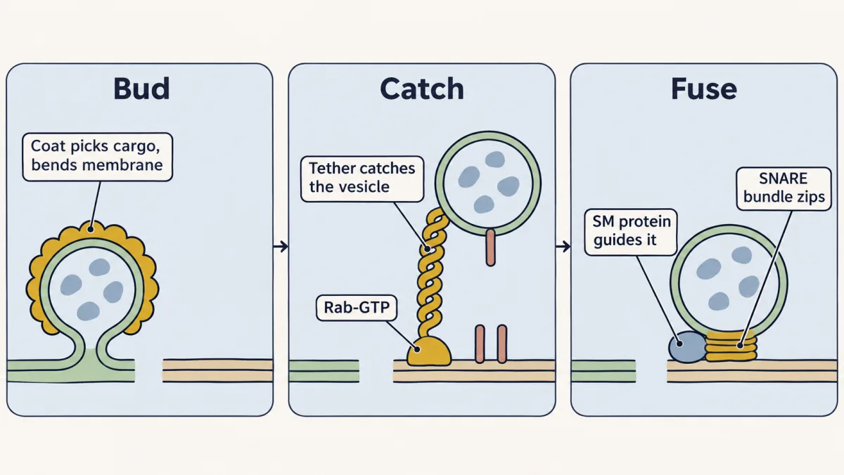
This picture fits the coat-dependent routes in this lesson. Some endocytosis uses other budding machinery, and tubular carriers and lipid transfer without a vesicle fall outside it.
A few terms. The cytosol is the cell’s fluid outside organelles; the lumen is the inside of a membrane-bound compartment. A vesicle is a small membrane sphere, often 50 to 100 nm across, that carries lumenal cargo and membrane proteins from a donor compartment to a target. Its lumen never mixes with the cytosol (see membrane topology), so every protein that steers it works on its cytosolic face. A coat grabs a membrane protein’s cytosolic tail directly. A soluble cargo, such as the secretory enzyme invertase measured below, cannot touch the coat: it is caught by a membrane-spanning receptor that the coat grabs (Erv29 in yeast, ERGIC-53 in mammals), or rides along in the fluid a bud encloses.
Step 1, budding: a coat picks the cargo and bends the membrane
A coat is a lattice of cytosolic proteins that assembles on a patch of membrane. The film’s cookie cutter with hands is a simplification: the cutter shapes the bud and the hands grab the cargo, but different parts of a coat often do each job.
Take COPII, the ER’s coat. It is started by a switch, Sar1, a small GTPase (see GTPase switches). Sar1-GTP grabs the ER membrane and pulls in an inner layer, Sec23/24, which holds cargo and in turn recruits an outer cage, Sec13/31. Cargo choice sits mainly in Sec24: yeast mutant studies support a model in which Sec24 has several independent cargo-binding sites. Sar1’s GTP is also a timer. When Sar1 hydrolyzes it, the coat begins to let go, and the vesicle can go on to be caught and fused.
| Coat | Started by | Where it works | Cargo signal it reads |
|---|---|---|---|
| COPII | Sar1-GTP | ER to Golgi | signals read by Sec24 |
| COPI | Arf1-GTP, recruiting coatomer (seven subunits) | within the Golgi, and Golgi back to ER | dilysine (KKXX) tails, read by α- and β′-COP |
| Clathrin with AP2 | the lipid PI(4,5)P2, which opens AP2 | plasma membrane | motifs read by AP2; clathrin binds no cargo |
A KKXX tail is a return label: COPI fetches escaped ER membrane proteins back from the Golgi. Crystal structures of recombinant AP2 show it locked, part of its β2 subunit covering both cargo sites; it opens on membranes with PI(4,5)P2, a phosphoinositide concentrated in the plasma membrane (see lipid anchors and addresses), so the lipid and the cargo together tell it where it is (compare the locked and open structures on this page). Dynamin, a GTPase, pinches the finished clathrin bud off.
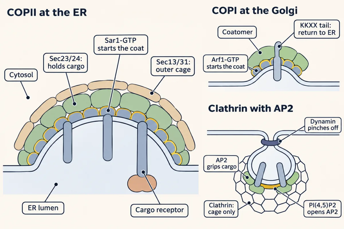
Predict. In 1994 Barlowe and colleagues made ER vesicles in a test tube with GMP-PNP, a GTP analogue that cannot be hydrolyzed. Do the vesicles bud? Can they fuse with the Golgi?
Check your prediction
They bud, but the timer never runs out. The vesicles kept their coat proteins and failed to target and fuse with the Golgi. Making a vesicle and delivering it are separate steps, and the coat must start to come off before the next handoff.
Real coats and ER exit sites are messier than this picture (Going deeper, “Coats that curve late, and tubes as well as spheres”).
Step 2, targeting: a Rab and a tether catch the vesicle
A budded vesicle must find its target among many membranes. Part of the answer is Rab proteins, a large family of small GTPases. A Rab has a lipid tail (prenyl groups) that can anchor it in a membrane. When the Rab carries GDP, a protein called GDI (GDP dissociation inhibitor) can wrap that tail and carry the Rab in the cytosol. At the right membrane the Rab leaves GDI, its tail enters the bilayer, and a guanine-nucleotide exchange factor (GEF) (an exchange factor) there loads GTP, which holds it on. Rab-GTP recruits proteins called effectors, which generally prefer the active, GTP-bound form. The film’s Rab “anchored in a membrane” and “held in the cytosol by GDI” are these two states, simplified: a Rab-GDP can also sit briefly on a membrane.
Among the effectors are tethers: long coiled-coil proteins or large complexes that capture a vesicle before its SNAREs engage. In yeast, the tether Uso1p works with the Rab Ypt1p on ER-to-Golgi vesicles. On the yeast vacuole, a six-subunit tether called HOPS binds the Rab Ypt7.
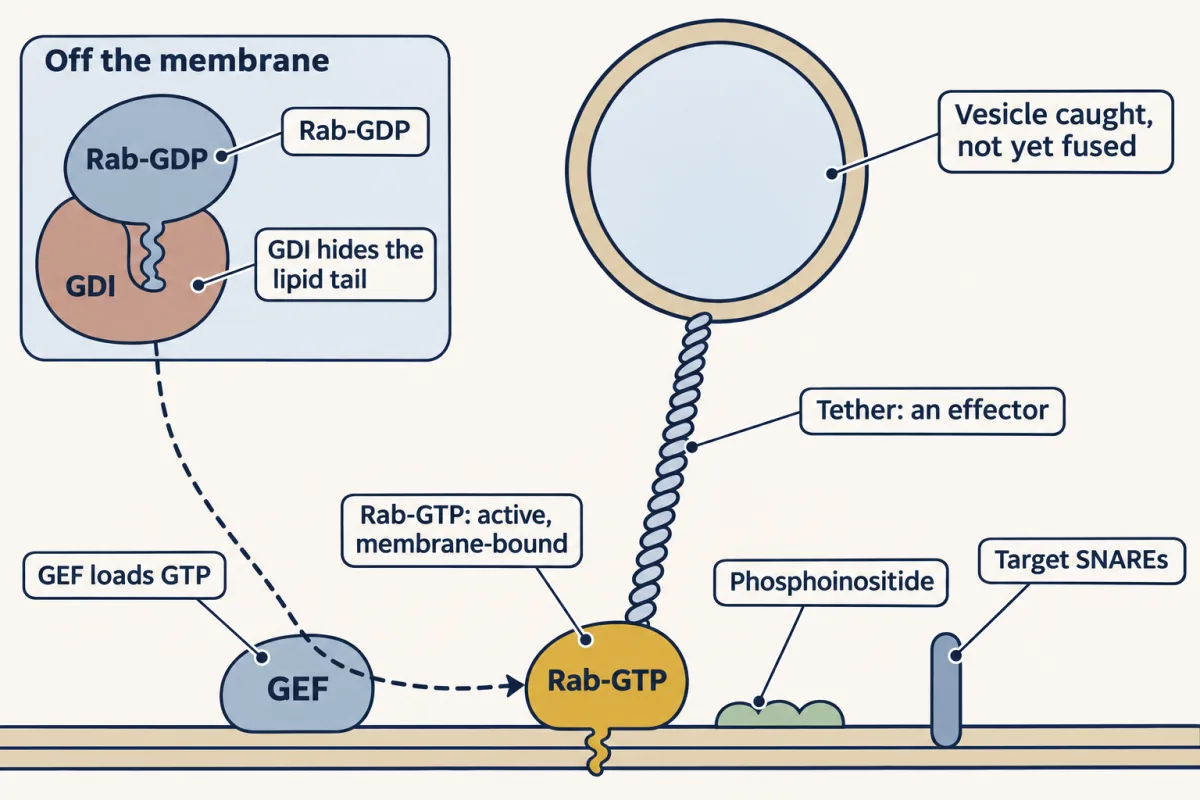
The film’s lit sign over a loading bay is one of several: the tether, the membrane’s phosphoinositide and the SNAREs also narrow the choice, and the evidence fits a combination better than any one label.
Step 3, fusion: SNAREs zip, an SM protein guides them, NSF resets them
A caught vesicle still has to merge, and bilayers resist merging: their water-coated surfaces repel at close range. SNAREs supply the pull. Most have a SNARE motif of about 60 to 70 amino acids next to a membrane anchor; v-SNAREs sit in the vesicle and t-SNAREs in the target. A lone motif is floppy. Four of the right kind wind into a parallel four-helix bundle. The strands are named by the amino acid each places at the bundle’s centre: three give a glutamine (Q) and one an arginine (R), so they are Qa, Qb, Qc and R. Often the vesicle gives R and the target gives the three Q strands, though not at every step. At a nerve terminal, syntaxin-1 gives Qa, SNAP-25 gives both Qb and Qc, and synaptobrevin on the vesicle gives R.
The film shows the zip, which runs from the N-terminal end toward the membrane anchors. Four strands are not four anchors: SNAP-25 gives two strands and attaches by lipid chains on the loop between them. While the strands come from two membranes, the bundle is a trans-SNARE complex. Afterwards the whole bundle sits in one membrane: a cis-SNARE complex.
Predict. Does the moment of fusion use ATP?
Check your prediction
No. Four floppy strands folding into one tight rope releases free energy, and the zip turns it into a pull on the membranes. The zip itself uses no ATP; ATP pays to get SNAREs ready. A cis bundle is stable and useless, since both ends are already in one membrane. NSF (Sec18 in yeast), an ATPase, binds cis bundles through its adaptor SNAP (Sec17) and spends ATP to pull the strands apart, after one fusion and before the next. NSF cocks the spring that the next zip releases.
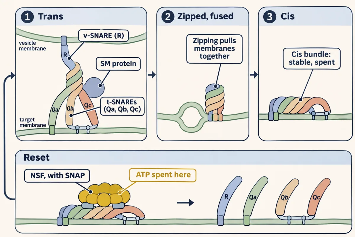
In a cell, SNAREs do not work alone. The main fusion steps that have been studied also need an SM protein, named for its first two members, Sec1 and Munc18, which binds SNAREs and helps the right four zip together. On the yeast vacuole the SM protein Vps33 is built into the tether HOPS, so one complex both catches and helps fuse.
That answers the opening puzzle. Sec1 is the SM protein for the last step of secretion: fusion of secretory vesicles with the plasma membrane. Upstream, invertase-laden vesicles still budded from the Golgi at 37 °C. They piled up, then fused after Sec1 worked again at 25 °C.
Sec1’s known job, not the pile, names fusion. The same pile of 80 to 100 nm vesicles appears when the secretory-vesicle Rab (Sec4), its tether (the exocyst) or a SNARE (Sec9, the yeast relative of SNAP-25) fails; a pile cannot tell a vesicle never caught from one caught but unable to fuse. Test tubes split them; imaging single vesicles in living cells can too. Before that, the steps had to be found and put in order.
The second film picks up here: how a pile of stuck cargo puts the handoffs in order, and how test tubes split catching from fusing.
Read the transcript of “Reading Where Cargo Stops”
Film transcript: Reading Where Cargo Stops
Chapter one followed a vesicle through three handoffs: bud, catch, fuse. But how did anyone put those handoffs in order? What can a pile of stuck cargo tell you? First, measure the block. Yeast secrete an enzyme called invertase. The fraction secreted is the enzyme outside the cell, divided by all of it. Is the trapped enzyme dead, or only waiting?
Return the cells to 25 °C, with new protein synthesis blocked. If the trapped enzyme moves outside, it was waiting to be delivered. At 37 °C, these secretion-blocked cells become dense. In 1980, Novick, Field and Schekman used that to enrich new mutants.
Electron microscopy then showed what each one piled up. Chapter one's rule applies here: on a linear route, cargo piles up in the last place it reached. In 1981, double mutants put the blocks in a broad order. Now a harder case. An enzyme called NSF resets SNAREs at many steps along the route. Where would a heat-sensitive NSF mutant pile up its cargo?
At its earliest blocked step. In classical sec18 experiments, ER and small ER-derived vesicles piled up, not late secretory vesicles. A pile shows where transport stops, not when or where else a protein acts. Test tubes add what a pile cannot. Cell-free assays mix donor and acceptor membranes with cytosol and ATP. Then each step can be timed, removed or rebuilt. Is catching enough to fuse? Yeast HOPS is a tether that binds the Rab Ypt7. Separate forms missing the beta propeller of Vps11 or Vps18 still tethered Ypt7-bearing lipid spheres. But they failed to promote fusion. So read stuck cargo in two ways. Where cargo stops gives the order; test tubes split catch from fuse.
From the film
- The fraction secreted is the enzyme outside the cell divided by all of it, so a block can be measured.
- If trapped enzyme leaves once the cells return to 25 °C with synthesis blocked, it was waiting to be delivered, not dead.
- Double mutants put the blocks in order, but a pile shows where transport stops, not when or where else a protein acts.
- Because NSF acts at several steps, a sec18 mutant reveals only its earliest blocked step.
- HOPS forms lacking the Vps11 or Vps18 beta propeller still caught Ypt7-bearing lipid spheres but failed to promote fusion.
Putting the handoffs in order: mutants
In 1980 the density screen from the second film used a Ludox gradient, and Novick, Field and Schekman sorted 188 clones that trapped invertase at 37 °C, the restrictive temperature, into 23 complementation groups (see complementation and pathways): the screen’s yield, not the number of genes secretion needs. The SEC numbers carry no information about order; order came from double mutants in 1981.
Now the numbers, made up for practice. Invertase is measured in arbitrary units after 1 h at 37 °C.
fraction secreted = external / (external + internal)
Step 1. Wild type: external 180, internal 20, total 200, so 180/200 = 90% secreted. Mutant secX: external 12, internal 188, total 200, so 12/200 = 6%. The block is real.
Step 2. The recovery test: shift secX back to 25 °C with cycloheximide to stop new synthesis. After 60 min external is 150 and internal 50, total still 200. The pool released is (188 − 50)/188 = 138/188 = 73.4%. External rose by the same 138 units, so the trapped material was deliverable.
Step 3. Electron microscopy at 37 °C shows secX accumulates ER, secY accumulates Golgi-like stacks (Berkeley bodies), and secZ accumulates 80 to 100 nm vesicles. The secX secZ double mutant accumulates ER. The secY secZ double accumulates Golgi-like membranes.
Predict. Put X, Y and Z in order. Which double mutant would test the one link the data leave open?
Check your prediction
Cargo piles up at the first block it meets, so a double mutant looks like the earlier single block: X acts before Z, and Y before Z. ER precedes Golgi on the secretory route, which places X before Y; a secX secY double accumulating ER would test that directly. Order: ER (X), Golgi (Y), vesicles (Z), surface.
The rule assumes a linear pathway with arrest states you can tell apart. It orders where transport is blocked, not when a protein acts: a gene product could still act early and be needed only later. A gene needed at several steps shows only its earliest blocked one. NSF resets SNAREs at many steps, and in classical sec18 experiments at 37 °C, ER and small ER-derived vesicles piled up, not late secretory vesicles.
Fraction secreted = external / (external + internal). In a double mutant on a linear route, the earlier block wins, so the compartment that piles up names the earlier gene. The numbers are the lesson’s made-up units: after 1 h at 37 °C, and for the secX shift-back after a further 60 min at 25 °C with cycloheximide. This is a sketch of the logic, not data.
Splitting catch from fuse: test tubes
Mutants show where cargo stops. Cell-free assays mix donor membranes, acceptor membranes, cytosol and ATP so that individual steps can be timed, removed or rebuilt. In the assays of the second film, docking means vesicles bound to the acceptor membrane, the tethered state.
Predict. In 1998 Cao, Ballew and Barlowe ran a yeast ER-to-Golgi assay and added GDI, which pulls Rabs off membranes, at different times. Added at the start, GDI blocked fusion. What happens to its effect if it is added after the vesicles have docked?
Check your prediction
It fades. Once vesicles had docked, fusion was no longer sensitive to GDI, so the GDI-sensitive step is passed by docking. That does not show the Rab left or stopped mattering: GDI pulls off GDP-bound Rabs, and an active Rab-GTP resists it. In general, losing sensitivity to a fast, specific inhibitor shows that an inhibitor-sensitive state has been passed; it times the end of a whole step only if the inhibitor can still reach and act on its target later, which is why budding, docking and fusion are measured separately. The authors proposed that this first docking needs the tether Uso1p and the Rab Ypt1p but not SNARE proteins.
Purified yeast HOPS shows that catching is not fusing: separate forms lacking the beta-propeller domain of Vps11 or Vps18 still tethered Ypt7-bearing liposomes but failed to promote fusion.
Design problem
You have this cell-free assay, with yeast ER as donor and Golgi as acceptor. You want to know whether GDI acts before or after vesicles dock, and whether a purified tether is enough for fusion. Design the experiment.
Saved in this browser only.
One way to do it
Use three readouts. Budding: vesicles released from ER, collected after spinning out large membranes. Docking: vesicles that sediment with the acceptor Golgi. Fusion: cargo that gains an acceptor-only sugar, or lumenal content mixing.
Start the reaction and add GDI at 0, 5, 10, 15 and 20 minutes. Plot fusion still blocked against time of addition, with docking measured on the same schedule. If GDI acts at docking, sensitivity is lost as docking completes, as Cao, Ballew and Barlowe found. If it acts at fusion itself, additions made after docking but before fusion still block. Measure when fusion itself happens on the same schedule, or hold vesicles docked but unfused before adding GDI, to tell the two apart (Going deeper, “Why the fusion time course matters”).
For the tether, run docking and fusion separately with the purified tether, matched SNARE proteoliposomes and an active Rab, since tethering without fusion is possible. Controls: no ATP, no cytosol, no tether, and a leakage check so that content released by broken membranes is not read as fusion.
In one line: coat buds, Rab and tether catch, SNAREs with an SM protein fuse, NSF resets; where cargo stops and when an inhibitor stops mattering point to the failed handoff.
Going deeper
A coat from three parts
With purified yeast proteins, Matsuoka and colleagues showed in 1998 that Sar1, Sec23/24 and Sec13/31 alone bud coated vesicles from chemically defined liposomes that contain PI4P or PI(4,5)P2, so the coat can bend membrane without help from other ER proteins. After Sar1 hydrolyzes GTP, parts of the coat can stay on long enough to help the vesicle be caught at the Golgi.
Coats that curve late, and tubes as well as spheres
In cultured BSC-1 cells, clathrin lattices began to curve only at around 70% of their final clathrin content, and raised membrane tension stalled the change, so a coat does not always curve from the start. And in mammalian cells, whole-cell imaging in 2021 found ER export through an interwoven tubular network, not only through separate small spheres.
HOPS up close
The purified six-subunit HOPS complex is about 43 nm tall, and a structural working model places its Vps39 and Vps41 subunits at the Ypt7 contacts. Its SM protein, Vps33, is held in the complex by Vps16 and binds SNAREs.
Paying for fusion
Assembly of one neuronal SNARE bundle releases about 65 kT, about 170 kJ/mol at body temperature (Gao and colleagues, 2012). In a single-molecule assay, NSF took one complex apart in one round of hydrolysis of the ATP already bound to it, at most six (Ryu and colleagues, 2015); other assays report more ATP per complex. As an illustration, six ATP at 50 to 57 kJ/mol each supply 300 to 342 kJ/mol, about 116 to 133 kT at 310 K, more than this construct’s assembly releases. Reversing the same assembly under the same conditions must cost at least what it released, but these separate experiments do not measure a matched reset cycle or a universal ATP budget.
Can SNAREs fuse membranes alone?
In 1998 Weber and colleagues put recombinant v-SNAREs and t-SNAREs into separate liposomes, and the docked liposomes mixed their lipids at physiological temperature, which the authors read as fusion. That shows SNAREs can merge those bilayers on their own, though lipid mixing alone cannot tell full fusion from hemifusion. It does not show they supply specificity or speed in a cell.
What NSF might do before fusion
NSF may do more than recycle SNAREs. In a 2024 yeast vacuole reconstitution, Sec18 still promoted fusion with Sec17 fixed to the membrane; whether this holds at other fusion steps is not yet known.
Why the fusion time course matters
Take a simple model: a batch of vesicles, all undocked at time zero, docks with a first-order half-time of 10 min (75% docked by 20 min), each docked vesicle then fuses with its own first-order half-time, and each inhibitor instantly and completely blocks its step. If the fusion half-time is about 2 min, a docking inhibitor and a fusion inhibitor added at 20 min block about 25% and 31% of eventual fusion: nearly the same. With a 15 min fusion half-time they block about 25% and 69%, and the steps separate clearly.
Your notes
Select any text in the lesson to highlight it or add a note to it. Your highlights and notes are saved in this browser and collected in your notebook.
This browser is not letting the site save anything (a private window or strict privacy settings), so highlights and notes will be lost when you leave the page.
How we measure it
Temperature-sensitive mutant screens and double mutants
Yeast mutants that work at 25 °C and fail at 37 °C are screened for secretory enzyme trapped inside the cell. Complementation sorts them into groups, and double mutants, read by electron microscopy, show which block comes first.
Cell-free transport assays
Donor membranes, acceptor membranes, cytosol and ATP are mixed in a tube. Cargo that reaches the acceptor gains a sugar only the acceptor can add, so transport becomes a number, and each ingredient can be removed, inhibited or added back.
Liposome and proteoliposome reconstitution
Purified proteins are added to synthetic lipid vesicles to ask what they can do alone: bud a coat, tether, or fuse. Fusion is read as lipid mixing and lumenal content mixing, with controls for leakage.
Electron microscopy of arrested cells
Thin sections of mutant cells at the restrictive temperature show which membrane piles up: ER, Golgi-like stacks or vesicles of a given size. The accumulated organelle marks where the pathway stopped.
Practice questions on this lesson (3) → MCAT-style, original, each with an explanation
Where this lesson fits
Before thisReview first
- Protein Sorting into CompartmentsThis course · Lesson 7
You need to know how signals send proteins into compartments, since the vesicles here carry that sorted cargo.
- Membrane Organization and TopologyThis course · Lesson 4
Vesicle lumen versus cytosol and the cytosolic face of each protein are used throughout this lesson.
- Experiments That Distinguish MechanismsThis course · Lesson 3
The sec-mutant and cell-free reasoning here applies the fractionation and ordering logic taught there.
After thisStudy next
- The Secretory PathwayThis course · Lesson 12
It follows cargo through the ER and Golgi using the budding, tethering and fusion steps learned here.
- Endocytosis, Recycling and DegradationThis course · Lesson 13
Clathrin, AP2 and dynamin budding from the plasma membrane are used again to take cargo into the cell.
- Molecular Motors and CargoThis course · Lesson 15
Motors move vesicles along tracks to the target that the Rabs and tethers then catch.
Elsewhere on the siteFilms, simulations and method films in other courses
- Counting failuresSimulation · Neuroscience
Vesicle fusion at a nerve terminal is a regulated SNARE-driven release step.
- Pulse-chase labellingMethod film
Following a labelled batch of secreted enzyme through compartments is the same approach used to time delivery steps.
Suggested by an AI model from the lessons themselves, then checked. How we use AI
Finish line
Test yourself
8 questions
Practice and MCAT-style, with explained answers.
Next · Lesson 12
The Secretory Pathway
Label a pancreas cell's new proteins for a few minutes, then watch them move: what route do they take, and how do you know?
Continue →← Lesson 10 · Nuclear Architecture and Membraneless Compartments