Cell Biology · Unit 3: Traffic and movement
Endocytosis, Recycling and Degradation
Rewritten on : new text, flashcards and film. Your notes and highlights stay, but the text around them may have moved.
Why can a patient's cells grip cholesterol-carrying LDL normally and still leave most of it on the surface?
Pause when the reading reaches a film, and offer to play it. Saved on this device.
Watch the film now, or keep listening
Builds on:Vesicle Budding, Targeting and FusionOrganelles as Distinct EnvironmentsProtein Sorting into CompartmentsWhy these?
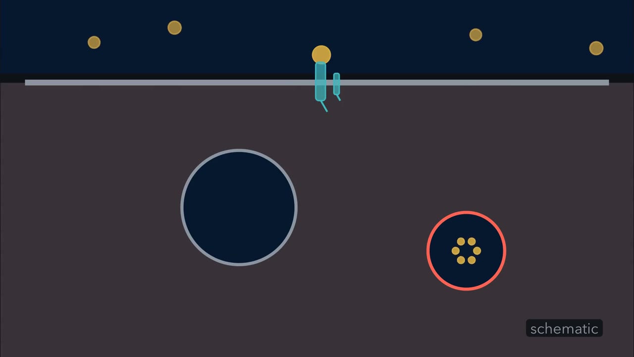 ▶ Watch A Loop and a One-Way Trip · 3:19
Read the transcript of “A Loop and a One-Way Trip”
Film transcript: A Loop and a One-Way Trip
This receptor brings LDL particles into the cell, where they are normally broken down. So how does the receptor hand its cargo to destruction, and escape, over and over? LDL, in gold, carries cholesterol in the blood. Its receptor, in teal, spans the membrane and grips it from outside. Gripping, though, is not entering. The receptor's tail, inside the cell, must be gathered into a pit. Adaptors, in violet, do the gathering. They hold the receptor tails and the inner face of the membrane. Then clathrin builds a cage of three-legged pieces beneath them. Clathrin holds neither membrane nor cargo; the adaptors do. As the cage grows, the membrane curves inward into a pit.
Dynamin, in orange, wraps the narrow neck like a collar and, using energy from GTP, cuts it. The vesicle pinches free, and the cage falls away to be used again. Look inside the vesicle. It still has the colour of the outside. The cargo is inside the cell, but no membrane has been crossed yet. Biologists say it is topologically outside.
The vesicle fuses with an early endosome, the cell's sorting station. Receptor and LDL arrived together, still bound. If the receptor is to escape, they must part here. What is different about this compartment? Think about it.
Its inside, the lumen, is mildly acid, kept that way by proton pumps. For LDL, at least, acid is the signal to let go of its receptor. This is the fork. Here the paths split. Empty receptors slide into a thin tubule. It pinches off and carries them back to the surface, ready to grip again. The LDL stays behind. The endosome matures and passes it to a lysosome, the cell's digestion compartment.
There, enzymes break it down and release its cholesterol. So the receptor runs a loop: bind, enter, release, return. Its LDL makes a one-way trip to the lysosome. The two part at one place, the acid early endosome. In this simplest picture, binding and entry happen in order. Follow one batch of receptors, and a complete block of either stops its later handling. Release defects can reduce return rather than abolish it, so test each transition directly. That is how the carrier escapes: it lets go in acid, and leaves before the end. But in a patient's cells, which step has broken, and how could you tell?
From the film
- For the LDL receptor, the receptor runs a loop and its LDL makes a one-way trip to the lysosome.
- Adaptors hold the receptor tails and the membrane; clathrin builds the cage, and dynamin cuts the neck.
- In the mildly acid early endosome, LDL lets go of its receptor, and their paths split.
- Empty receptors return to the surface in tubules; the LDL goes on to a lysosome to be broken down.
- Follow one batch of LDL receptors round the loop: losing binding or entry stops its later handling, but a release defect only reduces return.
The first film ended by asking which step of the loop breaks in a patient’s cells. In one patient with familial hypercholesterolaemia (FH), the low-density lipoprotein (LDL) receptors grip LDL normally, yet most of the LDL stays on the cell surface.
LDL, low-density lipoprotein, carries much of the cholesterol in human blood. In the 1970s Michael Brown and Joseph Goldstein grew skin fibroblasts (connective-tissue cells that grow well in a dish) from people with FH, an inherited disease of very high blood LDL. The homozygous cultures they first tested (two defective gene copies) lacked normal high-affinity LDL binding, the tight binding that fills normal cells’ receptors at low LDL levels. Cells from a patient known as J.D. bound LDL but took little of it in.
The rest of the page answers that question, and each part comes back to the loop: first an assay that measures binding and entry separately, then the first film’s steps (entry, the acid fork, return), each with a receptor that fails there, J.D.’s at entry. Last, with the second film, come receptors meant to be destroyed, which never finish the loop, and, for contrast, the other doors into a cell.
The one idea to carry through this lesson. The first film follows four assayable jobs: bind, enter, release and return. For one matched batch, loss of binding or entry prevents later handling of that batch; a release defect can reduce return and promote degradation, so each transition must be measured, not assumed all-or-none. So measure the steps in order in matched cells and find the earliest one that falls; then test that step directly, because a low reading tells you where, not why.
The record: binding, then uptake, read in order
First, the tool the rest of the page reads: one assay that measures the loop’s first two steps separately. Cells are given labelled low-density lipoprotein (LDL) at 4 °C, where it binds while membrane traffic is largely arrested: that is bound. They are washed free of unbound LDL and warmed to 37 °C for a set time, so the loop runs. An acid or heparin wash then strips LDL still on the surface; what resists the strip is internal.
binding relative to normal = bound (strain) ÷ bound (normal)
fraction internalized = internal ÷ bound at 4 °C
Predict. A receptor binds LDL normally but is never gathered into pits. Before reading the table, predict LDL bound at 4 °C, LDL still on the surface after warming, and LDL internal.
Check your prediction
Bound near normal, most of it still on the surface after warming, and little internal. That is strain Y below.
Worked problem: reading binding and entry
The numbers are made up for practice. Cultured fibroblasts bind LDL labelled with radioactive iodine (125I-LDL) at 4 °C, are washed, warmed to 37 °C for 10 minutes and stripped. Assume no labelled LDL is gained or lost while the cells are warm, the strip removes all surface LDL and none of the internal LDL, and nothing is degraded.
| Cells | Bound at 4 °C | Surface after warming | Internal after warming |
|---|---|---|---|
| Normal | 40 | 10 | 30 |
| Strain X | 1.0 | ||
| Strain Y | 38 | 35 | 3 |
(ng LDL per mg cell protein; because strain X binds very little LDL and no warm-uptake values are shown, its entry fraction is not estimated here)
Step 1. Strain X binds 1.0 ÷ 40 = 2.5 percent of normal. Binding is the earliest step that fails: a binding defect. Cold binding measures ligand (the molecule a receptor binds, here LDL), not receptor, so the receptor may be absent from the surface or unable to bind; an antibody to its outer part tells the two apart. Brown and Goldstein’s 1974 fibroblasts from five unrelated homozygous familial hypercholesterolaemia (FH) subjects showed this pattern.
Step 2. Normal cells internalize 30 ÷ 40 = 75 percent in 10 minutes. Strain Y binds nearly normally (38) but internalizes 3 ÷ 38 = 7.9 percent. Binding passes and entry is the earliest step that fails: an entry defect, like J.D.’s receptor. Check: 35 + 3 = 38, so no LDL went missing.
- Binding relative to normal
- Fraction internalized
Binding relative to normal = bound (strain) ÷ bound (normal); fraction internalized = internal ÷ bound at 4 °C. A low value of the first is a binding defect; normal binding with a low value of the second is an entry defect. The numbers are illustrative, made up for practice in the lesson.
Entry: adaptors choose, clathrin cages, dynamin cuts
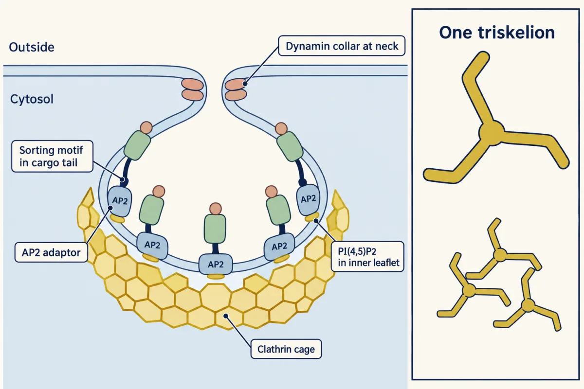
The first film built the coated vesicle, whose lumen is topologically outside the cytosol (membrane topology); here is what it leaves out, for mammalian cells.
- The cage. Clathrin is a coat, a protein lattice that assembles on one face of a membrane and bends it. The first film’s three-legged piece is the triskelion, three heavy and three light chains joined at a hub; the legs interlock into hexagons and pentagons, like a geodesic dome.
- The adaptors. AP2 and other clathrin-associated sorting proteins bind PI(4,5)P2, a phosphorylated lipid of the inner leaflet (lipids), and read short sorting motifs in cargo tails. AP2 anchors the coat and binds YXXΦ motifs (tyrosine, any two residues, then a bulky hydrophobic residue), such as the transferrin receptor’s. The low-density lipoprotein (LDL) receptor’s tail carries NPXY instead, which two other adaptors, Dab2 and ARH, can recognize; they also bind clathrin and AP2. The first film’s adaptors stand for the whole set.
- The cut and the uncoating. Dynamin is a GTPase, an enzyme that hydrolyses GTP; using that hydrolysis, its collar drives the neck to split. The chaperone Hsc70, recruited by its helper auxilin, then pulls the lattice apart, so the triskelia can be reused and the vesicle can fuse with an endosome.
J.D.’s receptors, seen and counted
In 1977 Richard Anderson, with Brown and Goldstein, bound LDL coupled to ferritin (an iron-storage protein that shows as a dot in the electron microscope) to cultured fibroblasts at 4 °C. In normal cells 60 to 70 percent of the label lay over coated segments covering no more than 2 percent of the surface, and on warming it entered through coated vesicles. Take 60 percent, and set total label and area to 1.
density in pits ÷ whole-surface density = 0.60 ÷ 0.02 = 30
uncoated density = 0.40 ÷ 0.98 = 0.408
density in pits ÷ uncoated density = 30 ÷ 0.408 = 73.5
Check: 0.02 × 30 + 0.98 × 0.408 = 0.60 + 0.40 = 1. Both are right; they differ only in the reference (whole surface or uncoated membrane), so say which you mean. These are calculated ratios, not binding constants.
Predict. Ferritin-LDL is bound to J.D.’s cells in the cold. If the receptors are spread at random over the surface, what fraction of the label lies over coated pits, and what is the density ratio over pits?
Check your prediction
About 2 percent of the label, so the density over pits matches the whole-surface average: a ratio near 1, not 30. The reported result fits: in J.D.’s cells the receptors that bound LDL failed to become incorporated into coated pits (Anderson, Goldstein and Brown, 1977).
The cause was inside the cell. In 1986 Davis and colleagues traced J.D.’s receptor to a single tyrosine-to-cysteine substitution in its short tail in the cytosol. Rebuilt in receptor-deficient hamster cells, it bound LDL normally but entered slowly.
The fork: acid parts receptor from cargo
The first film’s fork is the early endosome’s mildly acid lumen, roughly pH 6 (reported values run from about 5.6 to 6.5) and kept so by proton pumps (organelles as environments). For low-density lipoprotein (LDL), acid is the sorting signal: at this pH LDL comes off its receptor. A crystal structure of the receptor’s outer part at acid pH suggests one way (3D viewer): the arc of ligand-binding repeats folds back over a propeller-shaped domain, which binds two of them and so takes the place of LDL. The receptor grips itself and lets the cargo go.
The first film used the simplest case, one cargo that lets go; here is the fuller picture. Letting go does not by itself decide where a thing goes; the sorting machinery, reading each cargo and receptor, does. A cargo that holds on shows this.
Predict. Transferrin carries iron in the blood and enters cells on the transferrin receptor by the same route. In the acid endosome the iron comes off, but transferrin itself stays bound to its receptor. Where does transferrin go?
Check your prediction
Back to the surface, still on its receptor. Outside, at neutral pH, transferrin without iron binds its receptor only weakly, so it lets go and can pick up iron again. The iron, which let go in acid, stays in the cell.
Return: the loop closes, or runs down
The first film closed the loop; this section asks what happens when release fails, and how that fools the assay. Returning receptors reach the plasma membrane directly or by way of a recycling endosome. In the lysosome, enzymes digest LDL’s protein and hydrolyse its cholesteryl esters, releasing cholesterol, which damps the cell’s own synthesis through the enzyme 3-hydroxy-3-methylglutaryl coenzyme A (HMG-CoA) reductase (lipid synthesis).
Predict. A receptor engineered without its EGF-precursor domain (an outer stretch resembling the precursor of epidermal growth factor) still enters cells, but cannot let go of its bound ligand at acid pH. Follow it through several rounds of uptake. What happens to it, and to the amount of receptor on the surface?
Check your prediction
It returns inefficiently. Still carrying its ligand, much of it is rapidly degraded instead (Davis and colleagues, 1987), so surface receptor falls round by round. A release defect cuts return and raises degradation: the rule at work.
The first film’s picture was the simple case: one batch of receptors through one round. Over hours, though, the cell also makes and destroys receptor, so a late block can show up early. Suppose, for illustration, that with protein synthesis blocked half the receptors return each round: surface receptor falls to 0.5, 0.25 and 0.125 of the start over three rounds (a normal cell returning 99 percent keeps 0.99, 0.98, 0.97). Hours in, a cold-binding assay on the defective line reads like a binding defect, so measure binding before uptake starts.
One defect can also hide another. A patient receptor lacking exons 7 to 14 failed to recycle efficiently, but it also could not bind low-density lipoprotein (LDL), which hid the return defect until it was tested with beta-VLDL, another lipoprotein that it still bound. That is why the rule asks for the earliest step that fails, not the only broken one.
Does the LDL move, or the endosome change?
The first film said the endosome matures. How do we know? LDL appears first in early endosomes and later in late ones, and fixed cells cannot tell whether it moved between permanent compartments or its compartment changed. Compartments carry name tags, Rabs: small GTPases that, bound to GTP, sit on one membrane and mark its identity by recruiting the tethers and enzymes that belong there. In 2005 Marino Zerial’s laboratory imaged live human A431 cells with fluorescent Rab5 (early) and Rab7 (late) taking up LDL. On single endosomes carrying LDL, Rab5 was replaced by Rab7, which the authors read as Rab conversion: the compartment matures. Rab4-positive recycling tubules separated from Rab5-positive endosomes by fission, so maturation and carriers coexist.
Design problem
A patient’s receptor appears to reach the cell surface, but the cells lose surface receptor after repeated rounds of uptake. Design experiments to show a recycling defect.
Saved in this browser only.
One way to do it
Choose a ligand the mutant still binds (beta-VLDL if, like the exons 7 to 14 receptor, it cannot bind LDL). Measure surface receptor with an antibody to its outer part, surface biotinylation (tagging surface proteins with biotin) or Pronase sensitivity (an outside protease clips only surface receptor), and total receptor by immunoblot.
With protein synthesis blocked by cycloheximide, so no new receptor refills the surface, measure surface receptor after each round of uptake: a recycling defect predicts normal entry with surface receptor falling round by round, while normal cells hold it nearly steady. Faster destruction could also explain the loss, so label the receptor at the surface, let it enter, strip the surface, and measure how fast it returns and how much is degraded. Also measure acid release of bound ligand, since failure to release can keep the receptor from returning.
Controls: normal cells, mutant and normal cells matched for starting surface receptor, and a line whose receptor cannot bind the ligand used (it should show no ligand-driven loss).
Every receptor so far was meant to return. The second film picks up here: a receptor meant to be destroyed, and how the cell gets all of it digested.
Read the transcript of “The Receptor That Does Not Come Back”
Film transcript: The Receptor That Does Not Come Back
How can a cell destroy a membrane-spanning receptor without leaving part of it behind? In chapter one, the receptor escaped. It released its LDL in acid and left before the end. But some receptors are meant to be destroyed.
Many activated growth-factor receptors are like this, shown here in sky blue. Activation can add ubiquitin, a small protein shown in green. The tagged receptors enter an early endosome. Ubiquitin helps mark them for lysosomal degradation. Here is the problem. The receptor sits in the endosome's own membrane, with its tail out in the cytosol.
Suppose this endosome simply fused with a lysosome. Could the enzymes inside reach the whole receptor? Not all of it. The enzymes work in the lumen, so only the part facing the lumen is within their reach. The cell gets around this by moving the receptor off that outer membrane.
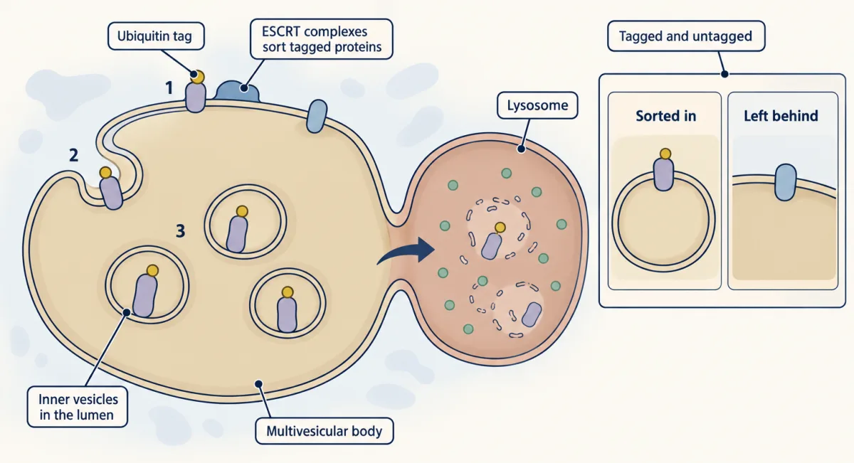
From the early endosome on, patches of its outer membrane, the limiting membrane, bud inward into the lumen. Each bud pinches off as a small vesicle inside. An endosome full of them is a multivesicular body. What decides which proteins go into the buds?
Complexes called ESCRT, in coral, recognize the ubiquitin tag. They sort the tagged receptors into the inner vesicles. In this comparison, an untagged protein stays behind. The multivesicular body then fuses with a lysosome. Each inner vesicle now floats whole in the lumen, receptor and all, and the enzymes digest it. This is how those receptors are switched off.
So the endosome sorts more than cargo. A receptor left in the limiting membrane can still return. A ubiquitin tag can select a receptor for inward budding. The lysosome can then digest all of it. Even a looping receptor can be pulled out of its loop.
PCSK9, in cyan, is a protein that cells secrete. It binds one small part of the LDL receptor's outer region. Cultured human cells show what happens next. A receptor bound this way goes to the lysosome instead of returning to the surface. Its loop has become a one-way trip. The receptor does not come back to grip again.
So a cell can destroy a receptor, not just its cargo. A ubiquitin tag can select the receptor for inward budding at the endosome. Inward budding puts the whole receptor within lysosomal enzymes' reach.
From the film
- Some receptors, such as many activated growth-factor receptors, are meant to be destroyed, not reused.
- A ubiquitin tag marks such a receptor, and ESCRT complexes at the endosome recognize the tag.
- ESCRT sorts tagged receptors into vesicles that bud into the endosome's lumen, making a multivesicular body.
- When it fuses with a lysosome, the inner vesicles are digested, receptors and all.
- In the cultured human cells tested, PCSK9 sends the LDL receptor to the lysosome instead of back to the surface.
When the receptor itself is the cargo
The second film showed this sorting sequence. Some receptors, such as many activated growth-factor receptors, are meant to be destroyed: tagged with the small protein ubiquitin, the receptor itself rides to the lysosome. From the early endosome on, patches of the endosome’s outer (limiting) membrane bud into the lumen; an endosome full of these inner vesicles is a multivesicular body. ESCRT complexes (endosomal sorting complexes required for transport), ESCRT-I among them, recognize the ubiquitin tag and sort the tagged protein into the inner vesicles. When the multivesicular body fuses with a lysosome, the inner vesicles are digested. This is how those receptors are switched off (turning signals off, receptor tyrosine kinases, signalling endosomes).
A recycling receptor can also be pulled out of its loop. PCSK9, the second film’s last example, is a secreted protein that binds the EGF-A part of the low-density lipoprotein (LDL) receptor’s EGF-precursor domain (the 3D viewer shows the pair). In the cultured human cells tested, it sends the receptor to the lysosome instead of back to the surface.
Predict. An antibody that blocks PCSK9 is given. What should happen to the LDL receptor on liver cells, and to blood LDL?
Check your prediction
More receptors finish the loop, so surface receptor rises, the liver takes up more LDL, and blood LDL falls. People who carry loss-of-function PCSK9 variants have lower LDL, and PCSK9-blocking antibodies are approved LDL-lowering drugs.
Other doors into a cell
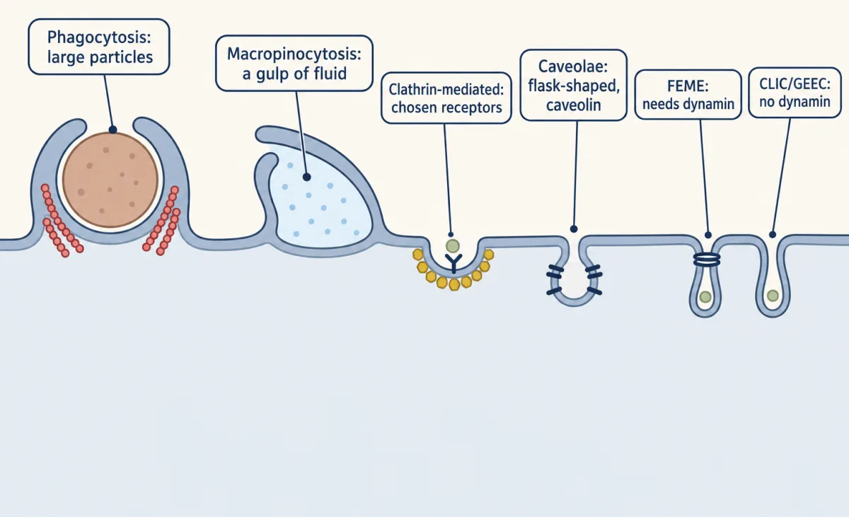
Both films followed one door, the clathrin-coated pit, the best-understood route for receptors. Cells have others, each with its own machinery.
| Route | Typical cargo or defining feature | Machinery |
|---|---|---|
| Phagocytosis | Large particles, such as bacteria or dead cells, into phagosomes | Actin; in animals mostly specialized cells, such as macrophages and neutrophils |
| Macropinocytosis | Gulps of fluid, in large vacuoles | Sheet-like projections fold back onto the surface; stimulated by growth factors in many mammalian cells |
| Clathrin-mediated | Chosen receptors, such as the LDL and transferrin receptors | Adaptors, clathrin and dynamin (this lesson) |
| Caveolae | Small flask-shaped pits; cargo disputed (Going deeper) | Caveolin |
| FEME and CLIC/GEEC | Distinct clathrin-independent routes | FEME (fast endophilin-mediated endocytosis) needs dynamin; CLIC/GEEC does not |
Going deeper
How the coat bends
This is still argued. Live imaging in human SK-MEL-2 cells found curvature appearing early in some selected events and only after much of the clathrin had arrived in others. A 2023 analysis of fixed coats in three cell lines inferred that coats, on average, gain about half their final area before bending, with a fitted preferred radius of about 72 nm in SK-MEL-2 cells. The vesicle traffic lesson treats coats in general.
Not every clathrin cargo leans on AP2
In HeLaM cells depleted of AP2, a chimeric protein carrying the low-density lipoprotein (LDL) receptor’s tail was still taken up, consistent with other adaptors reading its NPXY motif. That result is about one construct in one cell line; it does not make AP2 dispensable in general.
FH beyond the three defects
Familial hypercholesterolaemia (FH) is usually inherited as a dominant trait, and people with two defective copies are rarer and more severely affected (see pedigrees). Many FH mutations make little or no receptor, or a receptor held in the endoplasmic reticulum; a cold-binding assay reads these as binding defects. Some FH comes from changes in other genes, such as APOB, or gain-of-function changes in PCSK9. J.D.’s substitution is at residue 807, in the numbering of the 1986 paper.
Caveolae and the clathrin-independent routes
Caveolae are often drawn as a major entry route, but a 2020 review cautions that few if any cargoes are shown to rely on caveolae to enter. Their clearest job is mechanical: they flatten when the membrane is stretched, which buffers the tension. FEME is triggered when ligands activate selected surface receptors.
The ESCRT hand-off
ESCRT-0 binds the ubiquitinated cargo first and passes it to ESCRT-I and ESCRT-II, which also bind ubiquitin, and ESCRT-III cuts the inner vesicles free. These complexes were first worked out in yeast and also work in mammalian cells.
Your notes
Select any text in the lesson to highlight it or add a note to it. Your highlights and notes are saved in this browser and collected in your notebook.
This browser is not letting the site save anything (a private window or strict privacy settings), so highlights and notes will be lost when you leave the page.
How we measure it
Cold binding, warm uptake
Cells are given labelled ligand at 4 °C, where it binds while membrane traffic is largely arrested, then washed and warmed to 37 °C for a set time. An acid or heparin wash strips what is still on the surface, so binding and internalization are measured separately in the same cells.
Ferritin-LDL electron microscopy
LDL is coupled to electron-dense ferritin so each particle shows as a dot in thin sections. Counting dots over coated and uncoated membrane shows where bound ligand gathers. It reports where the ligand is, not the receptor directly.
Site-directed mutagenesis and expression
A patient's mutation is rebuilt in a cloned receptor and expressed in cells that lack their own receptor, such as receptor-deficient hamster cells. If the rebuilt receptor shows the patient's defect, that one change is enough to cause it.
Live-cell Rab imaging
Rab proteins tagged with fluorescent proteins are followed on single endosomes over time while the cell takes up a labelled cargo. A compartment that changes its Rab coat while keeping its cargo is maturing; cargo hopping between stable compartments is not.
Method films · the methods library
- Cell, primary and explant culture 3:58
- Electron microscopy 4:14
- Engineered mutants: site-directed, dominant-negative and constitutively active 4:25
- Gel electrophoresis, SDS-PAGE and western blot 4:01
- Live-cell time-lapse imaging 4:01
- Pulse-chase labelling 2:40
- Receptor binding and dose-response analysis 4:18
- RNAi and antisense knockdown 4:25
- X-ray crystallography 4:06
Practice questions on this lesson (3) → MCAT-style, original, each with an explanation
Where this lesson fits
Before thisReview first
- Vesicle Budding, Targeting and FusionThis course · Lesson 11
This lesson reuses coated vesicle budding, Rab identity and fusion with endosomes without re-teaching them.
- Organelles as Distinct EnvironmentsThis course · Lesson 6
You need proton pumps setting endosome and lysosome pH to see why acid makes LDL let go of its receptor.
- Protein Sorting into CompartmentsThis course · Lesson 7
Short tail motifs read by adaptors, like the NPXY motif here, build on how signals sort proteins.
After thisStudy next
- Cell Identity, Cell State and DiseaseThis course · Lesson 25
Classifying a receptor defect at binding, entry or return carries over to reading how cell state and mutations produce disease.
Elsewhere on the siteFilms, simulations and method films in other courses
- Shape is destinyFilm · Biochemistry
It covers cholesterol as a membrane lipid, the cargo LDL delivers to cells.
- The cholesterol gaugeFilm · Biochemistry
It shows how LDL import and cholesterol synthesis together control a liver cell's cholesterol supply.
Suggested by an AI model from the lessons themselves, then checked. How we use AI
Finish line
Test yourself
8 questions
Practice and MCAT-style, with explained answers.
Next · Lesson 14
Cytoskeletal Polymers and Dynamics
A microtubule in a dish of pure tubulin grows for minutes, then collapses many times faster. Why would a polymer be built to fail? Because a polymer that switches by itself lets a cell explore space, rebuild its scaffolding and organize division.
Continue →