Cell Biology · Unit 5: Cells in tissues
Adhesion and the Extracellular Matrix
Rewritten on : new text, flashcards and film. Your notes and highlights stay, but the text around them may have moved.
Take the calcium away and a sheet of epithelial cells comes apart. What was the calcium holding together?
Pause when the reading reaches a film, and offer to play it. Saved on this device.
Watch the film now, or keep listening
Builds on:Cytoskeletal Polymers and DynamicsMembrane Organization and TopologyThe Secretory PathwayWhy these?
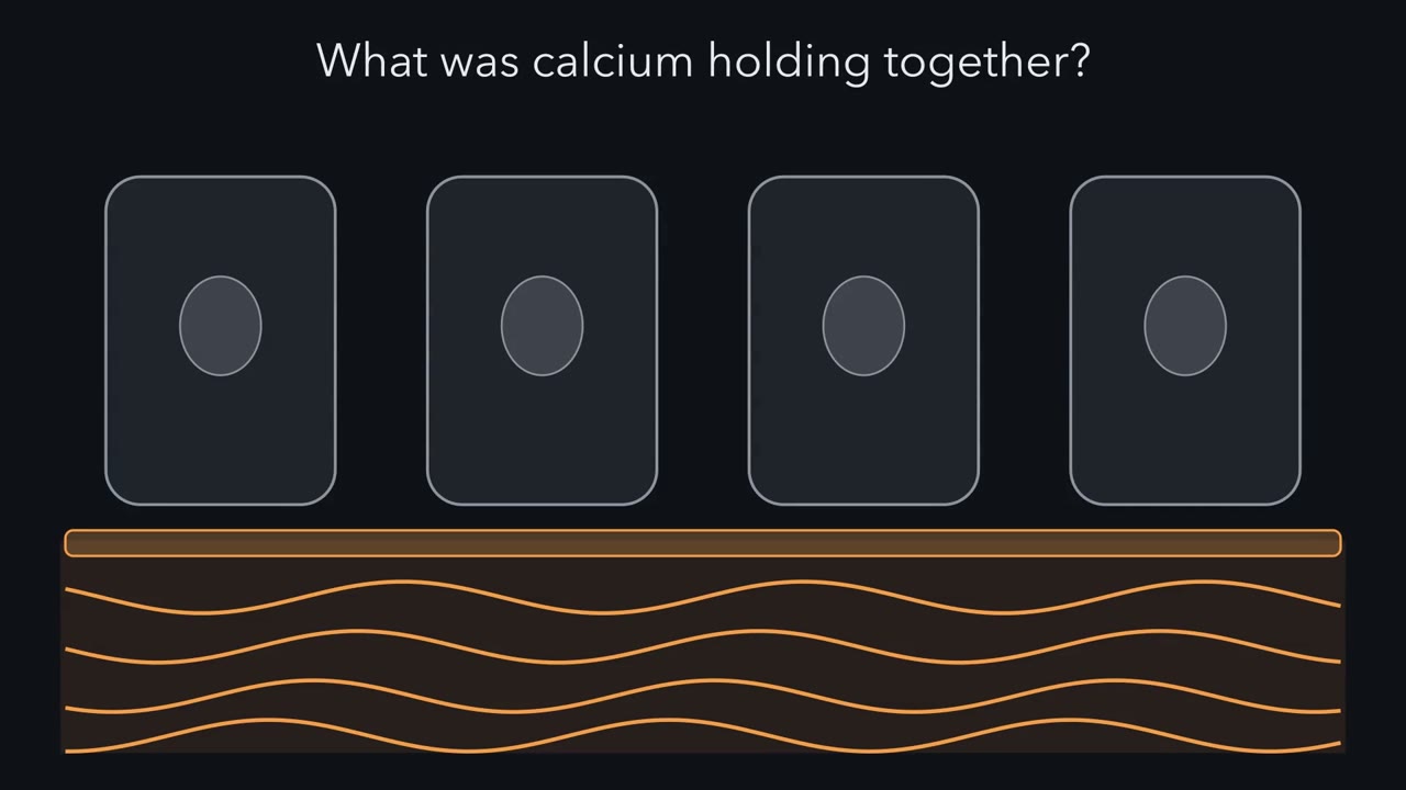 ▶ Watch One Chain, Four Links · 3:26
Read the transcript of “One Chain, Four Links”
Film transcript: One Chain, Four Links
Take the calcium away, and this sheet of cells comes apart. What was the calcium holding together? Put it back, and the cells knit together again. Each cell reaches across with arms called cadherins. Calcium sits at the joints of each arm and keeps it stiff. The arm on one cell grips the arm on the next. Follow one arm into the cell. Adaptor proteins grip its tail and pass the hold inward: beta β-catenin, then alpha α-catenin. They tie it to a cable, a filament of actin. This grip is an adherens junction. So, in its simplest form, one grip is a chain of four links. A partner outside, a receptor in the membrane, adaptors, and a cable inside. Now pull on the sheet.
In this simple picture, the pull runs from cable to cable, through every link. Break any one link, and this path is cut. Now take the calcium away. Which link gives way first? The arms outside, the adaptors, or the cable?
Choose one. Calcium leaves the joints, and the arms go floppy. The outer link lets go. The adaptors and the cable are still there, but this contact can no longer pass the pull across. One broken link, and the sheet comes apart. Now look down, at the matrix, the material cells secrete around themselves. Here the receptor is an integrin, a pair of subunits, α and β. It grips a matrix protein, fibronectin, at a short tag, RGD. Inside, talin ties the integrin's tail to actin, and vinculin adds extra ties to the same cable. Notice the pattern. The receptor has changed, but the four links have not. When the cell tugs on its cable, the pull runs down into the matrix.
Two more junctions build the same chain with a different cable. Desmosomes join cell to cell. Hemidesmosomes pin a cell to the basement membrane, the thin matrix sheet beneath. In epithelia, both end on keratin, an intermediate filament, not on actin. At the chain's far end is the matrix itself. Its collagen ropes straighten under a pull and resist it strongly. But squeeze the tissue, and the ropes buckle and go slack. Ropes are poor at pushing. So the matrix also holds a gel of proteoglycans, proteins with long sugar chains. In a first picture, the chains' negative charges draw in water, and the gel swells. Squeeze it, and it pushes back. The tissue is one web of chains, from matrix ropes to cell cables.
A real junction holds many chains side by side, sharing the load. One question is left. Does the pull change the links themselves?
From the film
- In its simplest form, each anchoring grip is one chain: partner outside, receptor, adaptors and a cable inside.
- In one simple chain, the pull passes through every link, so one missing link cuts that path.
- At adherens junctions, calcium keeps the cadherin arms stiff; without it, the outer link lets go.
- In epithelia, desmosomes and hemidesmosomes build the same kind of chain but end on keratin cables, not actin.
- Collagen ropes resist pulling far better than squeezing; water-holding proteoglycan gels resist the squeeze.
The first film followed one grip, from a neighbour’s cadherin to the cell’s actin. What it leaves out: the name of every link, where skin splits when one fails, the matrix’s working parts, and the experiments that found the links.
The one idea, in full. In the anchoring junctions of epithelia (the cell sheets that line and cover the body) and of cells on matrix, every grip is one chain of four links: a partner outside (a neighbour’s cadherin, or a matrix protein), an adhesion receptor, adaptor proteins, and a cable inside the cell. The first film drew the simplest case, one unbranched chain, where the pull passes through every link and a missing link cuts the path. A real junction is the fuller case: many chains, some branched, share and shift the load while bonds let go and rebind. So a missing or weak part weakens adhesion, but how a junction fails also depends on load sharing, bond kinetics (how fast bonds let go and re-form) and remodelling.
A cable is a filament inside the cell (actin, or keratin, an intermediate filament); collagen, outside, is a rope.
The chain, link by link
| Junction | Joins | Partner outside | Receptor | Adaptors | Cable |
|---|---|---|---|---|---|
| Adherens junction | cell to cell | the neighbour’s cadherin | classical cadherin | β-catenin and α-catenin (αE-catenin in epithelia); p120 near the membrane | actin |
| Desmosome | cell to cell | the neighbour’s desmosomal cadherin | desmogleins and desmocollins | plakoglobin and plakophilins, then desmoplakin | keratin |
| Focal adhesion (cultured cells) | cell to matrix | fibronectin and other matrix proteins | integrin (an α-β pair) | talin, reinforced by vinculin | actin |
| Hemidesmosome | cell to basement membrane | laminin-332 | integrin α6β4, with collagen XVII | plectin | keratin |
In the keratin rows, desmogleins and desmocollins are the desmosome’s own cadherins, held to keratin by a plaque of the three adaptors; laminin-332 is a basement-membrane laminin, and collagen XVII is a second membrane anchor beside the integrin.
Cadherins. A classical cadherin crosses the membrane once and usually binds the same kind of cadherin on the neighbouring cell (homophilic binding). Adherens junctions are belts of cadherin circling the top of an epithelial cell (epithelia builds the full sheet). p120-catenin binds the tail close to the membrane, the juxtamembrane region, and helps keep the cadherin at the surface. β-Catenin has a second job: outside junctions it is the transcriptional co-activator of the Wnt pathway (the Wnt lesson), and cells regulate how much goes to each job.
Integrins. Mammals pair 18 α and 8 β subunits into 24 integrins, each with its own preferred ligands. Eight, including the main fibronectin receptor α5β1, recognise the RGD tag (Arg-Gly-Asp); others, such as α6β4, bind by other sites. The grip is switched from inside. Bent with its head closed, or extended with its head still closed, an integrin binds its ligand weakly; extended with its head open, it binds strongly. Talin, helped by a second adaptor called kindlin, binds the β tail and shifts the integrin toward the open shape. That shift is integrin activation.
Fibronectin also ties the chain into the rest of the matrix: besides integrins, it binds collagen and heparan sulfate (one of the matrix’s sugar chains). Crawling cells make and break their focal adhesions constantly (migration and cilia); the adhesions’ signals through Rho GTPases belong to integrins and Rho GTPases.
Predict. Carcinoma cells (cancer cells of epithelial origin) that make E-cadherin but no α-catenin are put in calcium. Do they stick together, and how well?
Check your prediction
Poorly. In the PC9 cells studied by Hirano and colleagues (Cell, 1992), E-cadherin was present, but calcium-dependent aggregation was greatly reduced and the cells grew mostly singly; putting α-catenin back restored firm, compact aggregates. Binding outside is not enough for strong adhesion, though the experiment did not measure the force on one contact.
Where the chain breaks: blisters
Here the chain does real work: a missing link cuts the path, so the level of a split points to the layer whose chain failed. In skin, basal cells, the bottom layer of the epidermis, hold their neighbours at desmosomes and the basement membrane at hemidesmosomes, and collagen VII anchors that membrane to the dermis below.
Predict. Three children have inherited blistering of the skin (epidermolysis bullosa), from faults in keratin 14, in laminin-332, or in collagen VII. Where does each blister split? Then two adults make antibodies against their own skin proteins, one against desmoglein 3 and one against collagen XVII. Where does each of theirs split?
Check your prediction
- Keratin 14: inside the basal cells, because the cable itself fails (epidermolysis bullosa simplex).
- Laminin-332: within the basement membrane, where the hemidesmosome’s partner should be (junctional epidermolysis bullosa).
- Collagen VII: below the basement membrane, where it should anchor to the dermis (dystrophic epidermolysis bullosa).
- Anti-desmoglein 3: between epidermal cells just above the basal layer, as their desmosomes let go (pemphigus vulgaris).
- Anti-collagen XVII: below the epidermis, at the hemidesmosomes (bullous pemphigoid). Complement, inflammatory cells and protein-cutting enzymes add to the damage, so this is no simple snap of one link.
The split narrows the defect to a layer or adhesion complex; since several proteins can fail at one level, naming the protein takes genetic or antibody tests.
Where the rule stops. Some cell-cell contacts need no calcium. The Chinese hamster V79 cells that Masatoshi Takeichi studied in 1977 had a calcium-independent mechanism beside the calcium-dependent one, though it looked like simple clumping; immunoglobulin-family proteins such as NCAM are clearer cases. T-cadherin and one form of NCAM are held to the membrane by a lipid (GPI) anchor, so they bind with no adaptor and no cable. And not every integrin grips matrix: on white blood cells the integrin LFA-1 binds ICAM-1 on another cell.
The far end: what the matrix is made of
Every integrin chain ends in the matrix; here is that far end, taken apart.
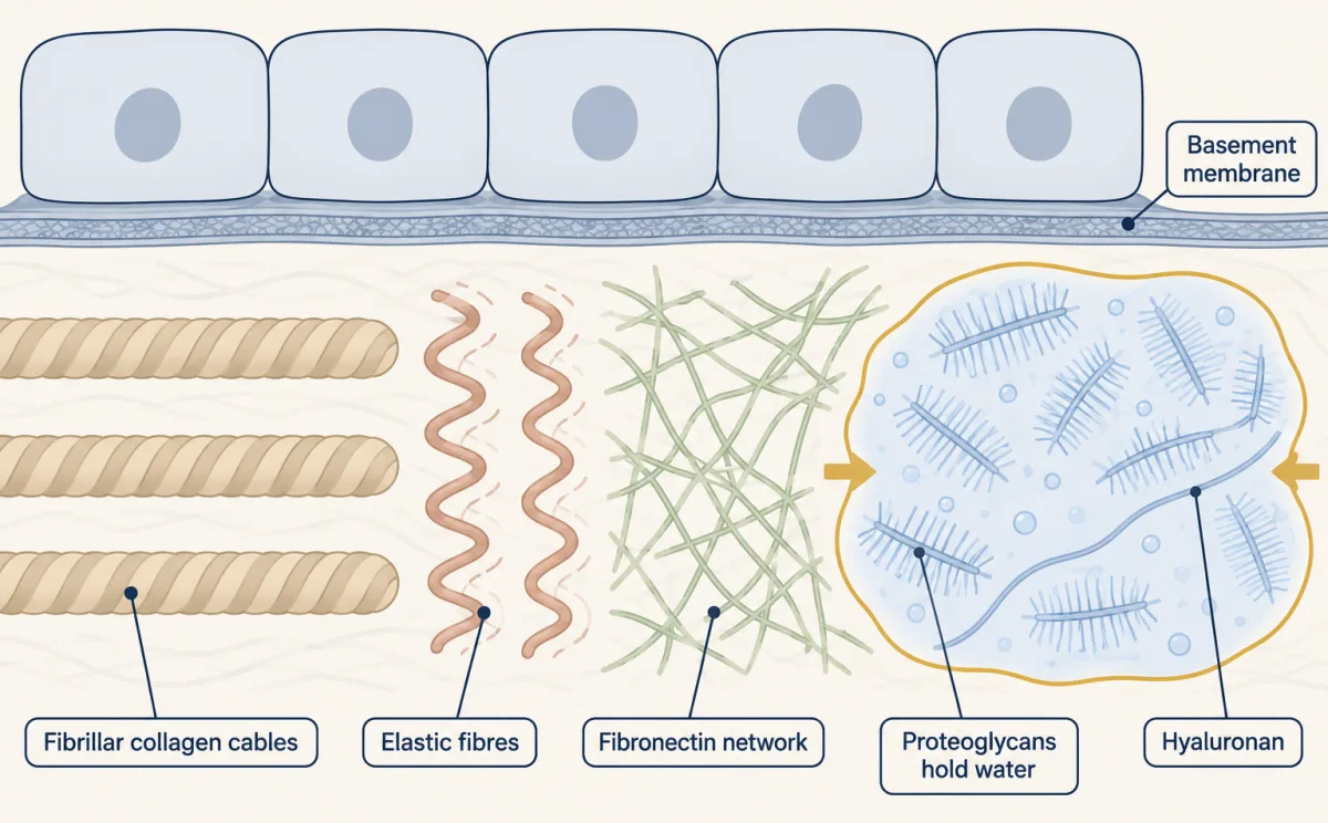
| Part | What it is | What it does |
|---|---|---|
| Fibrillar collagens | three chains wound into a triple helix; the helices pack side by side into fibrils | the ropes: strength under tension in bone and tendon |
| Elastic fibres | stretchy fibres | let artery walls and lung stretch and recoil |
| Fibronectin | fibrillar networks | a mesh cells grip through integrins; also binds growth factors and other signalling molecules |
| Basement membrane | two linked networks, one of laminins and one of type IV collagen (which forms networks, not ropes) | a thin sheet under almost every epithelium |
| Proteoglycans | core proteins carrying long glycosaminoglycan chains, sugar polymers studded with negative charges | a hydrated gel that resists compression, as in cartilage |
| Hyaluronan | a very long, unsulfated glycosaminoglycan with no protein core | a water-binding polymer, and the backbone for aggregates of hyaluronan-binding proteoglycans such as aggrecan |
Cells secrete fibrillar collagens as soluble precursors and trim them outside (the secretory pathway); the triple helix is in protein 3D structure, and the sugar chemistry in carbohydrates.
The first film used the simplest picture of the gel; here is the fuller one: the fixed negative charges attract mobile positive ions, and that excess of ions draws water in by osmosis, so the gel swells and pushes back when squeezed. Cartilage’s aggrecan binds along hyaluronan into aggregates large enough to stay trapped in the collagen net, which holds the swelling gel in.
The matrix is also a store. Growth factors held in it can be released when the matrix is cut or pulled, and the pull can come from cells tugging through their own integrin chains. Latent transforming growth factor beta (TGF-β) is the standard example (TGF-β and Smads). Neurons use basal lamina too: the agrin that organises the neuromuscular junction is deposited there.
Predict. A mutation puts alanine in place of one glycine in type I collagen’s triple helix (the collagen-like peptide in the 3D viewer carries this kind of swap). Which tissues fail, and under which load?
Check your prediction
Tissues that rely on collagen ropes for strength under tension, above all bone, which fractures easily. Glycine sits at every third position, where the strands pack closest, and a larger side chain loosens the helix there. Glycine substitutions in type I collagen are the commonest cause of severe osteogenesis imperfecta, brittle bone disease (Marini and colleagues, 2017).
So far each link has simply held or broken. The second film picks up here: can the pull change a link itself?
Read the transcript of “Links That Feel the Pull”
Film transcript: Links That Feel the Pull
Chapter one ended with a pull running through every link of the chain. Can that pull change a link itself? To find out, start where the pull begins, inside the cell. Here, a motor protein called myosin II grips the actin cables and slides them past one another. So the cell can keep its own chains under load. That pull reaches every link, down to the matrix. Look closer at the integrin chain's adaptor, talin. Its long rod contains 13 small bundles of alpha α-helices. Each bundle is folded up tight. Some of these bundles hide binding sites for vinculin, the second adaptor you met in chapter one. While a bundle stays folded, its site stays tucked away inside.
Now take a purified piece of the talin rod, outside any cell, and stretch it. A test piece from vinculin waits beside it. What happens to those hidden sites? Make a guess. The bundles unfold, and the hidden sites open. The vinculin test pieces bind to them. Back in a cell, activated vinculin can add another tie to actin. So pulling can recruit reinforcements. Magnetic tweezers pulled a bead tied to a single mouse talin rod.
Bundles unfolded at about 5 to 25 piconewtons, trillionths of a newton. They refolded as force fell. A model using those rates suggests the rod can buffer force. The cadherin chain has a similar trick. Alpha α-Catenin has a hidden vinculin site of its own, and tension opens it. So both chains can reinforce themselves under load. But is force the only key? Talin and vinculin each fold shut on their own, hiding their binding sites. This self-closing is called autoinhibition. Researchers tested this in mouse fibroblasts. Normal, autoinhibited talin and vinculin did not bind each other at mitochondria. Releasing either protein's autoinhibition enabled binding without force.
Those active forms still supported disorganized adhesions when myosin II was blocked. So force can expose binding sites, while engineered release from autoinhibition can bypass force. Whether a cell's own signals do that at the same steps in a tissue is not known. And a few piconewtons on one molecule are not the pull of a whole cell. One more link, where the catenins meet actin, behaves oddly under force. The text picks that up next. So a chain is more than fixed parts. Some links do not just hold or break; they respond to pull. Pulling can open sites and recruit ties. Engineered release from autoinhibition can bypass force.
From the film
- Myosin II can pull on actin cables, so a cell can load its adhesion chains from inside.
- Stretching a purified talin rod exposes hidden sites to a vinculin-head test piece; in cells, activated vinculin can add a tie to actin.
- α-Catenin has a hidden vinculin site that tension opens, so the cadherin chain can reinforce itself too.
- Normal talin and vinculin did not bind at mitochondria; releasing either protein's autoinhibition enabled force-independent binding.
- Active forms supported disorganized adhesions with myosin II blocked, but how normal signals bypass force in a tissue is unknown.
Which links feel the pull?
The second film showed a stretched piece of talin, and tensed α-catenin, opening hidden vinculin sites (for α-catenin, Yonemura and colleagues, 2010; Yao and colleagues, 2014). It then showed that engineered release of talin or vinculin autoinhibition lets the pair associate without force; whether normal signals bypass force at the same steps in a tissue is unknown. The film left out the link between the catenins and actin.
A puzzle: work in cells put α-catenin between the cadherin–β-catenin complex and actin, yet in 2005 purified α-catenin would not bind that complex and an actin filament at the same time. With no force applied, the link looked missing.
A bond’s lifetime is how long it holds before letting go; for an ordinary bond, a slip bond, pulling harder shortens it. The forces here are piconewtons (pN), trillionths of a newton.
Predict. Suppose the α-catenin–actin link is weak at rest but holds longer when pulled, up to a point. Sketch how its bond lifetime should change as the force rises from zero to about 20 pN. Why might a test tube with no force miss the link?
Check your prediction
Rise, then fall: short at almost no force, longer at a moderate force, short again once force tears the bond apart. A bond that lives longer as force rises, over a limited range, is a catch bond. On this hypothesis a zero-force assay mostly samples the weak state and might miss the link. In 2014 a reconstituted cadherin–catenin complex held against an actin filament in an optical trap (a laser that holds a bead and reads the force on it) behaved this way: some of its bonds lasted longer under a small force (numbers in Going deeper). That is one way to reconcile the 2005 result, not proof of why it came up empty.
A link’s lifetime can depend on the pull: a moderate force can steady the catenin–actin link compared with its own low-force state (the experiment did not compare it with the junction’s other links).
In culture, naive mesenchymal stem cells on soft, brain-like gels drifted toward neuronal-like cells, and on rigid, bone-like gels toward bone; blocking myosin II removed the effect (Engler and colleagues, 2006). One reading is that a cell feels stiffness by pulling on its chains and sensing how much the far end gives. This is a culture result, not a statement about fate in a body.
Reading adhesion assays
This section is a separate tool: the links were found by experiment, and reading one is how you would test a link yourself. Classic cadherin and integrin experiments used dissociation and reaggregation assays (separate cells, then measure re-clumping), attachment assays (count cells left on a coated well after washing) and antibody-blocking versions of them.
Worked problem (made-up data). Wells are coated with fibronectin or with bovine serum albumin (BSA) only, a protein cells do not grip. Each well gets 20,000 fibroblasts. After 30 minutes and a standard wash, the retained cells are:
- fibronectin, no antibody: 14,000
- fibronectin plus isotype-control antibody (same antibody class, irrelevant target): 13,600
- fibronectin plus integrin-blocking antibody: 3,200
- BSA only: 1,000
Viability is above 95% in every well, so lost cells were not dead cells.
Predict. The BSA well counts cells that stick to any surface. If you compared raw counts in the blocking and isotype wells without removing them, would the antibody look more or less effective than it really is?
Check your prediction
Less effective: background cells sit in both wells and dilute the drop. Raw counts give 1 − 3,200/13,600, about 76.5%, against 82.5% in Step 2.
Step 1: subtract the background. The BSA well shows cells that stick to plastic or protein nonspecifically. Specific attachment is 13,600 − 1,000 = 12,600 with the isotype control, and 3,200 − 1,000 = 2,200 with the blocking antibody.
Step 2: inhibition.
inhibition = 1 − (blocking well − background) / (isotype well − background)
1 − 2,200/12,600 = 1 − 0.175 = 0.825, or about 83%.
Step 3: why the isotype well. Of the 20,000 seeded, 70% attached with no antibody, 68% with the isotype control, 16% with the blocking antibody and 5% on BSA. An irrelevant antibody cost 2 points; comparing with the no-antibody well would credit that drop to the integrin.
Step 4: what this does not show. An antibody can block by sitting near the receptor, or by locking it in an inactive shape. Lower attachment implicates the integrin in attachment; it does not show that the integrin touches fibronectin. Horwitz and colleagues made that next step in 1985 with purified antigen in equilibrium gel filtration, and the antibody blocked the purified laminin association.
Then check each well in the reader.
Subtract the BSA background from both wells, then compare the blocking well with the isotype-control well, not with the well that has no antibody. Lower attachment shows the integrin is needed, not that it touches fibronectin. The counts are made-up data.
Design problem
Do cadherins sort cells into tissues? Design an experiment in the spirit of Nose, Nagafuchi and Takeichi (1988), using mouse L cells, which have little cadherin of their own.
Saved in this browser only.
One way to do it
Make two L-cell lines, one expressing E-cadherin and one expressing P-cadherin. Label them with different dyes, mix them, and let them aggregate in calcium.
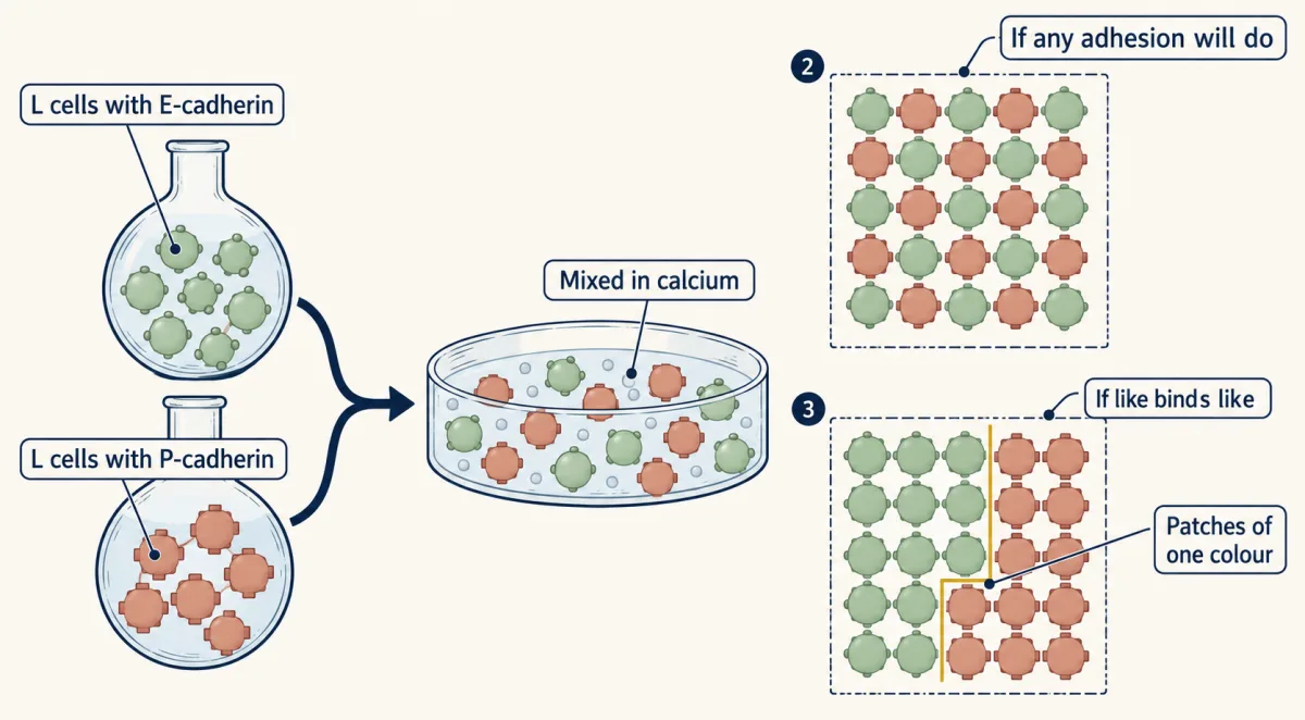
Predictions: as in the figure. Patches show a preference, not exclusivity: different classical cadherins can also bind one another. Nor do patches alone show how weakly the two cadherins bind each other, because cortical tension and cell movement also shape sorting.
Controls: untransfected L cells (little aggregation); EGTA, which removes calcium (aggregation should fail); a function-blocking antibody for each cadherin (it should break up only its target’s contacts); and matched surface expression, measured by flow cytometry (fluorescent antibody read cell by cell), because a difference in amount alone can drive sorting, as differential-adhesion arguments predict. Score images with a mixing index, such as the fraction of each cell’s neighbours sharing its colour, across many aggregates and independent experiments.
Limit: these are fibroblasts in suspension, not an embryo. The result shows cadherins can sort cells; whether they do in a tissue needs work in the embryo, such as the cadherin changes as neural crest cells leave the neural tube.
Going deeper
The measurements behind the force section
Talin unfolding. The rod has 13 helical bundles. In 2009 Armando del Rio and colleagues stretched a purified rod fragment at 12 pN and saw fluorescent vinculin-head binding events rise from one at most to as many as three. Magnetic-tweezer work (a magnet pulling on a bead tethered to the protein) on the mouse talin1 rod in 2016 found bundles unfolding at about 5 to 25 pN and refolding when force fell, and a model built on those rates suggests the rod can act as a force buffer.
The catch-bond numbers. Craig Buckley, Jiongyi Tan and colleagues (2014) built the complex with zebrafish αE-catenin and β-catenin. A subpopulation of bonds lasted up to 25 seconds at 5 to 10 pN, and a two-state catch-bond model fit the force-lifetime data better than the alternatives tested; 25 s is the long tail, not the typical lifetime.
Gel relaxation in culture. In 2024 Qiao and colleagues varied how fast gels relax under strain while holding initial stiffness fixed, and greater relaxation favoured astrocytes from rat and mouse neural stem cells. Like the Engler result, this is a culture system.
Your notes
Select any text in the lesson to highlight it or add a note to it. Your highlights and notes are saved in this browser and collected in your notebook.
This browser is not letting the site save anything (a private window or strict privacy settings), so highlights and notes will be lost when you leave the page.
How we measure it
Dissociation and reaggregation
Cells are separated with a chosen treatment (EDTA, trypsin with calcium, trypsin with EDTA), then shaken together and counted as they form clumps. Which treatments spare aggregation, and whether calcium is needed during re-aggregation, separates one adhesion mechanism from another.
Attachment assay with a function-blocking antibody
A known number of cells is put into wells coated with a matrix protein, unbound cells are washed off, and the rest are counted. An antibody that lowers the count, compared with a control antibody, implicates its target in attachment but does not show direct binding.
Equilibrium gel filtration
A purified receptor runs through a gel filtration column pre-equilibrated with its ligand, so a complex that falls apart and re-forms quickly stays occupied throughout. A complex that runs larger than the receptor alone is evidence of direct association.
Single-molecule force spectroscopy
One protein or one complex is held between a surface and a bead in magnetic tweezers or an optical trap and pulled with a known force of a few piconewtons. It shows whether domains unfold, binding sites open, or a bond lasts longer under load.
Practice questions on this lesson (3) → MCAT-style, original, each with an explanation
Where this lesson fits
Before thisReview first
- Cytoskeletal Polymers and DynamicsThis course · Lesson 14
The cable at the end of every chain is actin or keratin, so you need how these filaments are built.
- Membrane Organization and TopologyThis course · Lesson 4
Cadherins and integrins are single-pass membrane proteins whose tails face the cytosol, which this lesson assumes.
- The Secretory PathwayThis course · Lesson 12
Collagens and other matrix proteins are made and trimmed through this route before they reach the matrix.
After thisStudy next
- Epithelia and Tissue OrganizationThis course · Lesson 23
Sheets of cells held by adherens junctions, desmosomes and a basement membrane are put to work in tissue organization here.
- Stem Cells and Tissue RenewalThis course · Lesson 24
Niche attachment and matrix signals use these adhesion chains to hold stem cells and steer their fate.
- Designing a Cell Biology ExperimentThis course · Lesson 26
The blocking-antibody and control-well logic from the adhesion assays carries into designing a full experiment.
Elsewhere on the siteFilms, simulations and method films in other courses
- A Path Is Not a PumpFilm · Biochemistry
It shows how the membrane holds receptors like cadherins and integrins.
Suggested by an AI model from the lessons themselves, then checked. How we use AI
Finish line
Test yourself
8 questions
Practice and MCAT-style, with explained answers.
Next · Lesson 23
Epithelia and Tissue Organization
The gut lining is one cell thick. How does it keep pulling glucose into the blood, even when the gut holds less than the blood does?
Continue →