Developmental Neuroscience · Unit 7: Survival and competition
The Death Machinery in Neurons
Rewritten on : new text, figures, flashcards and film. Your notes and highlights stay, but the text around them may have moved.
Stop a starving neuron from making new proteins, and it lives longer. Why?
Pause when the reading reaches a film, and offer to play it. Saved on this device.
Watch the film now, or keep listening
Builds on:Too Many Neurons: Naturally Occurring Cell DeathThe Neurotrophic Hypothesis TestedNeurotrophins, Trk Receptors and p75Why these?
 ▶ Watch A Neuron Must Work to Die · 2:34
Read the transcript of “A Neuron Must Work to Die”
Film transcript: A Neuron Must Work to Die
When a young neuron loses its survival signal, does it simply run down? Or does it do something? Picture three wells of young rat sympathetic neurons. These neurons depend on nerve growth factor, NGF. The first well keeps its NGF. Those neurons live. The second well loses its NGF. Most of those neurons die within a day or two. The third well loses its NGF too. It also gets a drug that stops protein synthesis. What will that drug do? Will these neurons die sooner, later, or on the same schedule?
Later. The death is delayed. In 1988, Johnson's laboratory reported this result. Inhibitors of RNA synthesis, or of protein synthesis, kept deprived neurons alive. Why is that strange? A cell starved of what it needs should simply run down. Stopping protein synthesis should, if anything, make it run down faster. Instead, the neuron has to make something in order to die. Its death is something the cell does. It is a program. Pathologists describe two broad ways that cells die. The first is necrosis. In necrosis, after trauma or loss of blood supply, the cell swells. Its membranes fail. Its contents spill out, and the tissue around it becomes inflamed.
The second is apoptosis. In apoptosis, the cell shrinks. Its chromatin condenses into dense clumps. It breaks into membrane-wrapped pieces, and other cells swallow them. A nucleus condensed this way is called pyknotic. Early studies of neuron death in embryos counted these pyknotic nuclei. The neuron's program raises three puzzles. First, what must a deprived neuron make, in order to die? Second, which proteins carry out the death? Third, what keeps those proteins off while NGF is present? So hold on to one fact. A deprived young sympathetic neuron in culture does not just fade. It runs a program that needs new RNA and protein.
From the film
- A young sympathetic neuron deprived of NGF does not just run down, because it runs a program that needs new RNA and protein.
- Stopping protein synthesis delays the death of deprived neurons.
- Apoptosis shrinks the cell and condenses its chromatin, while necrosis spills the contents and inflames the tissue.
Take nerve growth factor (NGF) away from a young sympathetic neuron in a dish, and most such neurons die within a day or two.
Predict. Now add a drug that stops the neuron from making new proteins. Should the deprived neuron die sooner, later, or on the same schedule?
Check your prediction
Later. The death is delayed. In 1988 David Martin, Eugene Johnson and colleagues reported that inhibitors of RNA synthesis or of protein synthesis kept rat sympathetic neurons alive after NGF was withdrawn.
That answer is the strange one. A cell starved of what it needs should simply run down, and stopping protein synthesis should, if anything, make it run down faster. Instead the neuron has to make something in order to die. The previous lessons showed that developing nervous systems lose a large share of their neurons. This one asks what machinery carries out those deaths, where it was found, and what you learn when you take it away.
Two ways to die
Pathologists first described two broad ways cells die. In necrosis, after trauma or loss of blood supply, the cell swells, its membranes fail and its contents spill out, inflaming the tissue around it. In apoptosis, a term introduced by John Kerr, Andrew Wyllie and Alastair Currie in 1972, the cell shrinks, its chromatin condenses into dense clumps, and it breaks into membrane-wrapped pieces that other cells swallow. A nucleus condensed this way is called pyknotic, and pyknotic nuclei were what researchers counted in early studies of neuron death in embryos. Several other regulated forms of death, such as necroptosis and pyroptosis, have since been recognised; this lesson follows apoptosis.
The Martin experiment raises three puzzles, which the rest of the lesson answers in order.
- What must be made? Which genes must a deprived neuron switch on in order to die?
- What are the parts? Which proteins carry out the death?
- What keeps them off while nerve growth factor (NGF) is present?
The one idea to carry through this lesson. The killers are already built; a survival signal keeps brakes on them. In the worm’s core pathway there is one brake. A young sympathetic neuron that depends on NGF has two, and it dies by apoptosis only when both are off.
Cells marked with an X
Read the transcript of “Cells Marked with an X”
Film transcript: Cells Marked with an X
How do you find the genes for a death, when you do not know which proteins are involved? The worm C. elegans is about a millimetre long, and it is transparent. With Nomarski optics, its nuclei are visible in the living animal, without any staining. Researchers followed its cells as they divided, and drew the full family tree. Some branches end in a death. On the chart, each death is marked with an X. The same identified cells die in every animal, at the same place in the tree. Counting both lineages, 131 of the hermaphrodite's 1,090 somatic cells die during development. A death that happens to a named cell, on schedule, is a phenotype. And a phenotype can be screened for.
Horvitz's laboratory started from a mutant called ced-1. In it, corpses are not engulfed, so they stay visible. They searched for new mutations in which the corpses never appeared. In 1986 they reported two genes, ced-3 and ced-4. In strong mutants, the cells that should have died lived. Some took on features of neurons. Then came a gene that works the other way. It is called ced-9. A gain-of-function mutation in ced-9 kept doomed cells alive. Loss of ced-9 killed cells that should have lived. With severe loss of ced-9, the worms die as embryos. Put the results in a table, one genotype per row. Losing ced-3 or ced-4 saves doomed cells. Losing ced-9 kills extra cells.
Now remove both ced-9 and ced-3. Do the extra deaths still happen? No. Few cells die, and the embryos survive. Removing ced-4 instead of ced-3 gives the same result. If removing the killer cancels the effect of removing the brake, the brake works through the killer. So, in genetic order, CED-9 acts upstream. It is a brake on the two killers. Genetic order does not, by itself, show which proteins touch. One more gene sits above the brake. It is called EGL-1. EGL-1 carries only a short stretch, called a BH3 domain. It binds CED-9 and releases the brake. It is needed for most somatic cell deaths in the worm. Without it, most doomed cells live. So the worm's core pathway has one brake.
CED-9 holds back the killers, and EGL-1 releases it.
From the film
- In the worm, the same named cells die in every animal, so a death can be screened for as a phenotype.
- Loss of ced-3 or ced-4 saves doomed cells, while loss of ced-9 kills cells that should have lived.
- If removing the killer cancels the effect of removing the brake, the brake works through the killer.
The nematode Caenorhabditis elegans is about a millimetre long and transparent. With Nomarski optics, which make nuclei visible in a living animal without staining, John Sulston and H. Robert Horvitz followed the cell divisions after hatching, outside the gonad, and in 1977 published the postembryonic cell lineage, the full family tree of those cells. Sulston and colleagues completed the embryonic lineage in 1983. Some branches ended in a death, marked on the chart with an X, and the same identified cells died in every animal, at the same place in the tree. Counting both lineages, 131 of the 1,090 somatic cells produced in the hermaphrodite die during development.
A death that happens to a named cell on schedule is a phenotype you can screen for. Hilary Ellis and Horvitz started from a mutant, ced-1, in which corpses are not engulfed and so stay visible, then searched for new mutations in which the corpses never appeared. In 1986 they reported two genes, ced-3 and ced-4, that are needed for programmed cell death. In strong mutants the cells that should have died lived, and some took on features of neurons, such as making serotonin or dopamine; the best-documented case is the sister of the serotonergic NSM neuron. The animals looked grossly normal in the tests used.
Then came the gene that works the other way. In 1992 Michael Hengartner, Ronald Ellis and Horvitz reported that ced-9 protects cells: a gain-of-function mutation kept doomed cells alive, and loss of ced-9 killed cells that should have lived. Worms with severe loss of ced-9 die as embryos. Read the results as a table. “Loss” means the gene’s function is removed; “gain of function” means a mutation keeps the gene’s protein active when it would normally be switched off.
| Worm genotype | Cells that normally die | Cells that normally live |
|---|---|---|
| Wild type | die | live |
| Loss of ced-3 (or of ced-4) | live | live |
| Gain of function in ced-9 | live | live |
| Loss of ced-9 | die | many die |
| Loss of ced-9 and of ced-3 | ? | ? |
Predict. Fill in the last row. If CED-9 protects cells by holding back CED-3, what happens when both genes are lost?
Check your prediction
Few cells die, in either column, and the embryos survive. The same is true when ced-9 and ced-4 are both lost. Removing ced-3 or ced-4 stopped the extra deaths caused by losing ced-9.
That is a double-mutant argument, and it has a one-sentence rule: if removing the killer cancels the effect of removing the brake, the brake works through the killer. So in genetic order CED-9 acts upstream, as a brake on CED-4 and CED-3. Genetic order does not by itself show which proteins touch.
One more gene sits above the brake. In 1998 Barbara Conradt and Horvitz found EGL-1. Its only resemblance to the BCL-2 family is a short stretch called a BH3 domain; it binds CED-9 to release the brake, and is needed for most somatic cell deaths in the worm. Without EGL-1, most doomed cells live.
The same parts in us
Read the transcript of “The Same Parts in Us”
Film transcript: The Same Parts in Us
The worm has two killers, a brake, and a protein that releases it. Does a mammal use the same parts? Cloning the worm genes gave the answer. In 1993, ced-3 turned out to resemble a mammalian protease. That protease belongs to a family later named caspases. Caspases cut with a cysteine in their active site. They cut just after an aspartate in their target. Caspases are made as inactive precursors. Initiator caspases switch on when they are gathered onto a platform. They then cut and activate executioner caspases. Executioners cut hundreds of cellular proteins, and take the cell apart. In 1994, ced-9 turned out to be a relative of human Bcl-2.
Bcl-2 is a protein that keeps cells alive. It belongs to the BCL-2 family. Its members decide whether mitochondria release cytochrome c. Some members protect. Others, such as Bax, promote death. The BH3-only members, like EGL-1, promote death too. That short domain is their only shared family feature. And ced-4 proved related to a mammalian protein, Apaf-1. So each worm part has a mammalian counterpart. But read where the match fails, too. First, a question. Which mammalian part has no clear counterpart in the worm?
Bax. It is a gate in the outer mitochondrial membrane. The worm has no clear counterpart. Now read where the match fails. The worm brake holds CED-4 directly. The mammalian brake guards the mitochondria. Apaf-1 needs cytochrome c. There is no clear role for cytochrome c in worm apoptosis. EGL-1 resembles BH3-only proteins, such as Bim, only in that domain. So EGL-1 is best called a functional counterpart. And the worm's main caspase, CED-3, maps onto several mammalian caspases. Mammals split the initiator and executioner roles. In 2002, a Nobel Prize honoured discoveries concerning the genetic regulation of organ development and programmed cell death.
Here is the first part of this lesson's one idea. The killers are already built; a survival signal keeps brakes on them.
From the film
- Each worm death protein has a mammalian counterpart: caspases, Bcl-2 family proteins and Apaf-1.
- Bax is a gate in the outer mitochondrial membrane that the worm has no clear counterpart for.
- The match is imperfect: Apaf-1 needs cytochrome c, and the worm brake holds CED-4 directly.
Cloning connected the worm to mammals. In 1993 Junying Yuan, Horvitz and colleagues reported that ced-3 encodes a protein similar to mammalian interleukin-1β-converting enzyme, a protease of the family later named caspases.
Caspases cut with a cysteine in their active site, just after an aspartate in their target. They are made as inactive precursors. Initiator caspases switch on when they are gathered onto a platform, and they then cut and activate executioner caspases, which cut hundreds of cellular proteins and take the cell apart.
In 1994 Hengartner and Horvitz reported that ced-9 encodes a relative of human Bcl-2, a protein that keeps cells alive. Bcl-2 belongs to the BCL-2 family, whose members decide whether mitochondria release cytochrome c; released cytochrome c sets off the caspase cascade. Some family members protect, and some, such as Bax, kill. The BH3-only members, named for the one domain they share with the family, promote death, as EGL-1 does. CED-4 later proved related to mammalian Apaf-1, described by Xiaodong Wang’s laboratory in 1997; once bound to cytochrome c, Apaf-1 gathers and switches on initiator caspases (Step 4 below). The Cell Signaling course teaches this biochemistry in that lesson.
The match is real but imperfect, so read the last column as carefully as the others.
| Worm | Job | Mammalian counterpart | Where the match fails |
|---|---|---|---|
| EGL-1 | releases the brake | BH3-only proteins, such as Bim | the resemblance is confined to the BH3 domain |
| CED-9 | the brake | Bcl-2 and relatives, such as Bcl-xL | the worm brake holds CED-4 directly; the mammalian brake guards the mitochondria |
| CED-4 | gathers and activates the killer | Apaf-1 | Apaf-1 needs cytochrome c; there is no clear role for cytochrome c in worm apoptosis |
| CED-3 | the killer, a protease | caspases, including caspase-9 and caspase-3 | CED-3 is the main worm caspase in this pathway; mammals split initiator and executioner roles |
| (none) | a gate in the outer mitochondrial membrane | Bax (and its relative Bak) | the worm has no clear counterpart of Bax |
So EGL-1 is best called a functional counterpart of mammalian BH3-only proteins. In 2002 Sydney Brenner, Horvitz and Sulston shared the Nobel Prize in Physiology or Medicine for discoveries concerning the genetic regulation of organ development and programmed cell death.
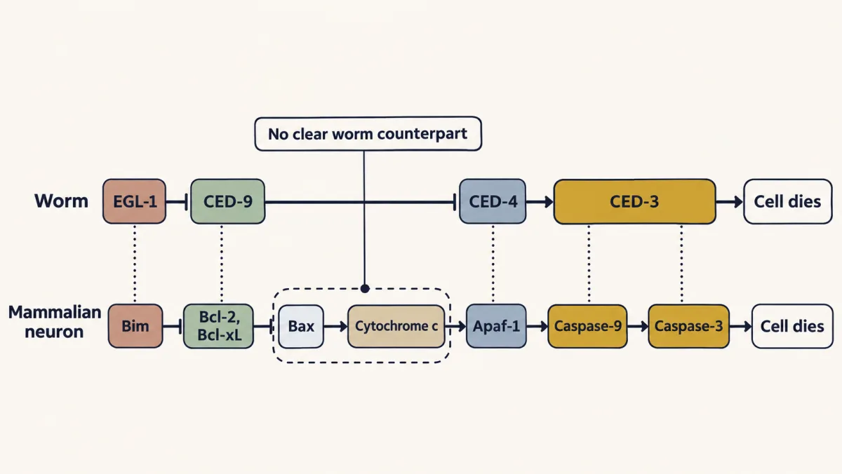
That answers the second puzzle: the parts are the worm’s, plus a gate the worm lacks.
The first brake: from a lost signal to cytochrome c
Read the transcript of “The First Brake: The Mitochondrial Gate”
Film transcript: The First Brake: The Mitochondrial Gate
A sympathetic neuron loses its NGF. What happens, step by step, before the killers switch on? Keep the worm in mind. There, EGL-1 takes the brake off the killers. The neuron's brake works one step earlier, as a gate at the mitochondria, which store cytochrome c. Step one. The survival signal falls. NGF from the target acts through its receptor, TrkA, on the axon terminals. The signal keeps kinases such as Akt active. Akt holds back several pro-death proteins. Without NGF, that restraint weakens. Step two. Pro-death genes are switched on. A stress pathway turns on. The kinase JNK becomes active. JNK adds phosphates to a transcription factor called c-Jun.
c-Jun rises in deprived neurons, and blocking its function protects them. Among the genes then induced are BH3-only proteins, such as Bim. This is part of the new gene expression that the 1988 drugs blocked. Step three. The gate opens. A BH3 domain is a short helix. It fits a groove on protective proteins, such as Bcl-2. That is what EGL-1 does to CED-9 in the worm. Protectors with filled grooves can no longer hold Bax back. Bim can also activate Bax directly. Inactive Bax sits in the cytoplasm, with its last helix tucked into its own groove. Active Bax moves to the mitochondria. It inserts that helix into the outer membrane. With other Bax molecules, it makes the membrane leaky.
Cytochrome c escapes into the cytoplasm. Step four. The killers assemble. Cytochrome c binds Apaf-1. Apaf-1 assembles into a wheel, called the apoptosome. The apoptosome activates caspase-9. Caspase-9 cuts and activates caspase-3. Caspase-3 takes the cell apart. Now delete Bax, and take NGF away. Which steps still happen? Does the neuron live?
Steps one and two should still happen. They lie upstream of Bax. Step three cannot happen. The gate stays shut, and cytochrome c stays inside. The neuron survives. But it survives as a shrunken cell, because NGF also supports its growth. That is the first brake. Brake one is the gate. NGF loss opens it through new gene expression.
From the film
- Brake 1 is a gate at the mitochondrion, and NGF loss opens it through new gene expression.
- Loss of NGF raises c-Jun and BH3-only proteins such as Bim, which free Bax and let cytochrome c escape.
- Without Bax the gate stays shut and the neuron survives, though as a shrunken cell.
Return to the sympathetic neuron that has lost nerve growth factor (NGF). In it, the counterpart of CED-9 is brake 1, a gate at the mitochondrion: protective BCL-2 proteins hold Bax shut, so cytochrome c, the partner the caspase activator needs, stays inside. A second brake, on the caspases themselves, has no counterpart in the core worm pathway; it comes in the next section. Follow NGF loss through the gate step by step. Each step has been tested by blocking it.
Step 1. The survival signal falls. NGF made by the target acts through its receptor TrkA on the neuron’s axon terminals (that lesson). The signal keeps kinases such as Akt active (that lesson). Akt adds phosphates to several pro-death proteins and so holds them back; among them are FOXO transcription factors. Without NGF, that restraint weakens and those proteins are freed.
Step 2. Pro-death genes are switched on. At the same time a stress pathway turns on. The kinase c-Jun N-terminal kinase (JNK) becomes active and adds phosphates to the transcription factor c-Jun (that lesson). In the mid-1990s groups including Johnson’s reported that c-Jun rises in deprived sympathetic neurons and that blocking its function protects them. Among the genes then induced are BH3-only proteins such as Bim; freed FOXO factors are thought to help switch Bim on. Other BH3-only proteins share the work, and neurons lacking Bim alone die more slowly rather than not at all. This is part of the new gene expression that the 1988 inhibitors blocked, and it is one major part of the answer to the first puzzle.
Step 3. The gate opens. A BH3 domain is a short helix. It fits a groove on protective proteins such as Bcl-2 and Bcl-xL, which is what EGL-1 does to CED-9 in the worm. Protectors with their grooves filled can no longer hold Bax back, and Bim can also activate Bax directly.
Inactive Bax sits in the cytoplasm with its last helix tucked into its own groove. Active Bax moves to the mitochondria, inserts that helix into the outer membrane and, with other Bax molecules, makes the membrane leaky. Cytochrome c, which normally carries electrons inside the mitochondrion, escapes into the cytoplasm. In 1996 Thomas Deckwerth, Johnson and colleagues reported that sympathetic neurons from mice lacking Bax survive NGF deprivation. Later work showed that in these neurons Bax is needed for cytochrome c to leave the mitochondria.
Step 4. The killers assemble. In the cytoplasm, cytochrome c binds Apaf-1, the counterpart of the worm’s CED-4. Apaf-1 assembles into a wheel-shaped complex, the apoptosome, which gathers and activates the initiator caspase-9. Caspase-9 cuts and activates the executioner caspase-3, which takes the cell apart.
Predict. Delete Bax and take NGF away. Which of the four steps still happen, and does the neuron live?
Check your prediction
Steps 1 and 2 lie upstream of Bax, so they should still happen: Akt activity falls, and c-Jun and Bim rise. Step 3 cannot happen. With no Bax the gate stays shut, cytochrome c stays in the mitochondria, step 4 never starts, and the neuron survives. It survives as a shrunken cell, though: Deckwerth and colleagues reported that such neurons are atrophic, because NGF supports growth as well as survival.
That is the first brake, and NGF keeps the genes that would open it switched off. Is it the only one?
The second brake: competence to die
Read the transcript of “The Second Brake: Competence to Die”
Film transcript: The Second Brake: Competence to Die
If the gate were the only brake, injected cytochrome c should kill these neurons, even in NGF. Does it? Mohanish Deshmukh tested this directly, with cultured sympathetic neurons. With a fine glass needle, he injected cytochrome c straight into the cytoplasm. The injection skips the gate. First, inject neurons kept in NGF. If the gate is the only brake, what should happen?
They should die. They survived. Now take NGF away for about a day. But keep each neuron's own gate shut. Deshmukh did that by blocking protein synthesis, or by using neurons lacking Bax. These neurons were alive when injected. Cytochrome c killed them within hours. A control injection did not. A caspase inhibitor prevented the death. So something below cytochrome c release also changes, when NGF is taken away. The change builds up gradually. Over about a day without NGF, more and more neurons respond. Deshmukh called this acquired susceptibility competence-to-die. What does the change need? Look again at how these neurons were kept alive. They made no new protein, yet they became competent.
So the change needs no new protein. Neurons lacking Bax became competent too. So it needs no Bax. So this change is a separate brake, and deprivation removes it. Later work identified that brake. It is a caspase inhibitor called XIAP. XIAP levels fall during NGF deprivation. XIAP can hold caspases off, partly because these neurons have little Apaf-1. Here is a test of that idea. Keep NGF present, and remove the inhibitor. What should cytochrome c do then?
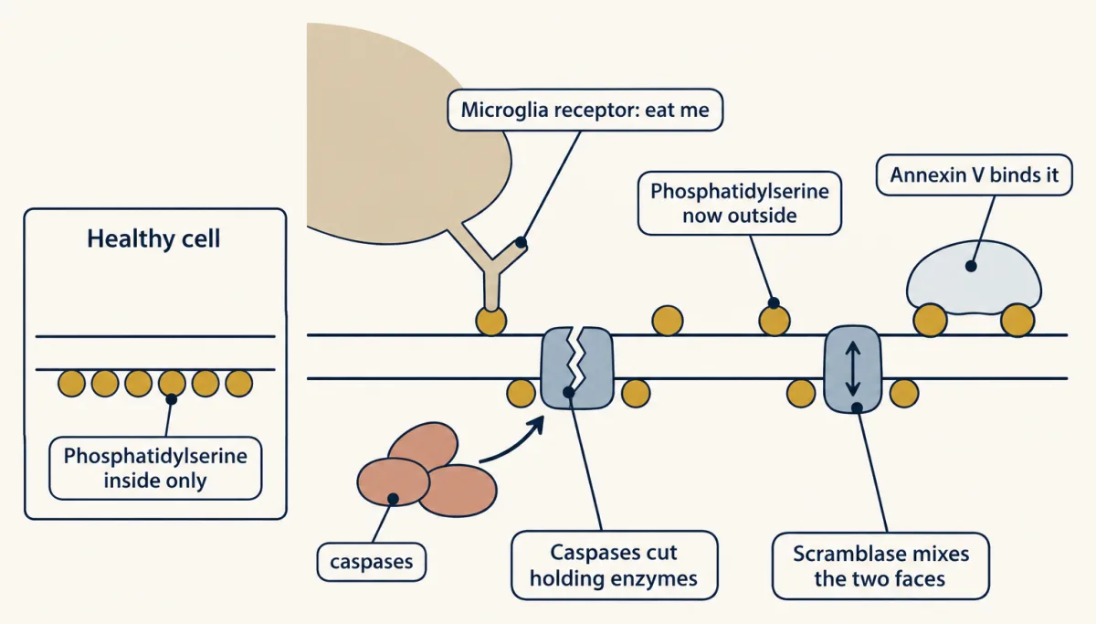
Smac is a mitochondrial protein that removes inhibitors from caspases. Cytochrome c injected along with Smac killed neurons kept in NGF. Put every result against the two brakes. A neuron dies by apoptosis only when the gate is open or skipped, and the caspase brake is off. Once the caspases run, the dying cell signals to be eaten. Where this has been studied, caspases expose a lipid, phosphatidylserine, on the membrane's outer face. It works as an eat me signal. Microglia and other phagocytes recognise it, and eat the cell. The cell is normally eaten before its membrane fails. So its contents do not spill, and the tissue is not inflamed. Brake two is on the caspases, and deprivation lifts it without new protein.
This apoptotic route needs both brakes off.
From the film
- Cytochrome c injected into neurons kept in NGF does not kill them, so the gate is not the only brake.
- Brake 2 is XIAP on the caspases, and deprivation lifts it without any new protein.
- A neuron dies by apoptosis only when the gate is open or skipped and the caspase brake is off.
Mohanish Deshmukh, working in Johnson’s laboratory, skipped the gate. With a fine glass needle he injected cytochrome c straight into the cytoplasm of cultured sympathetic neurons, with a marker dye to identify the injected cells, and published the results in 1998.
Predict. Inject cytochrome c into neurons kept in nerve growth factor (NGF). If the mitochondrial gate were the only step NGF controlled, what should happen?
Check your prediction
On the one-brake view they should die, because the injection skips the gate. They survived.
A second group had lost NGF for about a day. A deprived neuron opens its own gate and dies anyway, so Deshmukh kept each neuron’s gate shut, in two separate ways: by blocking protein synthesis, which stops the gate opening, or by using neurons lacking Bax. These neurons were alive when injected. Cytochrome c killed them within hours, a control injection did not, and a caspase inhibitor prevented the death.
So something below cytochrome c release also changes when NGF is taken away. Deshmukh called this acquired susceptibility competence-to-die. It built up gradually over roughly the first day without NGF: injection began to kill many neurons after about 15 to 20 hours, sooner in some than in others.
How the neurons were kept alive shows what the change does not need. Neurons making no new protein became competent, and so did neurons lacking Bax. It is a separate brake, and deprivation removes it.
Later work by Deshmukh and colleagues identified the brake. It is the caspase inhibitor X-linked inhibitor of apoptosis protein (XIAP), whose level falls during NGF deprivation. XIAP can hold caspases off in these neurons partly because they have little Apaf-1: with few apoptosomes, the caspase signal is small enough to block. When cytochrome c was injected along with Smac, a mitochondrial protein that removes inhibitors from caspases, neurons kept in NGF died too.
Now the whole rule can be stated.
The two brakes. Brake 1 is a gate at the mitochondrion: protective BCL-2 proteins keep Bax shut, and BH3-only proteins such as Bim, switched on by c-Jun when NGF is lost, open it. Opening it needs new gene expression. Brake 2 sits on the caspases: XIAP holds them back, and it is decisive partly because these neurons have little Apaf-1. Deprivation lifts it without any new protein. The neuron dies by apoptosis only when the gate is open (or skipped by injection) and the caspase brake is off.
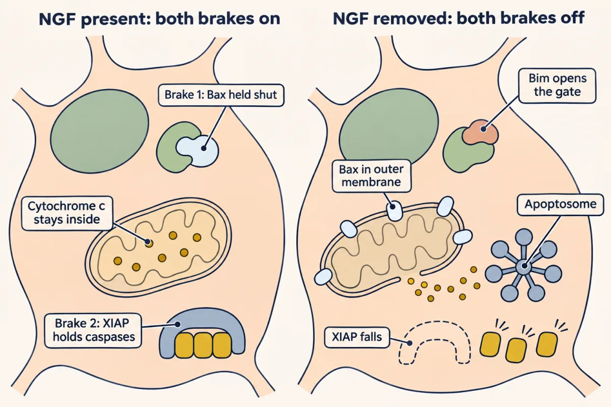
What the rule covers. Apoptosis after trophic deprivation in young NGF-dependent sympathetic neurons, where both brakes were measured. It may extend to other Bax-dependent peripheral populations, but brake 2 has been mapped mainly in sympathetic neurons. Other regulated deaths are outside it. The strength of brake 2 depends on how much Apaf-1 a cell has, and axon breakdown after injury and the local use of caspases inside axons are separate matters (Going deeper).
The table puts every result so far against the two brakes.
| Condition | Brake 1: the gate | Brake 2: XIAP on caspases | Outcome |
|---|---|---|---|
| NGF present | shut | on | lives |
| NGF removed | opens | off | dies |
| NGF removed, protein synthesis blocked | stays shut | off | death delayed |
| NGF removed, no Bax | stays shut | off | lives |
| NGF present, cytochrome c injected | skipped | on | lives |
| NGF removed for a day, gate held shut, cytochrome c injected | skipped | off | dies within hours |
| NGF present, cytochrome c and Smac injected | skipped | removed by Smac | dies |
A single-brake model gets the fifth row wrong, and it cannot explain why the injection kills only after many hours without NGF. The second brake is the simplest addition that fits the data, and it answers the third puzzle.
Cleaning up: the corpse sends a signal
A developing brain kills many cells, yet at any moment few dead cells are visible, because they are removed quickly. In the worm, neighbouring cells engulf the corpses. In the vertebrate brain and spinal cord the main phagocytes are microglia, the brain’s resident immune cells; in the developing dorsal root ganglia of mice, glial precursors, not macrophages, do most of the eating.
The dying cell helps. A healthy cell keeps the lipid phosphatidylserine on the inner face of its plasma membrane. In the cells where this has been studied, caspases cut the enzymes that hold it there and activate a scramblase that mixes the two faces, so phosphatidylserine appears outside, where receptors on phagocytes recognize it as an “eat me” signal. The same exposure lets researchers label apoptotic cells with annexin V, a protein that binds phosphatidylserine. Because the cell is normally eaten before its membrane fails, its contents do not spill, and the tissue is not inflamed. Microglia also remove synapses, which another lesson covers.
Take the machinery away
Read the transcript of “Take the Machinery Away”
Film transcript: Take the Machinery Away
Culture shows how the program runs. What happens in a whole animal when you delete its parts? Delete caspase-3, or caspase-9, from a mouse. Do the spinal and peripheral neurons that normally die survive? No. Oppenheim's group counted them. The same number died as in normal mice. But the deaths came later, and they did not look apoptotic. Chromatin condensation was absent or reduced. The cytoplasm filled with vacuoles. So deleting a caspase changes how these neurons die. Now delete Bax instead. In many neuron populations, naturally occurring death was largely eliminated. Bax deletion does not rescue every developing neuron. But where neurons depend on Bax, deleting it stops the death.
In those populations, caspases decide how. Bax decides whether. The saved neurons were not normal. In the spinal nerve roots and the facial nerve, myelinated axon counts rose by about 30 to 55 percent. Many extra axons were very small. Extra spinal motor neurons began to innervate their targets. Then a subset shrank, and lost contact. A neuron saved from death may survive as a smaller cell. That fits too little target support. Mice lacking NGF lose their sympathetic neurons. So you cannot ask what else NGF does for them. In mice lacking both NGF and Bax, the sympathetic neurons survive. If NGF did nothing but keep neurons alive, what would their innervation of organs look like?
It would look like the innervation in mice lacking only Bax. It did not. The axons failed to fully innervate many targets, and the failure differed by organ. The salivary glands had none, or almost none. The heart was greatly reduced. The trachea was normal. So NGF is needed for axons to grow into many targets, as well as for survival. Here is the general logic. Delete the death machinery, and look at what is still wrong once dying is no longer an option. The deleted gene may have other jobs too, so read such results with care. The killers are already built; a survival signal keeps brakes on them. Brake one is the gate at the mitochondrion. NGF loss opens it through new gene expression.
Brake two is on the caspases, and deprivation lifts it without new protein. The neuron dies by apoptosis only when both brakes are off. Which neurons get enough NGF in the first place is a question about targets, and about competition.
From the film
- Deleting caspase-3 or caspase-9 changes how these neurons die, not whether they die.
- In Bax-dependent populations, caspases decide how and Bax decides whether.
- Neurons saved by Bax deletion are not normal, and NGF is also needed for axons to grow into many targets.
Culture shows how the program runs. Whole animals ask what it does. Mice can be made lacking a caspase or lacking Bax (knockouts, in which a gene is deleted from the germ line).
Predict. Delete caspase-3 or caspase-9 from a mouse. Do the spinal and peripheral neurons that normally die during development survive?
Check your prediction
No. In 2001 Ronald Oppenheim and colleagues counted spinal interneurons and motor, sensory and autonomic neurons in these mice, and the same number died as in normal mice. The deaths came later and did not look apoptotic: chromatin condensation was absent or reduced, and the cytoplasm filled with vacuoles.
That is the first limit of the two-brake rule: it gates the apoptotic route, not every way a neuron can die. Deleting a caspase changes how these neurons die; in the populations that depend on Bax, deleting Bax stops the death. There, caspases decide how; Bax decides whether.
If Bax is the gate, what happens in an animal that lacks it? In 1998 Fletcher White, William Snider, Stanley Korsmeyer and colleagues reported that naturally occurring death was largely eliminated in many neuron populations of Bax-null mice. Across these and later studies, Bax deletion rescues many developing populations, among them sympathetic and sensory neurons, facial motor neurons and retinal ganglion cells. It does not rescue every developing neuron.
The saved neurons were not normal. In the lumbar dorsal and ventral roots and the facial nerve of Bax-null mice, myelinated axons were about 30 to 55 percent more numerous than in normal mice (put the other way, 24 to 35 percent of the Bax-null axons were the extra ones), and many were very small in cross section. Later work from Oppenheim’s laboratory found that the extra spinal motor neurons of Bax-null mice had begun to innervate their targets, after which a subset shrank and lost target contact. A neuron saved from death may survive as a smaller cell, which fits limited target support.
Natalia Glebova and David Ginty used Bax deletion to ask a different question. Mice lacking nerve growth factor (NGF) lose their sympathetic neurons, so you cannot ask what else NGF does for them. In mice lacking both NGF and Bax, reported in 2004, the sympathetic neurons survived.
Predict. Bax deletion keeps these neurons alive without NGF. If NGF did nothing but keep neurons alive, what would their innervation of organs look like?
Check your prediction
It would look like innervation in mice lacking only Bax, which keep their NGF. It did not. In embryos and newborns, the axons failed to fully innervate many targets, and the failure differed by organ.
| Innervation in NGF/Bax double nulls, compared with Bax-null mice | Example |
|---|---|
| absent or nearly absent | salivary glands |
| greatly reduced | heart |
| somewhat reduced | stomach, kidneys |
| normal | trachea |
NGF is therefore needed for axons to grow into many targets, as well as for survival.
The logic is general. A population that survives because the death machinery was deleted gives clues to what the death was for, by showing what is still wrong once dying is no longer an option, though the deleted gene may have other jobs too.
Design problem
Many surviving neurons in Bax-null mice are small, and some lose target contact. Design an experiment to tell whether that is because they lack trophic support or because Bax has a job in neurons that survive.
Saved in this browser only.
One way to do it
Delete Bax in only some neurons of a population, at a defined time, and compare them with their normal neighbours in the same ganglion. Run two versions, each on its own.
- Late deletion. Delete Bax only after the period of naturally occurring death, in neurons that have already survived with normal support. If they now shrink or lose target contact, that suggests Bax has a job in neurons that survive, to be confirmed by restoring Bax. If they stay normal, this test found no such job at that age; it cannot rule out an earlier one.
- Sparse early deletion. Delete Bax in a scattered few neurons before the death period, so that their neighbours compete normally. If limited support alone explains the size, roughly the share that would normally have died (about half in many peripheral populations) should be small and the rest should match their neighbours. If the neurons that would have survived anyway are smaller too, that points to a job for Bax itself; late deletion tests that group more cleanly.
In either version, adding trophic factor is a useful last check: if it restores size, limited support is a sufficient explanation. For rescued spinal motor neurons that check has been made. Oppenheim’s laboratory reported in 2003 that the trophic factor GDNF reversed their shrinkage in Bax-null mice and promoted axon regrowth. Whether Bax has other jobs in other populations is still open.
Putting it together
The three puzzles have one answer: the killers are already built, and nerve growth factor (NGF) keeps two brakes on them. What must be made includes c-Jun’s targets, BH3-only proteins such as Bim, which open the gate. The parts are the worm’s, found again as caspases, Apaf-1 and the BCL-2 family, with Bax as the mammalian gate. What keeps them off is the closed gate and X-linked inhibitor of apoptosis protein (XIAP) on the caspases. Delete the caspases and these neurons still die, another way; delete Bax and many live, smaller.
Which neurons get enough NGF in the first place is a question about targets, and how targets choose among their neurons is the subject of competition.
Going deeper
Caspase knockouts and the forebrain
Mice lacking caspase-3 or caspase-9 can have enlarged, malformed forebrains with extra cells. This reflects reduced death early in neurogenesis, largely among progenitors, not a block of the trophic deaths this lesson follows. In caspase-3 knockouts the severity depends heavily on the genetic background, the mouse strain the deletion is bred into; on the C57BL/6J strain the brain develops almost normally.
Apaf-1 as neurons mature
In fully mature neurons Apaf-1 is largely silenced, which is one reason brake 2 is a property of particular cells at particular ages, not of all cells. One prediction of the rule is that young sympathetic neurons lacking X-linked inhibitor of apoptosis protein (XIAP) and kept in nerve growth factor (NGF) should be killed by injected cytochrome c alone; later work from Deshmukh’s group reported this. In mature neurons, with little Apaf-1, removing XIAP need not be enough.
Caspases inside axons
Caspases are also used locally, inside axons. As the degeneration lecture puts it, “Caspases required for developmental degeneration but not injury.” The firm result is for axons that lose trophic support, which use caspases, including caspase-9 and caspase-3, to degenerate, while an axon cut by injury breaks down by a different route. Pruning in the living animal is less uniform. The point is taken up in Pruning Without Dying.
Your notes
Select any text in the lesson to highlight it or add a note to it. Your highlights and notes are saved in this browser and collected in your notebook.
This browser is not letting the site save anything (a private window or strict privacy settings), so highlights and notes will be lost when you leave the page.
How we measure it
Lineage tracing by direct observation
In a transparent animal, nuclei are followed under the microscope as they divide, move and die, and every event is recorded in a family tree. It tells you which identified cells die, and when, so that a missing death in a mutant can be pinned to one cell.
Forward genetic screen
Animals are treated with a chemical that causes random mutations, and their descendants are searched for a phenotype, here the absence of cell corpses. Finding the gene comes after finding the mutant, so the screen makes no guess about which proteins are involved.
Microinjection into cultured neurons
A fine glass needle delivers a protein, such as cytochrome c, straight into the cytoplasm of single neurons, with a marker dye to identify the injected cells. Skipping the upstream steps this way shows whether a cell can still respond to a later step in a pathway.
Knockout and double-knockout mice
A gene is deleted from the mouse germ line, and two deletions can be bred together. Deleting Bax keeps neurons alive, and adding a second deletion, such as NGF, shows what that factor does once survival no longer depends on it.
Practice questions on this lesson (3) → MCAT-style, original, each with an explanation
Where this lesson fits
Before thisReview first
- Too Many Neurons: Naturally Occurring Cell DeathThis course · Lesson 28
It shows that neurons are overproduced and die during development, which is the death this lesson explains mechanistically.
- The Neurotrophic Hypothesis TestedThis course · Lesson 29
You need the idea that target-derived NGF keeps neurons alive, since this lesson asks what happens when it is withdrawn.
- Neurotrophins, Trk Receptors and p75This course · Lesson 30
NGF acts through TrkA and Akt to hold back death, which is the survival signal the two brakes depend on.
After thisStudy next
- Competition: How Neurons Win a TargetThis course · Lesson 32
It takes the two-brake survival rule further to ask which neurons win limited NGF and which are lost.
- Pruning Without Dying: Axons That Cut Themselves BackThis course · Lesson 34
It builds on local caspase use in axons to show how a branch is removed while the neuron lives.
- Microglia and Complement Prune SynapsesThis course · Lesson 41
It follows the clearance step, where microglia remove dying cells and synapses.
Elsewhere on the siteFilms, simulations and method films in other courses
- Sealed or TornFilm · Cell Biology
It shows apoptosis as caspases taking the cell apart while its membrane stays sealed, then clearance of the corpse.
- The Safeguard Comes Before the CutFilm · Cell Signaling
It covers BCL-2 family control of cytochrome c release and the caspase cascade in signaling terms.
- Reading annexin V and PISimulation · Cell Biology
It lets you read annexin V and propidium iodide to tell apoptotic from necrotic cells.
Suggested by an AI model from the lessons themselves, then checked. How we use AI
Finish line
Test yourself
8 questions
Practice and MCAT-style, with explained answers.
Next · Lesson 32
Competition: How Neurons Win a Target
If the target makes too little NGF for every neuron, which neurons get to live?
Continue →