Cell Signaling · Unit 6: Channels, the nucleus, and life or death
Death Receptors, BCL-2 and Caspases
The protein that carries electrons in your mitochondria is also the order for a cell to destroy itself.
Pause when the reading reaches a film, and offer to play it. Saved on this device.
Watch the film now, or keep listening
Builds on:Stress Signals: p38 and JNK, p53, Unfolded Proteins and OxidantsAdaptors, Docking Proteins and ScaffoldsWhy these?
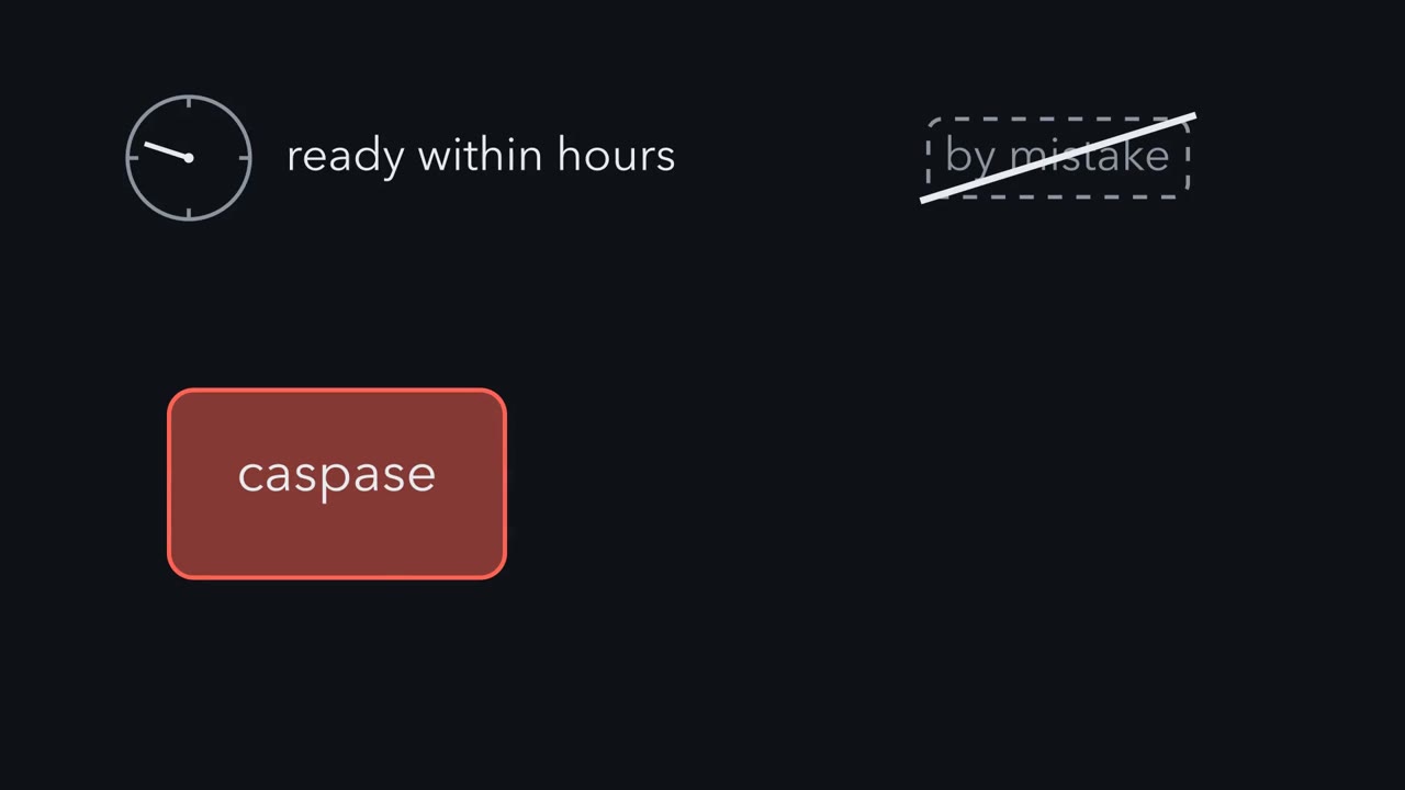 ▶ Watch The Safeguard Comes Before the Cut · 2:51
Read the transcript of “The Safeguard Comes Before the Cut”
Film transcript: The Safeguard Comes Before the Cut
How can a cell be ready to destroy itself within hours, and still never do it by mistake? The killer is an enzyme called a caspase. It cuts other proteins. Nothing undoes a cut. So where can the cell place its safeguard? Every caspase is made ahead of time, as an inactive precursor, so it is ready to act. An initiator, such as Caspace 9, starts as a single inactive chain. It turns on only when a platform gathers several copies, and they pair up. Then it cuts an executioner caspase, which is already paired but inactive. So the first step is a gathering, and every step after it is a cut. One part of the platform, Apaf -1, sits in the cytosol. The other part, cytochrome c, is kept behind the outer membrane of the mitochondrion, so no platform forms.
If that membrane opens, cytochrome c escapes. Cytochrome c and the nucleotide dATP then make seven copies of Apaf -1 assemble into a wheel. This wheel, the apoptosome, gathers Caspase -9. At that membrane, a family of proteins named for BCL-2 decides, and it has three classes. BACs and BAK, the effectors, open the outer membrane once they are activated. BCL-2 and its relatives, the guards, hold them in check. The sensors, such as BAD and BID, carry a short helix that fills a guard's groove. Some also switch on BACs.
In a healthy cell, guards and effectors are both present. Their amounts and binding set how hard the cell must be pushed. A stress must switch on enough sensors to tie up the guards. Then the guards are occupied, backs and BAK are free, and the membrane opens. Cytochrome c escapes from the mitochondrion. Now follow the whole chain, in order. Enough sensors tie up the guards, and backs and BAK open the membrane. Cytochrome c escapes, and the wheel assembles and gathers cast -paste 9. Then the cuts begin, one after another. Keep this rule: the balance of guards and sensors mostly decides before the first cut, since no cut is undone.
Cytochrome c, the small protein you met carrying electrons to complex IV, has a second job: once it leaves the mitochondrion, it is the order for the cell to take itself apart. A cell that can destroy itself within hours must be certain never to do so by mistake. This lesson asks what the killing enzyme is and what holds it off, and then how a signal from outside the cell or from inside it lets it go.
The idea: a cell can die by accident or by program
In the early 1960s John Kerr, a doctoral student in London, tied off blood vessels to part of a rat’s liver and studied how the starved cells died. Over the following years he found two deaths in one tissue. In the first, sheets of cells swelled, burst and spilled their contents. That is necrosis, death by injury, and the spill often causes inflammation. In the second, single cells scattered through working liver shrank, grew dense and broke into parcels wrapped in unbroken membrane. Neighbouring cells ate the parcels, and no inflammation followed.
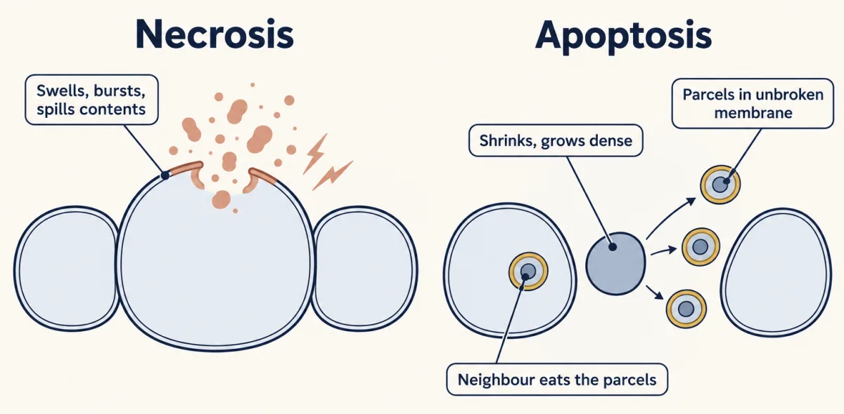
Kerr published the second death in 1971 as “shrinkage necrosis”. In 1972, with Andrew Wyllie and Alastair Currie in Aberdeen, he gave it its own name: apoptosis, from a Greek word for the falling of leaves. The name made a claim: this death is something a cell does, as an ordinary part of a tissue’s life.
Why would a cell carry a program for its own death? An animal has to remove cells as well as make them. Development builds cells that are later taken away, and an adult tissue keeps its size only if losses match births. An infected cell, or an immune cell aimed at the body’s own tissues, is safer gone. So there are two groups of apoptosis-involving diseases: too little cell death, as in cancers and autoimmune disease, and too much, as in some forms of neurodegeneration, where apoptosis is one of several ways neurons die.
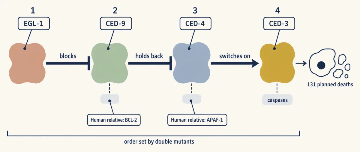
The proof that such deaths are planned came from a worm. A hermaphrodite, the self-fertilising form of the nematode Caenorhabditis elegans, makes 1,090 body cells as it develops, and the same 131 of them die every time. Robert Horvitz’s laboratory hunted for mutants in which the deaths fail. It found two genes needed for nearly all 131 deaths, ced-3 and ced-4, then a brake on them, ced-9, and a gene that releases the brake, egl-1. Putting two mutations into one animal and seeing which effect won set the genes in order: EGL-1 blocks CED-9, CED-9 holds back CED-4, and CED-4 switches on CED-3. Each has human relatives, which the rest of this lesson follows.
The killer is an enzyme kept as a precursor
CED-3 is a protease, and its relatives in your cells are the caspases, short for “cysteine-dependent aspartate-specific proteases”. A caspase uses a cysteine in its active site to cut other proteins just after an aspartate.
Every caspase is made as an inactive precursor, a zymogen, like the digestive proteases you know. The caspases that carry out apoptosis come in two kinds. (A third group, the inflammatory caspases such as caspase-1, drives a different, inflammatory death.) Initiator caspases, such as caspase-8 and caspase-9, are single inactive chains that become active when a platform gathers several copies and they pair up. Executioner caspases, which are caspases 3, 6 and 7, are already paired but stay inactive until an initiator, or another active caspase, cuts them. The first step is a gathering, and every step after it is a cut.
Active executioners cut hundreds of the cell’s proteins, in what is called the demolition phase. One target is the inhibitor of a DNA-cutting enzyme. Freed, that enzyme cuts DNA between nucleosomes, the protein spools that DNA is wound on, and leaves a ladder of fragments about 200 base pairs apart. Other targets support the nucleus and the cytoskeleton, so the chromatin condenses, the cell rounds up and its surface bubbles into blebs.
Compare this with a kinase cascade. A phosphate that a kinase adds, a phosphatase removes. No enzyme rejoins a peptide bond that a caspase has cut. Inhibitor proteins such as X-linked inhibitor of apoptosis protein (XIAP) can restrain an active caspase, and the mitochondrion, when it opens, releases a protein called SMAC that frees caspases from XIAP. But nothing undoes a cut. That is why the main safeguards act earlier, on whether initiators are gathered at all.
Killed from outside: a death receptor gathers caspase-8
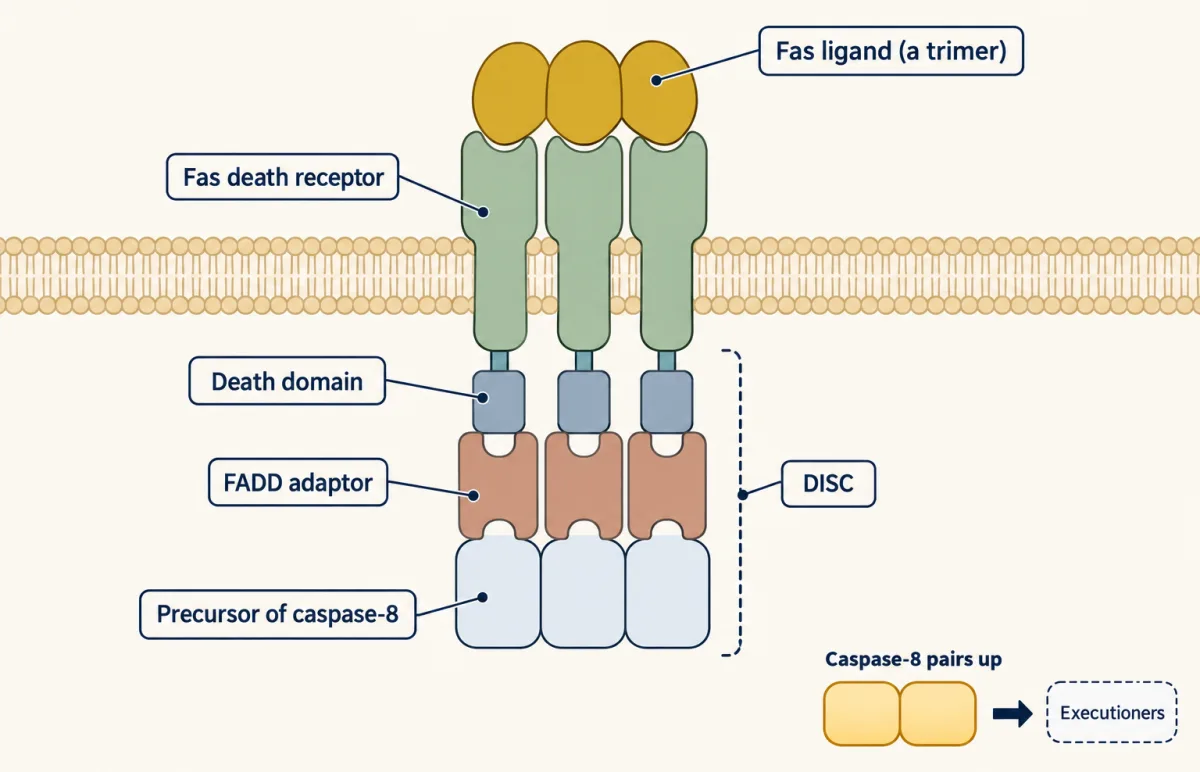
The outside route, called the extrinsic pathway, starts at a death receptor: a membrane protein whose tail inside the cell carries a death domain, a small fold that binds other death domains. The best studied is Fas. A killer T cell that has found an infected cell can present Fas ligand, which comes as a trimer and pulls Fas receptors into a cluster.
The clustered tails bind an adaptor, FADD. Like Grb2, FADD has no enzyme activity and two binding ends. Its death domain holds Fas, and its death effector domain holds a matching domain on the precursor of caspase-8. Receptor, adaptor and caspase together make the death-inducing signaling complex, or DISC. In it, copies of caspase-8 sit side by side, pair up and become active, and then cut the executioners. The receptor needs no second messenger and no kinase.
Mice showed that animals use this route. Shigekazu Nagata’s laboratory in Osaka cloned human Fas in 1991, starting from an antibody that killed the cells it bound. In 1992 the laboratory found that mice of the lpr strain, kept since the 1970s for their swollen lymph nodes and lupus-like disease, have a defective Fas gene. A second strain with the same disease, gld, proved in 1994 to have a mutation in Fas ligand. Lymphocytes that should have been cleared after an immune response were staying alive.
The receptor for tumor necrosis factor (TNF), TNFR1, has a death domain too, but its usual output is NF-κB and survival, and it kills only when that arm fails. That is “the anti-death, death pathway” of the NF-κB lesson.
Killed from inside: a series of checks and balances
Read the transcript of “Cytochrome c, Found in a Test Tube”
Film transcript: Cytochrome c, Found in a Test Tube
Can a cell's death be made to run in a test tube? In 1996, Xiaodong Wang's laboratory made it happen. They took the soluble contents of HeLa cells. They added the nucleotide dATP. And caspase-3 switched on. Which part of those contents was needed? The laboratory fractionated them. That narrowed the requirement to one small protein. Then came a test. An antibody removed that protein, and the reaction stopped. Then the laboratory added the pure protein back, and the reaction was restored. The protein was cytochrome c. It is the small protein that carries electrons to complex IV. Once it leaves the mitochondrion, it has a second job. The next year, the laboratory purified its partner, APAF-1, a relative of the worm's CED-4. Cytochrome c and dATP make seven copies of APAF-1 assemble into a wheel, the apoptosome.
It gathers caspase-9, as the death-inducing signaling complex gathers caspase-8. Take cytochrome c away, and the reaction stops. Give it back, and it is restored.
Many deaths start inside the cell, with damaged DNA acting through p53, as in the previous lesson, or with the loss of a survival signal. This inside route, the intrinsic pathway, is decided at the outer membrane of the mitochondrion by a family of proteins named for its first member, BCL-2.
BCL-2 was found at a chromosome break. In follicular lymphoma, a cancer of antibody-making B cells, a piece of chromosome 18 is joined to chromosome 14 next to an antibody gene. Yoshihide Tsujimoto in Carlo Croce’s laboratory, and then Stanley Korsmeyer’s laboratory, cloned the junction in 1984 and 1985 and found a gene beside it. In 1988 David Vaux, Suzanne Cory and Jerry Adams in Melbourne showed what the gene does. It does not make cells divide. It keeps them from dying.
In 1993 Korsmeyer’s laboratory found the opposition: BAX, which resembles BCL-2, binds to it, and speeds death. The family is a series of checks and balances with three classes.
- BAX and BAK are the effectors. Once activated, they open the outer mitochondrial membrane.
- BCL-2 and relatives such as BCL-xL are the guards. They hold BAX and BAK in check.
- BH3-only proteins, such as BIM, BID, PUMA, NOXA and BAD, are the sensors of stress. After DNA damage, p53 switches on the genes for PUMA and NOXA. They share with the family only one short helix, the BH3 helix, which fits into a groove on a guard and occupies it. Some, such as BIM and cut BID, can also switch on BAX and BAK directly.
Each sensor has its own control: BAD is switched off when a survival kinase such as Akt phosphorylates it. Korsmeyer read the family as a rheostat: guards and effectors are both present in a healthy cell, and their proportion sets how hard the cell must be pushed.
When BAX and BAK open the membrane, cytochrome c escapes. Xiaodong Wang’s laboratory found what it does next, in 1996, by making apoptosis run in a tube. They took the soluble contents of HeLa cells, added the nucleotide dATP, and saw caspase-3 switch on. Fractionation narrowed the requirement to one small protein. An antibody that removed it stopped the reaction, and adding the pure protein back restored it. It was cytochrome c. The next year the laboratory purified its partner, APAF-1, a relative of the worm’s CED-4. Cytochrome c and dATP make seven copies of APAF-1 assemble into a wheel, the apoptosome, which gathers caspase-9 as the DISC gathers caspase-8.
The two routes meet. Active caspase-8 cuts the sensor BID, and the cut piece goes to the mitochondrion and releases cytochrome c, as the laboratories of Junying Yuan and Xiaodong Wang reported in 1998. Later work showed that it does so by setting off BAX and BAK.
Design problem
A cell must be able to destroy itself within hours, and must never do so by accident. Design the safeguards. Say in what form the killing enzyme is kept, what holds it off, and why you would require two separate events before it is released. Then say what a tumour gains by making too much BCL-2, and how a small molecule shaped like a BH3 helix would take that gain away.
Saved in this browser only.
One way to do it
Speed means the enzyme must already be made, so store it as a zymogen: an executioner that is inert until it is cut. Give the cut to an initiator that works only when gathered on a platform, and keep the platform’s parts apart: APAF-1 in the cytosol, cytochrome c behind the outer mitochondrial membrane.
Then require two separate events. A stress must switch on BH3-only sensors, and there must be enough of them to tie up the guards, whose level is set separately by survival signals. Suppose noise alone produces each condition in one cell in a thousand. If the two are independent, both arise by chance in one cell in a million. Once the membrane opens in most of the mitochondria, caspases switch on and no cut can be reversed, so the outcome is usually all or none.
A tumour with extra BCL-2 has extra guards. The BH3-only proteins made by damage that would kill a normal cell are soaked up, and the cell lives. A small molecule shaped like a BH3 helix fills the groove on BCL-2 and displaces them. BAX and BAK are freed, and the tumour cell dies of the stress it was already under.
When the brakes are too strong, or the machinery is missing
The lymphoma has too strong a brake. A B cell with the translocation makes too much BCL-2 and outlives its time, and further mutations can then make it a cancer. The drug venetoclax, approved in the United States in 2016 for a form of chronic lymphocytic leukaemia, is the small molecule of the design problem. It sits in the BH3 groove of BCL-2 and spares the related guard BCL-xL.
Now take the machinery away, in a knockout mouse, one in which a chosen gene has been deleted. Most mice without APAF-1 die before or around birth, and their cells resist many kinds of death signal. Mice without caspase-9 have badly malformed brains with too many cells. So the inside route matters. Yet these embryos get through gastrulation, the early stage at which the body’s layers form, and go on to build organs, and many cells that should die still die. In the webbing between the toes of mice without APAF-1, cells die without the look of apoptosis, by a death that needs no known caspase.
A second backup, separate from that one, sits on the outside route. When TNFR1 is engaged and caspase-8 is blocked, as some viruses arrange, two kinases named RIPK1 and RIPK3 assemble, and RIPK3 phosphorylates a protein called MLKL. MLKL moves to the plasma membrane and breaks it. The cell swells and bursts like a necrotic cell, but on instruction, and the process is called necroptosis. Caspase-8 normally restrains it, in part by cutting RIPK1. A mouse embryo without caspase-8 dies, and it survives if RIPK3 is removed as well.
Your notes
Select any text in the lesson to highlight it or add a note to it. Your highlights and notes are saved in this browser and collected in your notebook.
This browser is not letting the site save anything (a private window or strict privacy settings), so highlights and notes will be lost when you leave the page.
How we measure it
Cell-free reconstitution with depletion and add-back
Break cells open, keep the soluble extract, and find conditions under which the process runs in a tube. Then fractionate the extract to find a single component the reaction requires, remove it with an antibody to show the activity is lost, and add the pure protein back to show it returns. This is how cytochrome c and APAF-1 were tied to caspase activation.
Double-mutant (epistasis) analysis
Combine two mutations with opposite effects in one animal and see which effect wins. The gene whose mutant effect shows through usually acts later in the pathway, when both are loss-of-function mutations in one linear pathway. In the worm this placed ced-9 before ced-4 and ced-3.
Electron microscopy of fixed tissue
Cut very thin sections of fixed tissue and image them with electrons to see membranes and organelles. Kerr used it to show that the fragments of an apoptotic cell are wrapped in intact membrane with recognisable organelles inside, which set this death apart from necrosis.
Co-immunoprecipitation
Pull one protein out of a cell extract with an antibody and identify what comes out with it. Pulling out BCL-2 brought down its partner BAX, an early sign that members of the family bind one another.
Method films · the methods library
- Binding assays: immunoprecipitation, pull-down and gel shift 3:43
- Cell-free extracts and reconstitution 4:01
- Centrifugation and subcellular fractionation 2:50
- Cryo-electron microscopy 4:06
- Electron microscopy 4:14
- Family linkage and positional cloning 3:52
- Forward genetic screens 4:07
- Gene cloning: vectors, libraries and screening 4:04
- Gene targeting: knockout and knock-in mice 4:13
- Protein purification and column chromatography 4:14
Practice questions on this lesson (3) → MCAT-style, original, each with an explanation
Where this lesson fits
Before thisReview first
- Stress Signals: p38 and JNK, p53, Unfolded Proteins and OxidantsThis course · Lesson 33
p53 switching on PUMA and NOXA after DNA damage is the starting signal for the inside route here.
- Adaptors, Docking Proteins and ScaffoldsThis course · Lesson 10
FADD and the DISC work as adaptors with two binding ends, as taught there.
After thisStudy next
- Endocytosis and Signaling EndosomesThis course · Lesson 36
Death receptors such as Fas can be internalised, and this lesson's DISC signal can then continue from endosomes.
- Hacking the Cell: Reporters, Actuators and Genetic ToolsThis course · Lesson 44
Reporters and tools like these are used to watch caspase activation and death in living cells.
- Oncogenes: When the Switch SticksThis course · Lesson 41
Excess BCL-2 in follicular lymphoma is a block on death that turns a cell toward cancer.
Elsewhere on the siteFilms, simulations and method films in other courses
- Cell death assaysMethod film
The lesson tells apoptosis from necrosis, which these assays do with annexin V, propidium iodide and TUNEL.
Suggested by an AI model from the lessons themselves, then checked. How we use AI
Finish line
Test yourself
8 questions
Practice and MCAT-style, with explained answers.
Next · Lesson 35
The Shape of a Signal: Size, Duration, Frequency and Place
EGF and NGF both switch on ERK in the same cell. How does the cell tell them apart? It reads the shape of the signal: how much, how long, how often and where.
Continue →← Lesson 33 · Stress Signals: p38 and JNK, p53, Unfolded Proteins and Oxidants