Cell Biology · Unit 2: Membranes and compartments
Membrane Organization and Topology
Rewritten on : new text, flashcards and film. Your notes and highlights stay, but the text around them may have moved.
A membrane has two faces that differ in their lipids and in the parts of each protein they carry. Who builds that difference, and why does vesicle traffic never flip it?
Pause when the reading reaches a film, and offer to play it. Saved on this device.
Watch the film now, or keep listening
Builds on:Crossing the MembraneOrganelles as a SystemWhy these?
 ▶ Watch Which Side Is Outside? · 2:53
Read the transcript of “Which Side Is Outside?”
Film transcript: Which Side Is Outside?
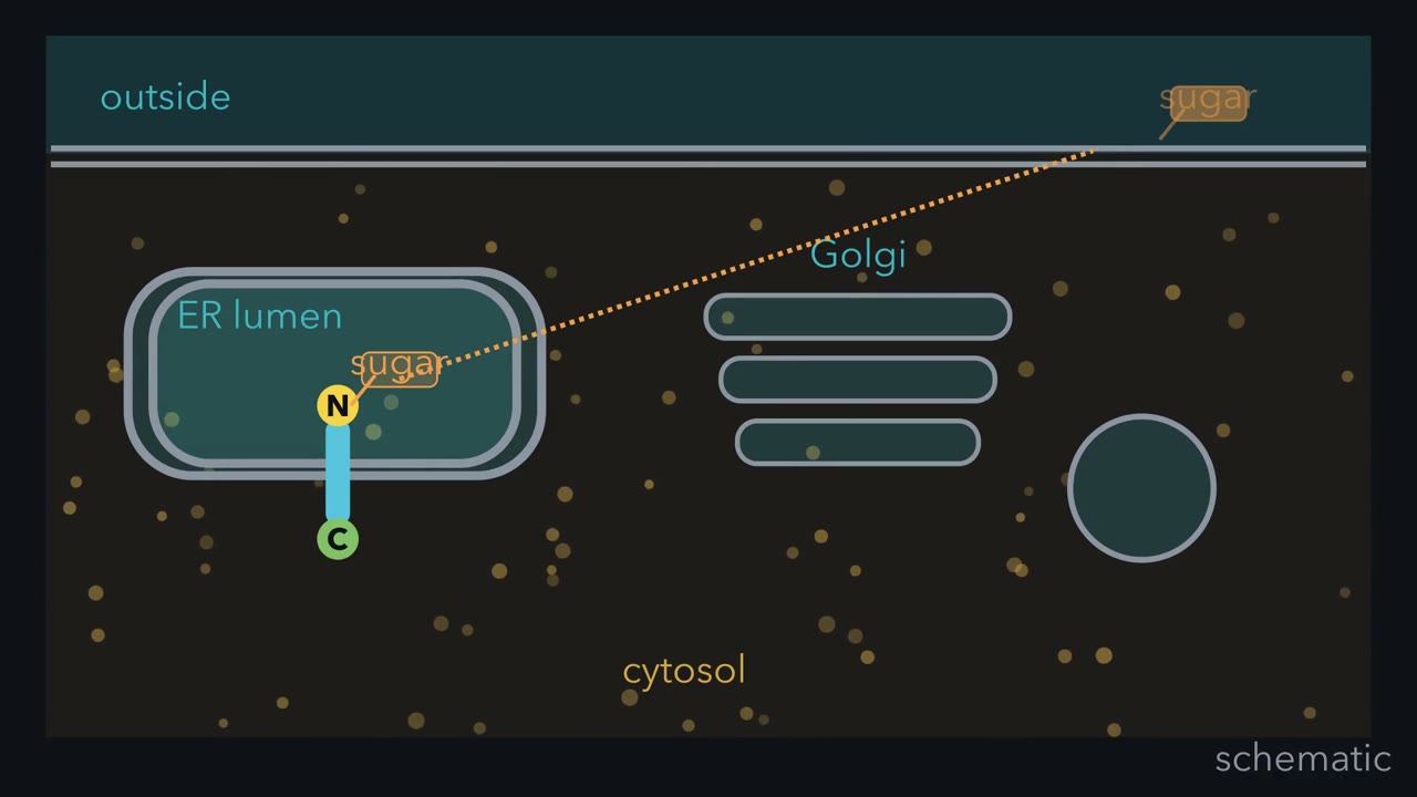
How does a sugar added to a protein inside its ER end up outside the cell? No vesicle ever carries it across a membrane. Every membrane here is two sheets of lipid, so it has two faces. One face always touches the cytosol, the fluid around the organelles. The other face touches either the outside of the cell, or a lumen, the space inside an organelle. Every space touching a cytosol face is gold. Every lumen on these routes, and the outside, is teal. On these routes, each membrane has one gold face and one teal face. A patch of the plasma membrane dimples inward and pinches off as a sac. Predict: what fills the sac, and which face touches the cytosol?
It holds a gulp of outside fluid, so its lumen is teal. The gold face still touches the cytosol. Nothing crossed a membrane. It only changed shape. Here is the rule. On these routes, lumens count as outside because budding and fusing connect them without crossing a membrane. So where does a protein get its direction? It is normally set once, at the ER, as the chain is made. A channel called the translocon threads one end into the lumen. Here the N terminus goes into the lumen, and the C terminus stays in the cytosol. That is called type I. In the lumen, an enzyme adds the sugar to the N-terminal region.
A vesicle buds from the ER and fuses with the Golgi. Its lumen joins first the ER lumen, then the Golgi lumen. The sugar never leaves teal. A secretory vesicle then fuses with the plasma membrane. Its lumen opens to the outside. The sugar now faces the fluid around the cell. It rode inside a lumen the whole way.
Endocytosis can bring the protein back in. Now the sugar faces the endosome lumen. At no step did the C terminus leave the cytosol. So the cell chooses a protein's sides once, usually at the ER. After that, traffic only carries it from lumen to lumen. Secretory and endocytic lumens count as outside; traffic never flips proteins.
From the film
- Membranes on the ordinary secretory and endocytic routes have two faces: one touches the cytosol, the other a lumen or the outside.
- Lumens on these routes count as outside, because budding and fusing connect them to the outside without crossing a membrane.
- A membrane protein's direction is normally set once, at the ER, as the translocon threads it in.
- A type I protein has its N terminus in the lumen and its C terminus in the cytosol.
- Vesicle traffic never flips a protein, so a sugar added in the ER lumen can face outside after the protein reaches the cell surface.
A cell adds a sugar to a protein deep inside its endoplasmic reticulum (ER), and later that same sugar sits on the outside of the cell, though no vesicle ever carried it across a membrane.
The answer is a rule about the two faces of every membrane, which proteins and lipids obey in different ways.
The one idea to carry through this lesson. Moving a protein or a phospholipid across a bilayer at a useful speed takes a dedicated machine. For most membrane and secreted proteins the cell uses that machine once, normally at the ER: as the chain is made, the translocon (the channel it threads through) decides which parts go into the lumen, the space inside the organelle. After that, membrane traffic carries it between compartments, and budding and fusion move nothing across a bilayer. So the face that met the cytosol always meets the cytosol, and the lumen of every compartment on the secretory and endocytic routes counts as “outside the cell.” Phospholipids the cell moves across all the time, on purpose: flippases pump some inward and scramblases let them mix, so the lipid difference between the faces is maintained, not frozen. Sliding sideways within a leaflet needs no machine, and a photobleaching experiment measures it.
The rule covers the ER and nuclear envelope, Golgi, secretory vesicles, plasma membrane, endosomes and lysosomes, not mitochondria, chloroplasts or peroxisomes. The few things that cross a membrane later on these routes each have their own machine (Going deeper, “Exceptions, and why they fit”).
Two faces of every membrane
The bilayer is two sheets of lipid, each sheet called a leaflet, with tails meeting in an oily core. Its chemistry and physics are covered in lipids and membrane structure and function. Here you need one consequence of that core: a polar head group, or a polar stretch of protein, crosses it only rarely. Phospholipids and proteins can slide sideways within their own leaflet, but on their own they cannot easily flip to the other one. (Cholesterol, ceramide and diacylglycerol, with small uncharged head groups, flip quickly on their own, so the lipid half of this lesson’s rule is about phospholipids with polar head groups.)
So every membrane has two faces, as the film above draws them: one touches the cytosol, the other the outside of the cell or a lumen, the space enclosed by an organelle such as the endoplasmic reticulum (ER) or Golgi. That second face is the exoplasmic face (or noncytosolic face): the one that never touches cytosol.
The rule: a lumen counts as outside
The first film above follows one protein from the endoplasmic reticulum (ER) to the cell surface and back. This section gives the reasoning behind the rule and the cases the film skips.
Start with the easy case. When a cell takes something in by endocytosis, a patch of plasma membrane dimples inward and pinches off as a small sac, an endocytic vesicle, which soon joins an endosome inside the cell.
Predict. What is in the fresh vesicle? And which face of its membrane touches the cytosol?
Check your prediction
A gulp of extracellular fluid: the sac closed around fluid that was outside the cell a moment earlier. The face that touched the cytosol still touches the cytosol, and the face that met the outside now faces the lumen. Nothing crossed the membrane. It just changed shape.
Textbooks say the endosome lumen is “topologically equivalent” to the outside of the cell. In plain words: you can get from one space to the other by budding and fusing alone, without ever passing through a membrane. The endosome lumen is equivalent to the outside because it was the outside.
Now run it backwards. When a sac fuses with the plasma membrane, its lumen opens to the outside and its contents spill out: exocytosis, the way cells secrete. Each vesicle step back through the Golgi to the ER works the same way, as the film shows. Within the Golgi, cargo can also stay in one sac while the sac itself matures, which keeps sides too. So the lumens of the ER, Golgi, secretory vesicles, endosomes and lysosomes all sit on the same side as the outside of the cell: in the film’s colors they are teal, and the cytosol (with the nucleus, which connects to it through pores) is gold.
Budding and fusion never swap the colors: when a vesicle fuses with its target, the two cytosol-facing leaflets merge, and the two lumens (or a lumen and the outside) join.
So where does a protein’s orientation come from? For a protein of the secretory pathway, it is normally set when the protein enters the ER membrane, usually through the translocon, the protein channel that threads new chains into or across the membrane and lets each membrane-spanning stretch slip sideways into the lipid.
Worked problem, part 1: trace one protein. A protein with one membrane-spanning stretch is made in the ER of a cultured mammalian cell, N terminus in the lumen and C terminus in the cytosol: the arrangement called type I. Its N-terminal domain gets an N-linked glycan (a sugar chain attached to an asparagine), added in the lumen because the enzyme that adds it, oligosaccharyl transferase, has its active site on the lumenal side.
- ER to Golgi. Vesicles bud and fuse. The sugar stays lumenal; the C terminus stays in the cytosol.
- Golgi to plasma membrane. A secretory vesicle carrying the protein fuses with the plasma membrane.
Predict. Where are the sugar and the C terminus now?
Check your prediction
The sugar faces the extracellular fluid, because the vesicle lumen it sat in opened to the outside. The C terminus is still in the cytosol. That solves the opening puzzle: the sugar’s part of the chain crossed once, in the ER, as it was made, and never again.
- Back in by endocytosis. The sugar and N terminus face the endosome lumen; the C terminus faces the cytosol.
A secreted protein, such as insulin, is threaded all the way into the ER lumen, so it ends up outside. Even viruses obey it: an influenza virus buds outward from the plasma membrane, so its spike proteins, made in the ER with their sugar-carrying ends in the lumen, stud the outside of the virus.
Four arrangements, one rule
A membrane protein’s topology is which of its parts face the cytosol and which face the other side. Because a lumen counts as outside, “in the lumen or outside” below is one side, not two.
- Single-pass type I: one membrane-spanning segment, N terminus in the lumen or outside, C terminus in the cytosol. Glycophorin A of the red blood cell (its structure is on this page) and the epidermal growth factor (EGF) receptor are type I.
- Single-pass type II: one segment, N terminus in the cytosol, C terminus in the lumen or outside. The transferrin receptor and many Golgi glycosyltransferases, the enzymes that build sugar chains, are type II.
- Multipass: several segments, with loops alternating between the two faces. The glucose transporter GLUT1 is one; the bacteriorhodopsin structure on this page shows the alternating loops.
- GPI-anchored: no spanning segment; the protein is attached by a glycolipid, glycosylphosphatidylinositol (GPI), to the lumenal leaflet, because the anchor is attached in the endoplasmic reticulum (ER) lumen. Prion protein and alkaline phosphatase are held this way. Proteins held by myristoyl or prenyl groups on the cytosolic face are a different class, covered in lipids as anchors and addresses.
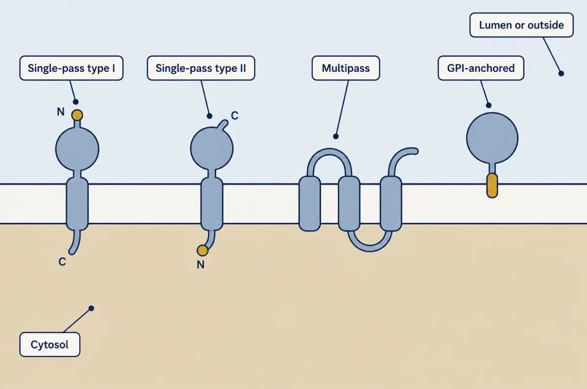
Predict. The transferrin receptor is type II. When it reaches the plasma membrane, which end is outside the cell? After endocytosis, which end faces the endosome lumen?
Check your prediction
The C terminus both times. Type II puts the C terminus in the ER lumen, and every lumen on the route counts as outside. The N terminus stays in the cytosol throughout.
Which arrangement a chain takes is normally decided at the translocon by signals in the chain itself, the subject of protein sorting. Reorientation normally happens during insertion, though a few proteins rearrange later in the ER membrane; a finished protein is not flipped by traffic.
How we know which way a protein faces
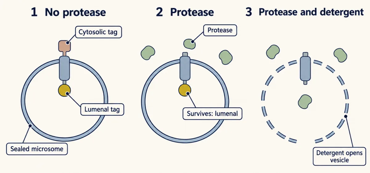
The standard test uses microsomes: sealed vesicles of endoplasmic reticulum (ER) membrane, made by breaking cells, with the lumen still inside. Tag the protein’s two ends with different epitopes, short tags an antibody recognizes, and add a protease. On the gel, an antibody to one tag still finds a protected fragment, while the other tag is gone. Add detergent before the protease and both tags are gone.
Now read it. A protease cannot enter a sealed vesicle, so the tag that survived was in the lumen. The detergent control shows that tag could be digested once the membrane was opened, so its survival was protection, not a protease-proof tag. A second test uses sugars: a glycosylation site placed in one end receives a sugar only if that end reaches the lumen (step 3 below).
Design problem
You have cloned T, a new protein with one predicted membrane-spanning segment. Determine which way it faces in the ER and plasma membrane of a cultured mammalian cell, and confirm that it is integrated in the membrane rather than stuck to it.
Saved in this browser only.
One way to do it
The quiz uses step 2; steps 3 and 4 are lab detail for a second pass. Write the predictions first. N-out (type I): N-terminal parts lumenal or extracellular, C terminus cytosolic. N-in (type II): the reverse.
- Tag both ends with different epitopes, and check that the tags do not change localization.
- Protease protection. Make sealed microsomes from transfected cells and treat with protease, with and without detergent, read as above.
- Glycosylation reporters. Place an N-X-S/T site (X not proline) in each end domain, one construct at a time, at least 12 to 14 residues from the membrane-spanning segment; a site closer than that is not used even in the lumen, and sites at the very C terminus can be skipped. A site becomes glycosylated only in the lumen. On the ER form of the protein, endoglycosidase H should remove the gel shift the sugar causes; sugars trimmed and rebuilt into complex forms in the Golgi resist endoglycosidase H, so use PNGase F for those. The construct whose site is used tells you which end crossed. A site that is not used does not by itself prove that end stayed in the cytosol.
- Antibody access in intact cells. Low digitonin permeabilizes the cholesterol-rich plasma membrane but leaves ER and Golgi membranes largely closed; Triton X-100 opens all membranes. An epitope seen after digitonin, but not on unpermeabilized cells, is cytosolic. One seen only after Triton is lumenal. On unpermeabilized cells, an antibody that labels the surface marks the extracellular end.
- Alkaline extraction. High pH strips peripheral proteins; an integrated protein stays in the membrane pellet.
If the methods disagree, suspect the tags or a mixed population of orientations, as Goder and colleagues found for engineered proteins.
The second film picks up here: fast crossing of a bilayer needs a machine, while sliding sideways does not.
Read the transcript of “Across or Along the Membrane?”
Film transcript: Across or Along the Membrane?
Chapter one showed why a membrane protein keeps its direction during traffic. But lipids are not locked in place. How does a cell keep different lipids on the two faces while molecules still move? A red blood cell gives the best-measured example. PS and PE sit mainly on the cytoplasmic leaflet. PC and sphingomyelin sit mainly outside.
Sideways drifting does not erase that pattern. Crossing is different. A phospholipid's polar head rarely enters the membrane's oily core unaided. Without help, most phospholipids change leaflets slowly. Slow is not zero, so cells spend energy to maintain asymmetry. An ATP-driven flippase selects lipids such as PS and PE and moves them inward. A scramblase does something different. When switched on, it lets lipids cross both ways without energy. The two leaflets then mix. Together, slow leakage, pumps, and regulated scrambling create a steady state, not a permanent arrangement. Flippases keep restoring selected lipids. Switching on a scramblase lets the pattern collapse.
Now predict. Suppose every phospholipid slides sideways, but none crosses to the other leaflet. Will the two faces become mixed? No. Each leaflet becomes well stirred, yet the lipids remain on their original face. Across and along are independent directions. Lateral diffusion is that motion along the membrane. Fluorescence recovery after photobleaching, or FRAP, measures lateral motion. Fluorescent membrane proteins begin bright. A brief laser makes one spot dark. Unbleached proteins then diffuse into it. The recovery curve answers two questions. How much fluorescence returns gives the mobile fraction. How quickly it reaches half of that return gives the half-time.
Dark molecules still move; bleaching only removed their fluorescence. Take the film's computed example. Fluorescence starts at one hundred, falls to twenty, then plateaus at eighty. Sixty units returned, but eighty were bleached. The mobile fraction is sixty divided by eighty, or zero point seven five. Half of the recoverable increase is thirty. Add that to twenty, making the halfway level fifty. The curve reaches fifty at four seconds, so its recovery half-time is four seconds. Under a stated diffusion model, spot size and half-time can estimate a diffusion coefficient. The estimate is approximate because the bleach and membrane must match that model. Binding or barriers can change recovery.
So a membrane can preserve two different faces while its components move. Fast crossing needs a machine; sideways motion does not. Pumps build lipid asymmetry, scramblases erase it, and FRAP measures lateral diffusion.
From the film
- In red blood cells, PS and PE sit mainly on the cytoplasmic leaflet, PC and sphingomyelin mainly on the outer leaflet.
- ATP-driven flippases move selected lipids inward; scramblases let lipids cross both ways without energy.
- Sliding sideways within a leaflet needs no transporter.
- FRAP recovery size reports mobile fraction, while recovery speed helps estimate diffusion under a stated model.
Lipids: the side the cell has to keep pumping
Proteins keep their sides with no upkeep; phospholipids keep theirs only with constant help: where each is made sets part of the pattern, and pumps keep restoring the rest.
In the red blood cell, the best-measured case, phosphatidylserine (PS) and phosphatidylethanolamine (PE) sit mainly on the inner, cytoplasmic leaflet, and phosphatidylcholine (PC) and sphingomyelin mainly on the outer leaflet. Other cells and organelles are less completely measured. The leaflets also differ in how unsaturated their tails are.
Left alone, a phospholipid moves between leaflets slowly, on a scale of hours or longer for most species. Slow is not zero: over the months a red blood cell lives, leakage alone would wear away any pattern set up at the start. Three kinds of protein move lipid across the bilayer, and their names differ by a letter: flip is in, flop is out, scramble is both ways.
| Protein | Moves lipid | Uses ATP? | Example |
|---|---|---|---|
| Flippase | Outer leaflet to cytoplasmic leaflet, against the gradient; chiefly PS and PE | Yes | Most are P4-type ATPases, a family of lipid pumps; in human cells, for example ATP11A or ATP11C with their partner CDC50A |
| Floppase | Cytoplasmic leaflet to outer leaflet | Yes | ABC transporters, a family of ATP-powered transporters, appear to do this job |
| Scramblase | Both ways, down the gradients | Generally no | TMEM16F, switched on by calcium |
So plasma-membrane asymmetry is a steady state: pumps push one way, slow leakage and regulated scramblases push the other, and switching on a scramblase collapses it far faster than spontaneous flip-flop would.
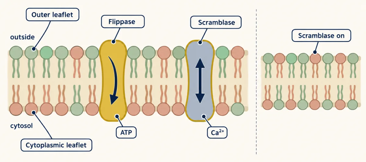
TMEM16F is the best-studied calcium-activated scramblase. Its activity can expose PS on the surface of activated platelets, where the negatively charged lipid supports blood clotting. A different route exposes PS on dying cells, where it serves as a signal for engulfment; that pathway belongs to death receptors, BCL-2 and caspases.
Predict. Two red blood cells lose their pattern in different ways. In one, ATP runs out and nothing else changes. In the other, calcium floods in while ATP stays normal. Which puts PS on its surface faster, and why?
Check your prediction
The calcium-loaded cell, by far. With no ATP the flippases stop, but PS can still only leak outward by spontaneous flip-flop, so it creeps out over hours or longer. Calcium opens a scramblase, which carries PS across within minutes, and high calcium also inhibits the flippases, so nothing pumps it back. Pump off is slow; a hole opened is fast.
The endoplasmic reticulum (ER) membrane is thought to be much closer to symmetric, because lipids there are largely scrambled between the leaflets. Cholesterol, which flips quickly, is a harder case, and how it divides between the leaflets of the plasma membrane is still argued.
Along the membrane, freely: FRAP
In 1972 Singer and Nicolson’s fluid mosaic model predicted the split this lesson has followed: proteins keep a fixed orientation across the membrane but are free to move within its plane. Fluorescence recovery after photobleaching (FRAP) measures the sideways motion. A laser destroys the fluorescence of labelled molecules in a small spot. If they are mobile, unbleached ones diffuse in and the spot brightens. The bleached molecules are still there and still moving; they are simply dark. The imaging side of the method is covered in microscopy and quantitative images. The second film worked the mobile fraction and half-time of the data set below; the page adds the diffusion coefficient, its assumptions and its limits.
Worked problem, part 2: read a FRAP curve. In this made-up data set, a fluorescent protein in the plasma membrane of a cultured cell is bleached in a circular spot of radius w = 2.0 µm. Assume the bleach is instantaneous and uniform, the membrane is flat, the unbleached reservoir is large, and recovery is by two-dimensional diffusion alone. The values are already corrected for background and for bleaching during imaging.
- Before the bleach, Fi = 100. Immediately after, F0 = 20. At 4.0 s, 50. Final plateau, F∞ = 80, not 100.
Step 1, mobile fraction:
mobile fraction = (F∞ - F0) / (Fi - F0) = (80 - 20) / (100 - 20) = 60 / 80 = 0.75
The denominator is 80, not 100: only what was bleached can recover. The immobile fraction is 1 - 0.75 = 0.25. That number does not say why a quarter stayed dark. Anchoring to the cytoskeleton, bleaching a large share of the cell’s total pool, or bleaching during imaging can each lower the plateau.
Step 2, half-time. Half the recoverable increment is 60 / 2 = 30, so half recovery is reached at 20 + 30 = 50. The curve reads 50 at 4.0 s, so t½ = 4.0 s. Half the prebleach value is also 50 here, by coincidence: with a plateau of 70, half recovery would be at 20 + 25 = 45, not 50. Always halve the recovered increase, not the prebleach value.
Step 3, diffusion coefficient, valid only for this model:
In words The diffusion coefficient is about 0.224 times the spot radius squared, divided by the half-time of recovery.
- D“D”
- the diffusion coefficient: how fast molecules spread in the plane of the membraneµm²/s
- w“w”
- the radius of the bleached spotµm
- t½“t half”
- the time to reach half of the recoverable increases
Sanity check Units: µm² divided by s gives µm²/s, the units of a diffusion coefficient.
Try one number
For the curve above: D ≈ 0.224 × (2.0 µm)² / 4.0 s = 0.224 × 4.0 / 4.0 = 0.22 µm²/s.
Where it comes from optional
Diffusion time grows with distance squared, not distance, so t½ is proportional to w² / D. The factor 0.224 comes from solving the recovery curve for an instantaneous, evenly bleached disk (Soumpasis, 1983).
Need a refresher? powers and roots · units and prefixes
Predict. The same cells are bleached again, this time with a spot of radius 4.0 µm. If recovery is pure diffusion, about how long is the new half-time?
Check your prediction
About 16 s. The radius doubled, so w² grew four times, and with D unchanged t½ grows four times: 4 × 4.0 s. If the half-time had stayed near 4.0 s, that would point to recovery limited by binding and release rather than diffusion, so bleaching at two spot sizes tests the model.
- recovery curve (shape: in this sketch)
- half of the recoverable increment
- half of the prebleach value
- time cursor
- At the cursor
- Mobile fraction
- Immobile fraction
- Right level
- Wrong level
- t½ read from the chosen level
- Diffusion coefficient (w = 2.0 µm)
Rule to keep: mobile fraction = (F∞ − F0)/(Fi − F0), and the half-time is the time to half of the recoverable increment, not to half of the prebleach value. D ≈ 0.224 w²/t½ assumes an instantaneous, uniform bleach, a flat membrane, a large unbleached reservoir and two-dimensional diffusion alone. The curve is a sketch of the logic, not a fit.
A D of 0.22 µm²/s sits in the range tabulated for membrane-bound molecules, more than a hundred times slower than a protein free in the cytoplasm. In a table by Day and colleagues, cholera toxin B bound to the plasma membrane of COS-7 and mouse embryonic fibroblast cells at 37 °C diffuses at about 0.2 µm²/s, and EGFP free in the cytoplasm of COS-7 cells at about 40 µm²/s.
Sideways freedom has limits. The plasma membrane has domains, regions that differ in composition or mobility. Tight junctions separate the apical membrane, which faces the free surface or the cavity of an organ such as the gut, from the basolateral membrane facing neighbours and underlying tissue in epithelia. An actin meshwork under the membrane can slow some molecules. Lipid rafts, proposed ordered domains rich in cholesterol and sphingolipid, are where care matters most: reviews describe their existence, size and composition in living cells as unresolved, not as one settled universal structure (Going deeper).
Going deeper
Exceptions, and why they fit
The rule is about a membrane’s own parts: small uncharged molecules such as oxygen, carbon dioxide and water cross bilayers without machines. The nuclear envelope is part of the endoplasmic reticulum (ER), and its lumen is the space between its two membranes; the nucleoplasm is on the cytosol side, joined to it through pores. Mitochondria and chloroplasts import most of their proteins from the cytosol across their own membranes and are not joined to the secretory pathway by the ordinary vesicle routes. Peroxisomes receive some membrane proteins from the ER in vesicles, but they import their matrix proteins, already folded, straight from the cytosol, so the lumen rule does not describe their contents.
On the vesicle routes, whatever crosses a membrane after the ER is moved by a dedicated machine, which fits the rule’s first clause. ER-associated degradation (ERAD) pulls misfolded proteins back to the cytosol to be destroyed. Chaperone-mediated autophagy threads cytosolic proteins into lysosomes. Some toxins escape to the cytosol. Hyaluronan synthase pushes its sugar chain out across the plasma membrane. And a few proteins change topology after insertion, while still in the ER membrane, aquaporin-1 being the textbook case. So the first film’s “orientation is set once, at the ER” is the usual case, not a law, though vesicle traffic itself still never flips a protein.
Wrapping is not an exception. Autophagosomes, the vesicles that bud into multivesicular bodies, and enveloped viruses budding from the cell each enclose a pocket of cytosol, and the membrane wrapped directly around each pocket keeps its cytosolic face toward it.
Sugars follow the same logic: where a sugar is added decides where it ends up. N-linked and mucin-type O-linked sugars face outside because they are added in the ER or Golgi lumen; O-GlcNAc added in the cytosol and nucleus stays there. So “glycans always face outside” is wrong.
Other names for single-pass proteins
This lesson sorts single-pass proteins by orientation only. Some textbooks add “type III” (N terminus outside, no cleaved signal sequence) and tail-anchored proteins, which are inserted after translation without the translocon; hence “usually” through the translocon.
How close the topology decision can be
Positive charges are statistically enriched on the cytosolic side of membrane segments, a bias first described for bacterial inner-membrane proteins; it is a tendency, not a law. Goder, Bieri and Spiess showed in COS-1 cells that engineered chains with competing signals can reorient within the translocon, and that glycosylation can shift the outcome (details on the Martin Spiess card). Marginally hydrophobic segments can also fail to insert or can reorient, which is why “the first segment sets the rest” holds only in the simplest multipass case.
The red-cell lipid measurements
The leaflet pairs in the main text come from human erythrocytes, in studies summarized in a recent review. Lorent and colleagues then digested away the outer leaflet’s lipids of intact human red blood cells and analysed what was left. The two halves of the membrane differed: the outer half’s phospholipids carry about 1.6 double bonds each, the inner half’s about 3.4. These 2020 erythrocyte numbers are one recent addition to the old finding that lipid composition differs between the leaflets, an active area of work on how that difference changes membrane physical properties.
How the transporters were found
In a haploid genetic screen in human cells, Segawa and colleagues found ATP11C and its partner CDC50A required for inward movement of lipid. Alvadia and colleagues purified the mouse TMEM16F protein and showed that, in artificial vesicles, it scrambled both labelled phosphatidylserine (PS) and labelled phosphatidylethanolamine (PE) when calcium was added. That reconstitution shows the protein can scramble lipid on its own. Its role in exposing PS on activated platelets is a separate, physiological finding.
When the FRAP formula fails
The 0.224 w²/t½ relation holds only under the assumptions of worked problem part 2. A scanning confocal bleach lets molecules move during the bleach and needs a different analysis, and binding-limited recovery breaks the w² scaling, as the spot-size Predict box showed.
Part of that slowness in the plasma membrane is the cell’s doing. In a FRAP study of cholera toxin B on cultured cells, Day and Kenworthy found its slow diffusion was impeded by actin- and ATP-dependent processes, as expected if an actin meshwork restrains it (PLoS ONE 2012).
What is and is not known about rafts
Membrane material that resists cold detergent was long called raft material, but a detergent-resistant fraction is not a native raft; detergent can create or rearrange domains.
The best support comes from particular systems. Stone and colleagues used super-resolution imaging in the CH27 mouse B-cell lymphoma line. A peptide probe preferring ordered membrane gathered near clustered B-cell receptors and one preferring disordered membrane was excluded, in live and fixed cells; fixed primary mouse B cells agreed. That supports phase-like sorting in that system. It does not give a raft’s composition, and it does not show that every cell has them.
Your notes
Select any text in the lesson to highlight it or add a note to it. Your highlights and notes are saved in this browser and collected in your notebook.
This browser is not letting the site save anything (a private window or strict privacy settings), so highlights and notes will be lost when you leave the page.
How we measure it
Fluorescence recovery after photobleaching (FRAP)
A laser bleaches the fluorescent label in a small spot of membrane, and the spot's brightness is followed as unbleached molecules move in. After correction for background and for bleaching during imaging, the plateau gives the mobile fraction and, with a stated model of the bleach and of diffusion, the half-time gives an approximate diffusion coefficient.
Protease protection of sealed microsomes
Sealed ER vesicles (microsomes) carrying the protein are treated with a protease, with and without detergent. Parts in the lumen survive unless the detergent opens the vesicles, so the epitope that survives tells you which side each end faces.
Glycosylation mapping with endoglycosidase H
An N-X-S/T site becomes glycosylated only if it reaches the ER lumen, where the oligosaccharyl transferase works. A shift on a gel that endoglycosidase H removes identifies the added sugar and so marks that segment as lumenal.
Leaflet lipidomics by enzyme digestion
Intact cells are treated with phospholipases that reach only the outer leaflet, and mass spectrometry compares the digested and protected lipids. In human erythrocytes this gave the unsaturation of each leaflet separately.
Practice questions on this lesson (3) → MCAT-style, original, each with an explanation
Where this lesson fits
Before thisReview first
- Crossing the MembraneBiology · High school
It teaches the bilayer's oily core and why polar molecules cross it slowly, which this lesson assumes.
- Organelles as a SystemBiology · High school
It introduces the ER, Golgi and vesicle routes whose lumens this lesson treats as outside the cell.
After thisStudy next
- Protein Sorting into CompartmentsThis course · Lesson 7
It shows how signals in the chain make the translocon set a protein's orientation in the ER.
- Vesicle Budding, Targeting and FusionThis course · Lesson 11
It details budding and fusion, which keep each membrane face on its own side.
- Endocytosis, Recycling and DegradationThis course · Lesson 13
It follows membrane and receptors inward, where the lumen is the former outside, as the rule predicts.
Suggested by an AI model from the lessons themselves, then checked. How we use AI
Finish line
Test yourself
8 questions
Practice and MCAT-style, with explained answers.
Next · Lesson 5
Transport Across Membranes
Which way will potassium flow if you hold a cell at −70 millivolts? Its concentrations alone will not tell you.
Continue →