Cell Signaling · Unit 4: Receptors with enzymes attached
Ras to MAPK: The Kinase Cascade
A growth factor at the cell surface switches on Ras, and a relay of three kinases carries the message to the cell's proteins and genes. The same relay, stuck on, is found in many cancers, and in PC12 cells how long it stays on seems to decide what the cell does.
Pause when the reading reaches a film, and offer to play it. Saved on this device.
Watch the film now, or keep listening
Builds on:GTPase Switches: The TimerAdaptors, Docking Proteins and ScaffoldsReceptor Tyrosine KinasesWhy these?
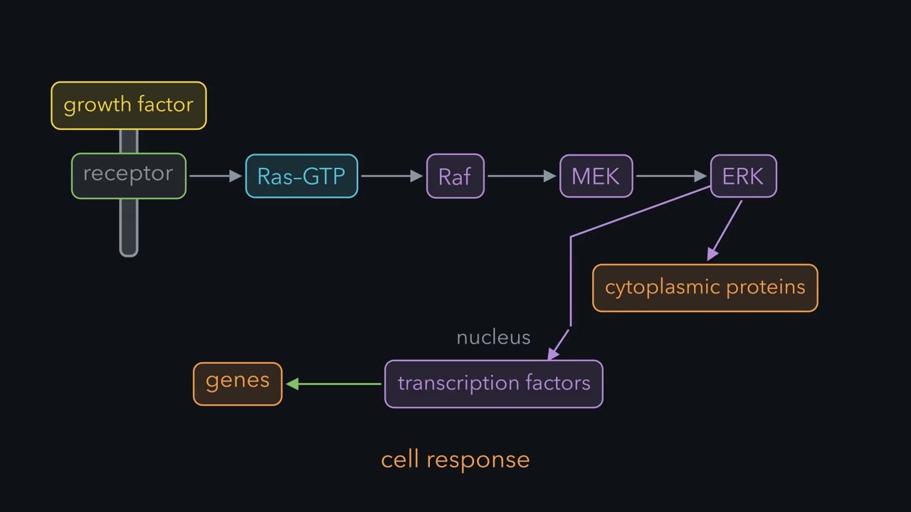 ▶ Watch Ras to MAPK: From Membrane to Genes · 2:48
Read the transcript of “Ras to MAPK: From Membrane to Genes”
Film transcript: Ras to MAPK: From Membrane to Genes
A growth factor binds a receptor at the cell surface. Ras switches on, then passes the signal through three kinases: Raf, MEK and ERK. ERK acts on proteins in the cytoplasm and transcription factors in the nucleus. That changes gene expression and cell behaviour. This is how a signal outside reaches the cell’s proteins and genes. Mutations that hold the relay on help drive cancer. So first learn what each step does to the next. The active receptor recruits Grb2 and SOS. SOS makes Ras release GDP so GTP can bind. Ras is not a kinase. It catches Raf and brings it to the membrane. Binding alone does not open Raf. Contact with membrane lipids and further steps help it open.
Two Raf molecules then pair, which is needed for normal activation. Raf adds phosphates to two serines on MEK. MEK adds one to a threonine and one to a tyrosine on ERK. ERK needs both. The narrow relay ends in a wide fan of targets. In the nucleus, ERK phosphorylates Elk-1, which helps switch on the immediate early gene c-fos. In cancer, mutations can hold Ras or Raf on. B-Raf V600E can signal without Ras-GTP. A Raf inhibitor can block it. But with normal B-Raf and active Ras, drug-bound Raf can activate a drug-free partner. ERK activity then rises. A MEK inhibitor holds MEK inactive below Raf. The drug’s effect depends on the pathway’s state.
The relay also limits itself. ERK weakens SOS and Raf by phosphorylation. It turns on genes for phosphatases that remove ERK’s activating phosphates. That takes longer, because a protein must be made. Signal duration also depends on the stimulus, cell type and dose. Timing helps shape the response. Three kinase tiers give amplification and separate control points. Other MAP kinase pathways use the same plan with different kinases. Scaffolds hold a module’s kinases together, helping them signal mainly to each other. They also help decide where the cascade runs. Milder over-activation also matters. Mutations inherited or new at conception cause developmental syndromes called RASopathies. Their effects differ between tissues.
The relay carries the signal, but its control and the cell’s context shape the answer.
A growth factor lands on a receptor at the cell surface, and a relay of four proteins, Ras, Raf, MAPK/ERK kinase (MEK) and extracellular signal-regulated kinase (ERK), carries the message to proteins in the cytoplasm and to genes in the nucleus. This lesson follows that relay step by step, because the same relay, stuck on, is found in many cancers, and mildly over-active it causes the RASopathy syndromes. One chain also does more than one job. In PC12 cells, a line grown from a rat adrenal tumour, epidermal growth factor (EGF) makes the cell divide, while nerve growth factor (NGF) makes it stop dividing and grow long, nerve-like fibres. Both growth factors send their signal through the same four proteins. You need the chain first: what each protein physically does to the next. After that, a large part of the answer seems to be a matter of time.
The idea: Ras-GTP catches Raf and holds it at the membrane
The first steps are review. An active receptor tyrosine kinase carries phosphotyrosines on its tail. The adaptor Grb2 binds one with its SH2 domain and holds SOS with its SH3 domains, so SOS arrives at the membrane, where Ras sits on its lipid anchors (see adaptors and scaffolds and lipid anchors and addresses). The next step, in one line: SOS is a guanine exchange factor. SOS makes Ras let go of GDP, and GTP, which is more plentiful in the cell, takes its place. Ras stays on until it cuts that GTP to GDP, the timer covered in GTPase switches.
Now think of “Ras as Pacman.” Picture Ras stuck to the inner face of the membrane with its mouth open. The mouth is a pair of surface loops, switch I and switch II. With GTP in the pocket they hold a shape that other proteins can grip. With GDP they go slack. What Ras-GTP catches in this lesson is Raf.
Raf is a kinase that phosphorylates serines and threonines. In a resting cell it floats in the cytosol folded shut, with its front half, which carries a Ras-binding domain, held against its kinase domain. Ras is not a kinase. It works by contact. Binding brings Raf from the cytosol to the membrane, which is well established. Binding alone does not open Raf; at the membrane, contact with the lipids and further steps help release the closed shape. At the membrane two Raf molecules then pair, kinase domain beside kinase domain, and pairing is needed for normal activation. Phosphates are also added to and removed from Raf along the way, and the exact order of those events is still being worked out.
Three kinases in a row, with a two-phosphate lock at the end
Read the transcript of “Who Switches ERK On?”
Film transcript: Who Switches ERK On?
ERK was found by following an activity. Melanie Cobb's laboratory followed a kinase activity that insulin switched on. They ran it through eight chromatography columns, cloned its gene, and named the enzyme ERK. Its sequence resembled two yeast kinases that respond to mating pheromone. Then Natalie Ahn, in Edwin Krebs's laboratory, asked what switches ERK on. In 1991 she found two activities in extracts of cells treated with growth factor. Both switched on inactive MAP kinase in a test tube. Was ERK switching itself on, or did something else do it? Here is the reasoning, step by step. The activator and ERK run down a column. If the activator comes apart from ERK there, it is a different protein.
Then ERK was not switching itself on. Ahn purified the activator with Rony Seger. It phosphorylates ERK on both threonine and tyrosine. Several laboratories reached this enzyme at about the same time. It is now called MEK. MEK is the kinase that sits between Raf and ERK. ERK needs both phosphates. Take either one off, and ERK goes quiet. An activator that comes apart from ERK is a different protein.
Active Raf phosphorylates the next kinase, MAPK/ERK kinase (MEK), on two serines in its activation loop, the flexible stretch beside the active site that you met in the phosphorylation switch. MEK then does something unusual. Most kinases phosphorylate either serine and threonine or tyrosine. MEK phosphorylates a threonine and a tyrosine, one residue apart, in the activation loop of extracellular signal-regulated kinase (ERK). ERK needs both phosphates. Take either one off with a phosphatase and ERK goes quiet.
Purifying the activator of ERK (extracellular signal-regulated kinase) from growth-factor-treated cells showed that it is a separate kinase, now called MEK, which phosphorylates ERK on both threonine and tyrosine. An activator that comes apart from ERK on a column is a different protein, so ERK was not switching itself on.
What do three kinases in a row give the cell? Signaling circuits covered two answers: each tier can amplify, and the cascade can turn a graded input into an all-or-none output. The two-phosphate lock on ERK is one ingredient of that sharpness. A third answer is control. Each tier is a separate place where a phosphatase, a feedback loop or a scaffold can act.
MEK has almost no known substrates other than ERK, so the middle of the cascade is a narrow pipe. ERK is the opposite. It phosphorylates serines and threonines that are followed by a proline, and many proteins carry such a site. Some are in the cytoplasm. Part of the active ERK also moves into the nucleus and phosphorylates transcription factors. One of them, Elk-1, helps switch on c-fos, the best-known immediate early gene. An immediate early gene turns on within minutes of a signal with no new protein made first, because the proteins it needs are already present. Its product is often a transcription factor that turns on later genes.
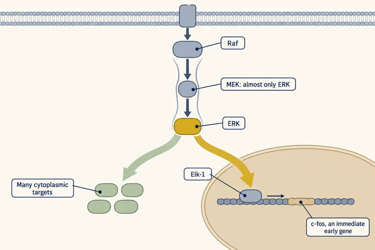
A piece of advice here: don’t memorize the downstream pathways. Learn the logic: a narrow relay that ends in a wide fan. “Putting it all together”:
growth factor → receptor → Grb2 and SOS → Ras-GTP → Raf → MEK → ERK → transcription factors → immediate early genes
One design, several cascades
Raf, MAPK/ERK kinase (MEK) and extracellular signal-regulated kinase (ERK) are one example of a general plan. A MAP kinase kinase kinase turns on a MAP kinase kinase, which turns on a MAP kinase by phosphorylating a threonine and a tyrosine in its activation loop. MAP kinase, or mitogen-activated protein kinase (MAPK), is the family name, and ERK is the member you have met. Animal cells run at least two more modules built this way, ending in the MAP kinases c-Jun N-terminal kinase (JNK) and p38. They use their own kinases at each tier, some shared between JNK and p38, and they answer mainly to stress, such as ultraviolet light or inflammatory signals.
Several look-alike cascades in one cytoplasm create a wiring problem, and scaffolds are part of the answer. A scaffold holds the kinases of one module side by side so that they pass the signal mainly to each other rather than to a neighbour, as Ste5 does in yeast and KSR does for Raf, MEK and ERK (see adaptors and scaffolds). A scaffold can also decide where the cascade runs: “Scaffolds can localize ras-mapk.” Two examples are paxillin, which gathers Raf, MEK and ERK at focal adhesions, the spots where a cell grips its surroundings, and MP1, which holds MEK and ERK on endosomes.
How long is the signal on?
Back to the PC12 cell. A signal has a size and also a duration. Negative feedback is one thing that sets the duration, alongside how long the receptor stays active, and extracellular signal-regulated kinase (ERK) supplies its own. Active ERK phosphorylates proteins above it in the chain, among them SOS and Raf, in ways that weaken them. It also turns on genes for dual-specificity phosphatases, enzymes that take both phosphates back off ERK. The second kind is slower, because a protein has to be made.
Different stimuli leave a pathway on for different lengths of time before feedback closes the window. How long the window stays open depends on the stimulus, the cell type, the dose and which readout is measured.
In PC12 cells, ERK activity after epidermal growth factor (EGF) rises and falls back within about an hour. After nerve growth factor (NGF) it climbs to a plateau that lasts for hours. In 1992 Christopher Marshall and Philip Cohen’s group in Dundee proposed that the difference in duration is itself the instruction: a brief ERK signal goes with division, and a sustained one with differentiation. Marshall set the idea out more widely in a 1995 review. The duration rule comes from this one cell line, and how widely cells read their kinases by the clock is an open question.
When the cascade is stuck on
In 1994 Sally Cowley, Christopher Marshall and colleagues gave PC12 cells a permanently active MEK1, and the cells grew neurites with no nerve growth factor (NGF) in the medium. They then put the same always-on MEK1 into mouse fibroblasts and got a different result. The cells piled up into foci, the disordered heaps that mark a cell transformed toward cancer. Unending extracellular signal-regulated kinase (ERK) activity means “differentiate” to a PC12 cell and “keep dividing” to an NIH 3T3 fibroblast.
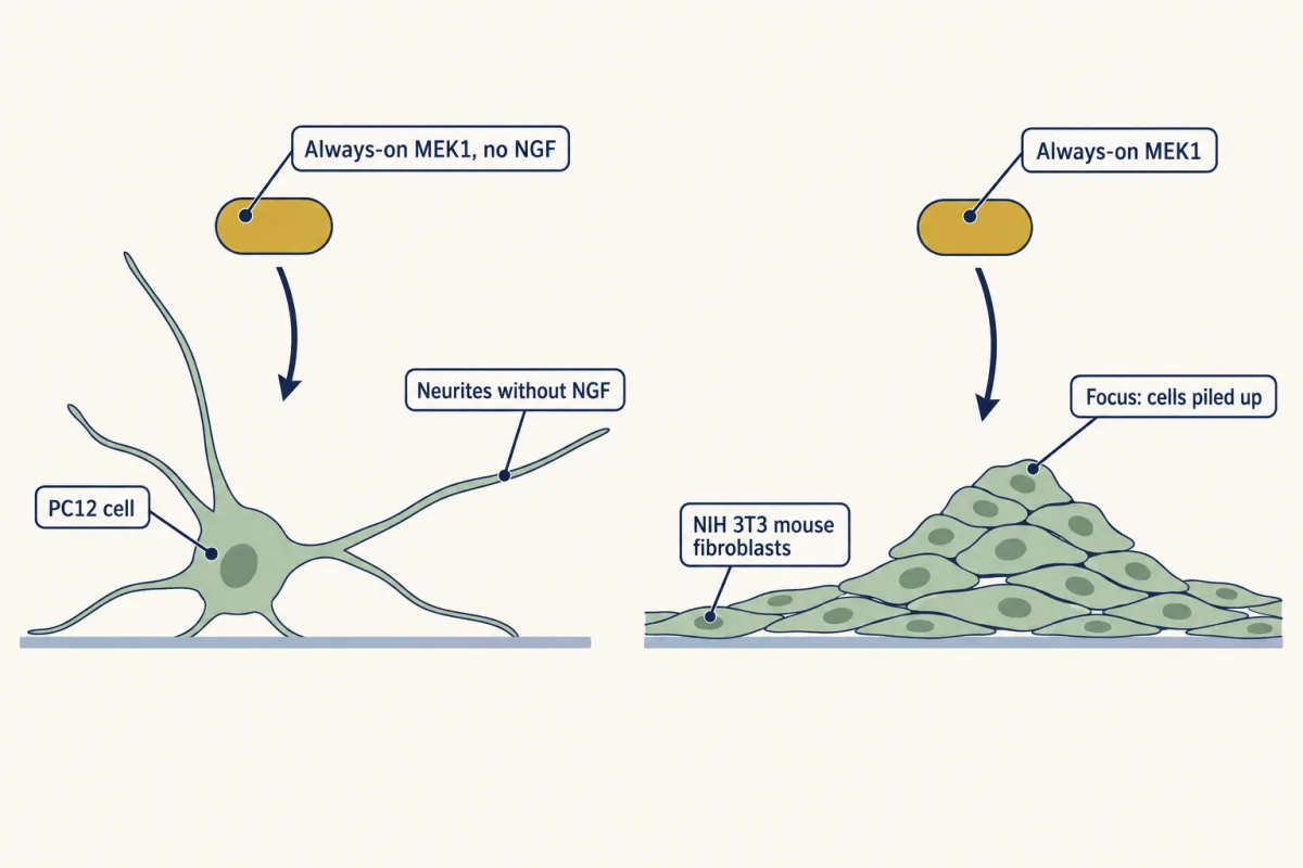
Human cancers reach the same state from higher up. In 1982 the laboratories of Robert Weinberg, Mariano Barbacid and Michael Wigler each compared a ras gene cloned from one bladder-tumour cell line with the normal gene. The two differed at a single base, which changed glycine 12 to valine: the G12V mutation, whose effect on the GTP timer you saw in GTPase switches. Mutant RAS genes are common in human cancers, and how common varies widely by tissue: a 1989 survey found them in roughly nine of ten pancreatic tumours and about half of colorectal cancers.
One tier down, roughly half of cutaneous melanomas, the common skin form, carry a mutation in B-Raf, one of three Raf kinases in human cells, most often V600E. The glutamate sits in the activation loop and makes B-Raf active by itself, with no Ras-GTP needed. It is often described as active without a partner, although mutant B-Raf can also pair, and whether it truly works alone is debated. Vemurafenib fills the ATP pocket of the mutant kinase, and trametinib binds MAPK/ERK kinase (MEK) in a pocket beside its ATP site and holds it inactive. Pairing explains a surprise that came with the Raf drugs. In a cell with normal B-Raf and active Ras, a drug-bound Raf can still pair with a drug-free Raf and switch it on, so the same drug raises ERK activity there. A MEK inhibitor blocks the cascade below that point, one reason the two kinds of drug are commonly given together.
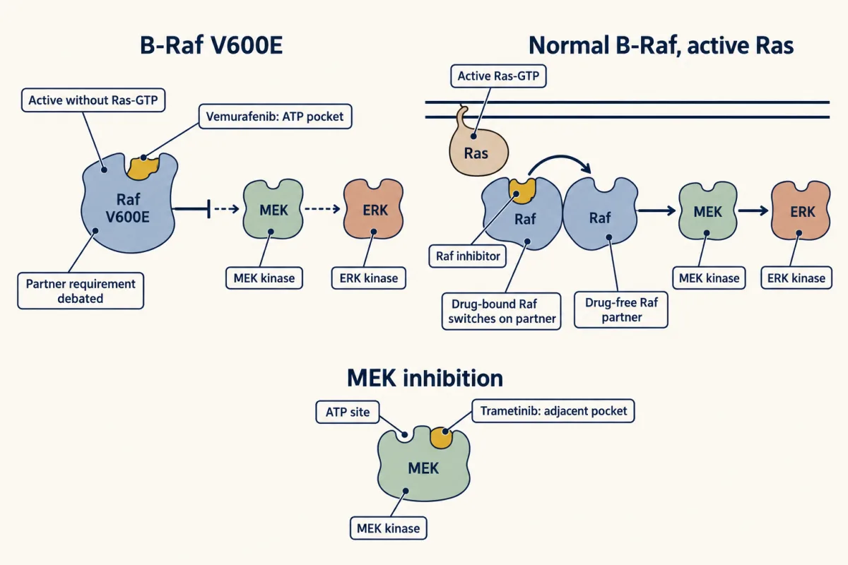
The RASopathies are developmental syndromes caused by mutations, inherited or new at conception, that mildly over-activate this pathway. The mutation is carried in every cell, but its effects differ from tissue to tissue, and rarer mosaic forms arise later and affect only some cells. They include Noonan syndrome, Costello syndrome, cardio-facio-cutaneous syndrome and neurofibromatosis type 1. Most of the genes are this lesson’s cast, and one example shows the logic: in neurofibromatosis type 1 the Ras GAP neurofibromin is lost, so the timer that turns Ras off is missing. Affected children share features of the face, heart and growth.
Raf is not the only protein that Ras-GTP can catch. PI 3-kinase is another, and it is the next lesson.
Design problem
In a PC12 cell the same Ras-to-ERK cascade answers epidermal growth factor (EGF) with division, and answers NGF with an end to division and the growth of nerve-like fibres. Design a way for one cascade to give two answers. Then predict what an always-on MEK would do in this cell with no growth factor at all, and compare your prediction with what Cowley and Marshall found.
Saved in this browser only.
One way to do it
Put the message in the duration, and build two parts. One sets the duration. Let ERK’s negative feedback close the EGF response quickly, and let the NGF response resist it, for instance through a receptor that stays active longer or a positive feedback that keeps Raf on. The other reads it: a gene product that appears after a delay and survives only if ERK is still active. One such reader has been described in fibroblasts. The immediate early protein c-Fos is short-lived unless ERK and RSK, a kinase that ERK switches on, phosphorylate it, so only a sustained signal makes c-Fos that lasts long enough to act on later genes.
The prediction follows. An always-on MEK keeps ERK on without end, which to a duration reader looks like a long NGF signal, even though NGF also switches on other pathways. The PC12 cell should stop dividing and grow fibres with no NGF present.
That is what Sally Cowley, Christopher Marshall and their colleagues reported in 1994. PC12 cells given a permanently active MEK1 grew neurites, the fibres a nerve cell extends, with no NGF in the medium. A matching MEK1 that could not be activated blocked the differentiation that growth factor normally causes. Forced on, MEK was enough. Forced off, nothing else could stand in for it.
One caution: the duration rule comes from this one cell line, and how widely cells read their kinases by the clock is an open question.
Going deeper
How the activator of ERK was found
Melanie Cobb’s laboratory followed an insulin-stimulated kinase activity through eight chromatography columns, cloned its gene and named the enzyme ERK, for extracellular signal-regulated kinase. Its sequence resembled two yeast kinases that respond to mating pheromone. Natalie Ahn, in Edwin Krebs’s laboratory, then asked what switches ERK on. In 1991 she found two activities in extracts of growth-factor-treated cells that switched on inactive ERK, a MAP kinase, in a test tube. An activator that comes apart from ERK on a column is a different protein, so ERK was not switching itself on. Purifying it with Rony Seger showed that it phosphorylates ERK on both threonine and tyrosine. Several laboratories reached this enzyme, now called MAPK/ERK kinase (MEK), at about the same time.
How common are RAS mutations?
Beyond the 1989 tissue figures above, reviews have long quoted up to about 30% for all human tumours. A 2020 analysis that cross-checked the large public cancer-mutation databases put it nearer 19% of patients, and the share varies widely by tissue.
Genes behind the RASopathies
Most of the genes are this lesson’s cast: PTPN11, the gene for the phosphatase SHP2, in about half of cases of Noonan syndrome and SOS1 and RAF1 in some others, HRAS in Costello syndrome, BRAF and the MEK genes in cardio-facio-cutaneous syndrome, and loss of the Ras GAP neurofibromin in neurofibromatosis type 1.
Your notes
Select any text in the lesson to highlight it or add a note to it. Your highlights and notes are saved in this browser and collected in your notebook.
This browser is not letting the site save anything (a private window or strict privacy settings), so highlights and notes will be lost when you leave the page.
How we measure it
DNA transfection and the focus-formation assay
DNA from tumour cells is precipitated with calcium phosphate and added to a flat, single layer of mouse NIH 3T3 fibroblasts. Cells that take up a transforming gene stop respecting their neighbours and pile into foci that can be counted. The assay shows that a piece of DNA is enough to change how a cell grows, with the caution that NIH 3T3 cells are already part of the way to transformed.
Activity-guided protein purification
A cell extract is passed over a series of chromatography columns, and every fraction is tested for one activity, here the ability to phosphorylate a test protein or to switch on ERK. Following the activity until a single band remains ties a behaviour to one protein that can then be sequenced and cloned.
Always-on and interfering mutants
A kinase is mutated so that it stays active with no signal from above, or so that it blocks the normal enzyme. For MEK the active form was made by replacing the serines that Raf phosphorylates with acidic residues that imitate phosphate. The active form tests whether a step is sufficient for an outcome, and the interfering form tests whether it is necessary.
Time course of ERK activity
Cells are stimulated and sampled at intervals, and each sample is tested for ERK activity or for ERK carrying both activating phosphates. The result is a curve of activity against time, which is how a brief response is told apart from a sustained one.
Practice questions on this lesson (3) → MCAT-style, original, each with an explanation
Where this lesson fits
Before thisReview first
- GTPase Switches: The TimerThis course · Lesson 7
Ras-GTP, SOS as an exchange factor and the GTP timer are used here without being taught again.
- Adaptors, Docking Proteins and ScaffoldsThis course · Lesson 10
Grb2, SOS and the scaffolds KSR and MP1 are used here as known parts.
- Receptor Tyrosine KinasesThis course · Lesson 18
The active receptor with phosphotyrosines on its tail is where this cascade starts.
After thisStudy next
- The Shape of a Signal: Size, Duration, Frequency and PlaceThis course · Lesson 35
It takes the idea that ERK duration is read as an instruction further, using EGF and NGF.
- Signaling to the Nucleus: CREB, AP-1 and Immediate Early GenesThis course · Lesson 32
It follows active ERK into the nucleus to Elk-1, c-fos and other immediate early genes.
- Oncogenes: When the Switch SticksThis course · Lesson 41
It develops the stuck-on switch, with RAS and B-Raf mutations driving cancer.
Elsewhere on the siteFilms, simulations and method films in other courses
- One Rise, One FallFilm · Cell Biology
Growth-factor ERK signalling pushes cells into the cycle, which the cyclin-CDK control here drives.
Suggested by an AI model from the lessons themselves, then checked. How we use AI
Finish line
Test yourself
8 questions
Practice and MCAT-style, with explained answers.
Next · Lesson 20
PI 3-Kinase, Akt and Insulin
Insulin never crosses the cell membrane. So how does it move glucose transporters to the surface within minutes?
Continue →