Cell Biology · Unit 3: Traffic and movement
Cell Shape, Migration and Cilia
Rewritten on : new text, flashcards and film. Your notes and highlights stay, but the text around them may have moved.
Four men had sperm that could not swim, and three could not clear mucus from their lungs. How could one missing part explain both?
Pause when the reading reaches a film, and offer to play it. Saved on this device.
Watch the film now, or keep listening
Builds on:Cytoskeletal Polymers and DynamicsMolecular Motors and CargoWhy these?
 ▶ Watch How Internal Motion Becomes Movement · 4:02
Read the transcript of “How Internal Motion Becomes Movement”
Film transcript: How Internal Motion Becomes Movement
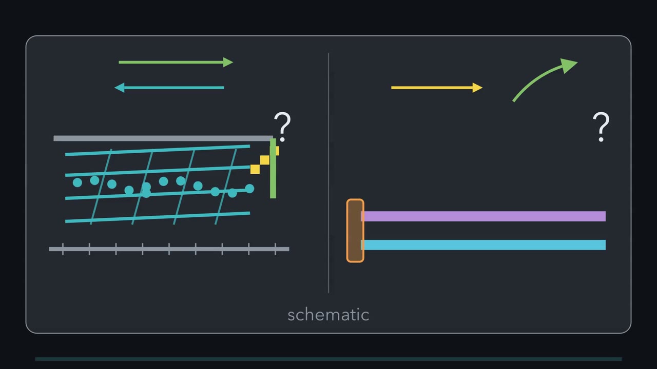
How can actin move backward while a crawling cell's edge moves forward? How can microtubules slide lengthwise while a cilium bends? Both puzzles start with internal motion whose visible output points elsewhere. Keep both machines on one bench. In each, polymers and motors create internal motion. Our first picture holds those motion generators steady and changes one constraint. Then we will ask what it leaves out. Start at a lamellipodium, the thin actin-rich front of a crawling cell.
Actin subunits extend filaments relative to the network behind. The substrate is the surface beneath the cell; the edge moves forward relative to it. Small fluorescent markers called speckles reveal the actin network moving backward over the substrate. That motion is retrograde flow. At a lamellipodial edge, polymerization adds forward speed, while retrograde flow subtracts it. Growth is measured against the network; edge and flow against the substrate. An integrin is a membrane receptor that grips extracellular matrix and connects through linker proteins to actin. Together they act like a clutch: polymerization is the engine, and adhesion transmits its motion.
Integrin coupling can reduce backward slip in this simple crawling model, so more growth appears as edge advance. Now move to a motile cilium, a hairlike projection that beats. Its axoneme is the internal microtubule core. In the classic 9+2 layout, nine doublets surround two central microtubules. Each doublet contains a complete A-tubule fused to an incomplete B-tubule. In motile axonemes, dynein motors slide neighbouring doublets. Their arms anchor on one doublet's A-tubule and grip the neighbour's B-tubule. A head steps toward that neighbour's base. Seen from the other side, the same action pushes the neighbour toward its tip. In the simplest picture, focus on two neighbouring doublets.
Free doublets would pass one another. Relative sliding creates shear, a lengthwise offset. In motile axonemes, links that resist sliding, with base anchoring, turn sliding into bending. Here the motors stay unchanged. A real bend needs more: dyneins on opposite sides must not be equally active.
Motor control selects which side is more active. The constraint shapes the output, while regulation helps choose where the bend forms. Predict both outcomes while the motion generators stay unchanged. Release the crawling clutch, and cut the axonemal links. What happens to edge advance and to the doublets?
With the clutch released, more actin motion becomes retrograde flow, so the edge advances less in this simple case. With links cut, dynein makes doublets slide apart instead of bending. The motors still work; the changed constraint reveals a different output. The two machines share a logic, not every detail. Polymers and motors create internal relative motion. With motors unchanged, a mechanical constraint helps decide whether motion slips, advances, slides apart, or bends. In these two systems, constraints and motor control shape the visible output of internal motion.
From the film
- At a lamellipodial edge, polymerization adds forward speed and rearward flow subtracts it.
- Integrin coupling can reduce backward slip in this simple crawling model.
- In motile axonemes, dynein motors slide neighbouring doublets.
- In motile axonemes, links that resist sliding, with base anchoring, turn sliding into bending.
- In these two systems, constraints and motor control shape the visible output of internal motion.
The first film built both machines in their simplest form. It left out the numbers, the rest of a crawling cell, and what happens when a part breaks; this page adds them in that order.
The one idea to carry through this lesson. In these two systems, polymers and motors create internal relative motion, while mechanical constraints and control of which motors are active shape its visible output. The first film used the simplest case of each; the sections below give the fuller picture. The rule does not cover every kind of cell movement.
For background, review the underlying cytoskeletal polymers and molecular motors.
A crawling edge: measure the hidden motion
Put numbers on the balance, with polymerization measured against the actin network and retrograde flow and the edge against the substrate:
edge speed = polymerization speed − retrograde-flow speed
This one-dimensional relation predicts the leading edge, not automatically the cell body.
Predict. Filaments extend at 40 nm/s relative to the network while the network flows rearward at 25 nm/s. How fast does the edge advance, and how far in 120 s?
Check your prediction
Take forward as positive. Edge speed = 40 + (−25) = 15 nm/s. In 120 s the edge advances 15 × 120 = 1,800 nm = 1.8 µm. Check the same result from the two contributions: polymerization adds 4,800 nm and flow subtracts 3,000 nm.
Only the flow value comes from the experiment behind this example. In PtK1 cells—a rat-kangaroo epithelial line—on flexible, fibronectin-coated gels, Gardel and colleagues measured distal actin flow at about 25 nm/s. The 40 nm/s polymerization rate is an illustrative assumption, and the calculation holds both rates constant.
Clusters of integrins and linker proteins form focal adhesions, substrate anchors that the adhesion lesson develops. In the first film’s analogy, polymerization is the engine and adhesion is the clutch.
Predict. Keep polymerization at 40 nm/s, but let stronger clutch engagement reduce rearward flow from 25 to 10 nm/s. By what factor does the edge’s 120-second advance change?
Check your prediction
Edge speed becomes 40 − 10 = 30 nm/s. The edge advances 3,600 nm, or 3.6 µm, in 120 s. Here, cutting flow from 25 to 10 nm/s doubles edge advance, from 1.8 to 3.6 µm.
- Edge speed = polymerization − flow
- 40 − 25 = 15 nm/s
- Distance in 120 s
- 15 × 120 = 1,800 nm = 1.8 µm
- Check: forward gain − rearward loss
- 4,800 − 3,000 = 1,800 nm
Starting case.
Rule to keep: edge speed = polymerization speed − retrograde flow speed. One-dimensional sketch: only the 25 nm/s flow is measured (PtK1 cells); the 40 nm/s polymerization is an illustrative assumption, as are the slider ranges and any flow other than 25 nm/s (10 nm/s is the lesson’s supposed case). Stronger adhesion is not always faster movement: in PtK1 cells traction stress and actin speed were related biphasically, switching near 8 to 10 nm/s, and in CHO cells strong adhesion slowed migration because the rear could not detach. Edge advance is not movement of the cell body. Speckle positions are arbitrary, and 120 s of model time plays in 8 s, so this is a sketch of the logic.
From edge advance to whole-cell crawling
The first film stopped at edge advance; a whole crawling cell also has to move its body and release its rear.
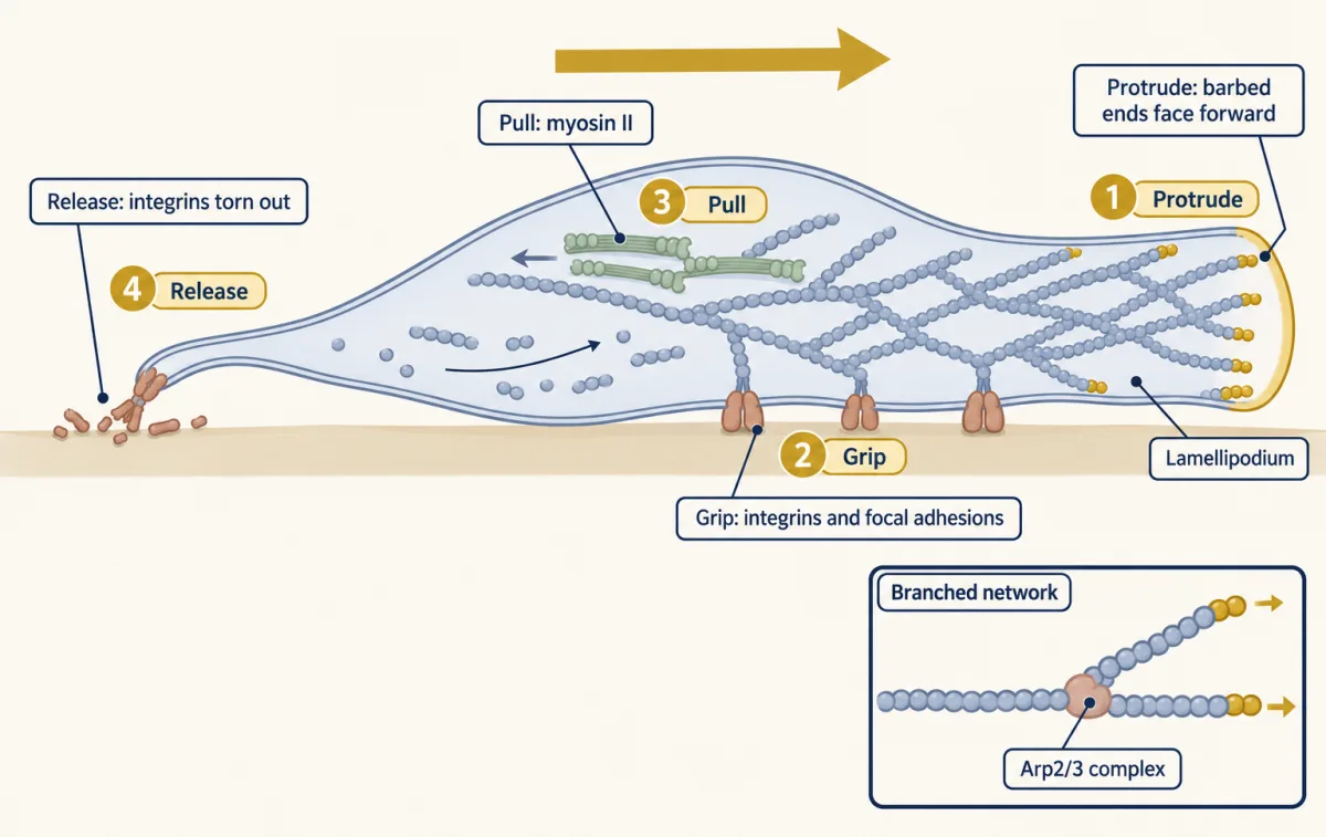
The actin-rich lamellipodium is a sheet of cytoplasm a fraction of a micrometre thick. At its front, actin subunits add at barbed ends, the fast-growing filament ends, and push the membrane. The Arp2/3 complex, a seven-protein assembly, starts a branch from an existing filament. Rac, a small signalling switch, helps activate Arp2/3 there; integrins and Rho GTPases develops that control. Behind the front, non-muscle myosin II—a two-headed relative of muscle myosin—forms small filaments and contracts actin. In fish keratocytes, evidence supported contraction where the lamellipodium meets the cell body as a driver of body movement.
The membrane pushing back on growing filaments and myosin II pulling the network rearward both contribute to retrograde flow.
Together, protrusion, grip, contraction and release form the crawling cycle. These four processes run at the same time in a smoothly gliding keratocyte; in cells such as fibroblasts they are more cyclic. At the rear, adhesions must release. Separately, filaments that come apart a short way behind the leading edge return monomers for growth at the front.
The clutch remains a starting model. In the PtK1 cells, traction stress—the force per area exerted on the substrate—and local actin speed had a two-part relationship that changed near 8 to 10 nm/s, so faster flow does not always mean more traction. Stronger adhesion can also slow migration by resisting rear release.
Predict. CHO cells engineered to express integrins locked in a high-affinity state grip fibronectin strongly but migrate slowly. Which crawling process is most likely limiting?
Check your prediction
Rear release. In CHO cells on matrix-coated glass, rear retraction often tore integrins from the cell and left them behind. At intermediate and high adhesiveness, rear retraction appeared to limit migration speed. Strong coupling at the front cannot help if the back cannot let go.
That cell failed because one part worked too well. The second film picks up here: what protease treatment or a missing part reveals, first in a dish and then in the body.
Read the transcript of “Reading a Broken Machine”
Film transcript: Reading a Broken Machine
In chapter 1, we cut the links in a drawing, and the doublets slid apart instead of bending. Now turn the question around. When a real cilium stops, which part broke, and where in the body does it show? Summers and Gibbons tested that idea in 1971.
They took axonemes from sea urchin sperm. They briefly treated them with trypsin, a protein-cutting enzyme, then added 0.1 mM ATP. Under dark-field microscopy, the axonemes did not beat. Groups of doublets slid out of the bundle. That dish test became a standard way to ask whether a flagellum's motors work. Now the body. In 1976, Björn Afzelius described four men whose sperm did not swim.
Electron microscopy indicated that the dynein arms, small projections on each outer doublet, were missing from their cilia. Sperm tails and the cilia lining the airways are built on the same basic axoneme. So predict: if the dynein arms are missing, what else should fail?
Three of the four men also had frequent bronchitis and sinusitis. Their airway cilia were not moving mucus, a sweep called mucociliary transport. One missing part disrupted motion in two organs. The match is not perfect: sperm and airway cilia differ in some dynein parts. And one mutation need not act the same everywhere. In mice lacking a protein called Cfap53, cilia on the embryonic node stopped completely. Windpipe and brain-ventricle cilia still beat, but abnormally. The instrument matters too.
Patients with DNAH11 mutations had normal-looking nasal cilia by conventional electron microscopy. Three-dimensional electron tomography found less outer-arm volume near the base. Crawling cells break the same way. Chapter 1's clutch needed integrins to grip. Neutrophils without a working integrin of the beta β2 family grip the vessel wall poorly. They largely fail to leave the blood, so infections are poorly controlled. Read a phenotype backward, tissue by tissue. Ask which machine failed, which part is defective, which tissue you examined, and with what instrument. Motors and constraints from chapter 1 become the places to look.
From the film
- After brief trypsin treatment, sperm axonemes given ATP slid apart instead of beating.
- Afzelius's four patients had immotile sperm; three had no detectable mucociliary transport.
- Sperm tails and airway cilia share a basic axoneme, though some dynein parts differ.
- One mutation can stop cilia in one tissue and only disturb the beat in another.
- Neutrophils without a working β2-family integrin largely fail to leave the blood.
The axoneme: what the model predicts
The dish test in the second film disrupted the axoneme’s resistance; here is the fuller machine it acted on.
In the classic 9+2 layout, nine outer doublets, each a complete A-tubule fused to an incomplete B-tubule, surround a central pair of single microtubules. The ring grows from a basal body, which anchors the axoneme in the cell. Dynein arms sit in outer and inner rows on each A-tubule; their motor heads grip the neighbouring B-tubule and walk toward its minus end, at the base, so each doublet pushes its neighbour toward the tip. Protein assemblies called radial spokes reach toward the central-pair complex and help regulate dynein. Between neighbouring doublets, the nexin-dynein regulatory complex links the pair, resists sliding and helps regulate the motors.
To feel how resisted shear can make curvature, hold two card strips side by side, joined at one end and tied loosely along their length. Push one strip lengthwise: because the pair cannot freely slide apart, it curves. In the axoneme, interdoublet resistance and base anchoring similarly turn dynein-driven shear into bending, while regulated motor activity helps determine where the bend forms. This is a model, not a full waveform.
Summers and Gibbons tested whether dynein changes doublets’ relative positions or shortens microtubules. They briefly treated sea-urchin sperm axonemes with trypsin, a protein-cutting enzyme that disrupted structures normally resisting sliding, then added 0.1 mM ATP. Under dark-field microscopy, groups of doublets slid out of the bundle. Disrupting the resistance exposed ATP-driven sliding as disintegration rather than bending.
Design problem
A Chlamydomonas mutant, or a patient’s sperm, has paralysed flagella. Design a test of whether its dynein motors themselves work. Isolate the axonemes first, taking the membrane and the cell’s own ATP supply out of the experiment.
Predict. After brief trypsin treatment and ATP, the mutant doublets slide apart like wild type. What has the result ruled in, and what has it not ruled out?
Saved in this browser only.
One way to reason through it
Sliding apart shows that at least some dynein arms can bind the neighbouring B-tubule, use ATP and generate motion. It does not show that both arm rows are intact; sliding can survive loss of one row. If both rows are present, a paralysed but sliding axoneme probably has a defect in motor regulation or in converting sliding into bending, such as in the radial spokes, central pair or nexin-dynein regulatory complex.
If doublets do not slide apart, suspect missing or inactive dynein arms. Confirm structures by electron microscopy, using three-dimensional electron tomography if conventional images look normal, as they did in some DNAH11 cases.
Controls are wild-type axonemes treated the same way, mutant and wild-type axonemes without ATP, and axonemes given ATP without trypsin. Sliding apart is expected only with ATP after the resistance has been cut. Intact wild-type axonemes given ATP should still slide internally but bend or beat rather than fall apart.
Even a single bend needs dyneins on one side to be more active than those on the other; to make a wave, the sides must take turns.
From a broken part to a cellular phenotype
Constraints and motors shape the output, so a broken part changes it. In 1976 Björn Afzelius described four men whose sperm could not swim. Three also had frequent bronchitis and sinusitis and no detectable mucociliary transport, and electron microscopy indicated that their cilia lacked dynein arms. One missing part disrupted motion in two organs because sperm tails and airway cilia share the same basic axoneme, though some of their dynein parts differ. Mucociliary transport is the sweep of mucus out of the airways, and airway cilia that do not beat severely impair it. The node is a small pit on the early mouse embryo, and nodal cilia that do not rotate cannot drive the leftward flow over it.
Predict. Mice lack the kinesin motor KIF3B and therefore have no cilia on the embryonic node. What happens to left-right organ placement?
Check your prediction
It becomes randomized because the node cannot make its normal leftward flow. Some people with immotile but present cilia have mirror-image organs; inherited disorders of motile cilia like theirs are called primary ciliary dyskinesia. KIF3B loss is different: it removes cilia altogether.
One mutation need not have one effect everywhere. In mice lacking Cfap53, nodal cilia were completely immotile, whereas tracheal and brain-ventricle cilia still beat, abnormally. The instrument matters too: conventional electron microscopy of nasal cilia from patients with DNAH11 mutations looked normal in one study, but electron tomography found reduced outer-arm volume near the base.
Migration defects follow the same logic. In leukocyte adhesion deficiency type I, neutrophils without functional β2-family integrins grip the vessel wall poorly and largely fail to leave the blood, so infections are poorly controlled.
Going deeper
Going deeper adds exceptions and methods.
Other ways to migrate
Not every cell uses lamellipodial clutch-based crawling. Some form blebs, rounded membrane bulges driven by pressure from contraction of the actin cortex under the membrane. Confined leukocytes can move without transmembrane force coupling, using actin flow that follows the texture of their surroundings (Reversat and colleagues, 2020). Developing neurons and neural crest cells use versions with their own features; see the growth cone, neural crest, radial migration and tangential migration.
What the leading-edge images showed
In extracted fish keratocytes, myosin-fragment decoration showed that barbed ends predominantly face forward. Svitkina and Borisy imaged extracted lamellipodia of Xenopus keratocytes and fibroblasts by electron microscopy and found extensive branching, with Arp2/3 at Y-shaped branch points. In keratocytes thinned with cytochalasin D, branches met their mother filaments at 67 ± 12 degrees.
Cilia that bend the simple categories
Not every cilium is 9+2 or motile. Primary cilia are single, non-motile 9+0 sensory organelles on many vertebrate cell types; they lack dynein arms and act as signalling antennae (see hedgehog at the primary cilium). Cilia of the mouse embryonic node are mostly 9+0. Those near its centre move by rotating rather than waving; those at its edge are thought to be non-motile and to sense the flow.
Repeats and the switching proposal
Cryo-electron tomography of Chlamydomonas axonemes identified the nexin link as the dynein regulatory complex, which supplied the combined name used above. In Chlamydomonas, the set of arms, spokes and links repeats every 96 nm along a doublet.
Cryo-electron tomography of rapidly frozen, beating sea-urchin sperm flagella found asymmetric dynein states across bends, with most dyneins in an active conformation. The authors suggested that the sides switch by selectively inhibiting one set of motors. This is a proposal; the switching signal remains open.
Your notes
Select any text in the lesson to highlight it or add a note to it. Your highlights and notes are saved in this browser and collected in your notebook.
This browser is not letting the site save anything (a private window or strict privacy settings), so highlights and notes will be lost when you leave the page.
How we measure it
Sliding-disintegration assay
Isolate axonemes, digest them briefly with a protease to cut the links between doublets, add ATP and watch by dark-field microscopy. If doublets slide apart, at least some dynein arms work; if not, and wild-type controls do slide, the motors are likely missing or inactive.
Fluorescent speckle microscopy
Label a small fraction of actin so the network appears as speckles, then track the speckles over time. The result is a map of how fast and in which direction the actin network moves at each point, such as the rearward flow in a lamellipodium.
Traction-force microscopy
Grow cells on a soft gel containing fluorescent beads and measure how far the cell displaces the beads. From the displacements and the gel's stiffness you calculate the stresses the cell exerts on the substrate.
Cryo-electron tomography
Freeze cilia or flagella rapidly, tilt them in the electron microscope and reconstruct them in three dimensions; averaging the repeating 96-nm unit shows dynein arms, spokes and links, and their shapes in a beating flagellum.
Practice questions on this lesson (3) → MCAT-style, original, each with an explanation
Where this lesson fits
Before thisReview first
- Cytoskeletal Polymers and DynamicsThis course · Lesson 14
You need actin and microtubule polymerization from there to understand protrusion and the axoneme's tubules.
- Molecular Motors and CargoThis course · Lesson 15
Dynein walking toward a minus end and myosin pulling actin are used here without re-teaching.
After thisStudy next
- Adhesion and the Extracellular MatrixThis course · Lesson 22
Integrin focal adhesions, which act as the clutch here, are developed in full there.
- Cell Identity, Cell State and DiseaseThis course · Lesson 25
It takes defects in a cellular part further, linking altered cell state to disease.
Elsewhere on the siteFilms, simulations and method films in other courses
- Optical trap (optical tweezers)Method film
Single-motor force measurements show how dynein and myosin generate the sliding and pulling used here.
Suggested by an AI model from the lessons themselves, then checked. How we use AI
Finish line
Test yourself
8 questions
Practice and MCAT-style, with explained answers.
Next · Lesson 17
Cyclin-CDK Control of the Cell Cycle
From yeast to humans, one kinase family drives every cell cycle, and it works only while a partner protein is present. Why would a cell destroy that partner every time it divides?
Continue →