Genetics and Molecular Biology · Unit 3: DNA: copying, changing, repairing
New to this? Review it at high school level: DNA: Structure and Copying
DNA Replication: Semiconservative Copying and Its Machinery
Rewritten on : new text, flashcards and film. Your notes and highlights stay, but the text around them may have moved.
One generation after a switch from heavy to ordinary nitrogen, all of E. coli's DNA sat halfway between heavy and light. Why halfway?
Pause when the reading reaches a film, and offer to play it. Saved on this device.
Watch the film now, or keep listening
Builds on:DNA Is the Genetic MaterialWhy these?
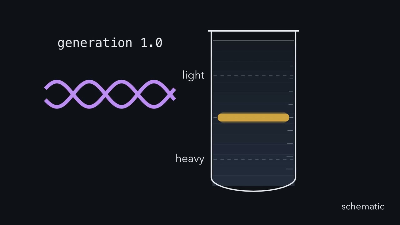 ▶ Watch What the Halfway Band Means · 3:46
Read the transcript of “What the Halfway Band Means”
Film transcript: What the Halfway Band Means
One generation after heavy-labelled E. coli switched to ordinary nitrogen, all of its DNA formed one band. That band sat halfway between heavy and light. Why did none remain fully heavy? To follow old DNA, Meselson and Stahl grew cells with heavy nitrogen. Old strands appear purple; strands built after the switch appear cyan. In a caesium chloride gradient, heavy DNA settles lower than light DNA.
Hybrid DNA settles between them. Three models start from the same purple duplex. Conservative copying keeps both old strands together and builds a separate cyan duplex. Semiconservative copying separates the old strands, then gives each a new partner. Uniform dispersive copying breaks the old material into segments and mixes those segments through both daughters. A band is not a picture of a helix. It marks many DNA molecules with the same buoyant density. One intermediate band could mean every molecule is half old. It cannot reveal whether that half is one intact strand or many segments until another generation separates their predictions. Watch each model make its initial daughters.
Which models can put every molecule at the hybrid position, with no heavy or light band? Conservative copying makes one heavy duplex and one light duplex. A single hybrid band after one generation rules out conservative copying. Semiconservative copying makes two purple-cyan hybrids.
Uniform dispersive copying can also put equal old and new material in every molecule. Both survive the initial test. Now let the survivors copy again in ordinary nitrogen. Semiconservative copying keeps each purple strand in one hybrid. The other daughters are fully cyan. Its tube gains separate hybrid and light bands. Uniform dispersive copying halves the old fraction inside every molecule again. Its single band shifts toward light. Semiconservative copying After two generations, the extracted DNA formed hybrid and light bands in equal amounts. That matches the semiconservative distribution, not one uniformly mixed band. Hybrid and light bands after two generations rule out uniform dispersive copying. The old material never disappears.
It only spreads among more molecules, and where it sits is decisive. The experiment tracked inherited material, not enzymes. Heating the hybrid DNA separated heavy and light subunits, consistent with old and new strands. Meselson and Stahl still stated their limit carefully. Their bands did not alone show that each conserved subunit was one polynucleotide chain. Each daughter molecule keeps one old strand and builds one new strand. That logic predicts later band patterns and gives replication its name: semiconservative. Keep the rule for chromosome replication in this lesson.
From the film
- Each daughter molecule keeps one old strand and builds one new strand.
- A single hybrid band after one generation rules out conservative copying.
- Hybrid and light bands after two generations rule out uniform dispersive copying.
- Band positions show how old label is distributed among DNA molecules.
One generation after Matthew Meselson and Franklin Stahl switched heavy-nitrogen E. coli to ordinary nitrogen, the extracted DNA formed one band halfway between heavy and light. Why halfway?
This lesson builds on DNA as the genetic material and the dNTPs from nucleotide metabolism.
The one idea. Each daughter molecule keeps one old strand and builds one new strand.
Scope: chromosome replication. Meselson and Stahl showed in E. coli that each daughter inherits one parental subunit, not which enzymes copy it. Heating fitted those subunits being the two strands, but their experiment alone did not prove that.
Three models, one experiment
The first film ran three models: conservative replication keeps the parental duplex whole, semiconservative gives each daughter one parental and one new strand, and dispersive mixes old and new segments through both strands.
Meselson and Stahl grew E. coli B for many generations with heavy nitrogen-15, then added a tenfold excess of ordinary nitrogen-14 ammonium chloride. Equilibrium density-gradient centrifugation in concentrated caesium chloride then separates heavy, hybrid and light DNA by buoyant density.
Predict. Start with one heavy-heavy duplex. Assume complete synchronous rounds, equal-length molecules, fully light new strands and no recycled heavy nitrogen. Under semiconservative replication, what molecules appear after a third round in light medium?
Check your prediction
Each original heavy strand stays in one hybrid; the other six molecules are light: 25% hybrid and 75% light.
| Model | After generation 1 | After generation 2 | After generation 3 |
|---|---|---|---|
| Conservative | 1 heavy + 1 light (50% heavy) | 1 heavy + 3 light (25% heavy) | 1 heavy + 7 light (12.5% heavy, 87.5% light) |
| Semiconservative | 2 hybrid | 2 hybrid + 2 light | 2 hybrid + 6 light |
| Dispersive, even mixing | one band 1/2 of the way from light to heavy | one band 1/4 of the way | one band 1/8 of the way |
Observed. A single hybrid band after one generation rules out conservative copying, though not dispersive. Hybrid and light bands after two generations, in equal amounts, rule out uniform dispersive copying.
Predict. Does the average heavy fraction per generation differ among the three models?
Check your prediction
No. In every model it is one-half, one-quarter, then one-eighth. The distribution of label among molecules, not its average, distinguishes the models.
Only the distribution of label among molecules tells the models apart: the average heavy content is 1/2, 1/4, 1/8 in all three. This is a sketch of the logic under the lesson’s assumptions (even mixing, synchronous rounds), not a trace of the gradient.
Autoradiography of bean (Vicia faba) chromosomes by J. Herbert Taylor and colleagues in 1957 was separate evidence.
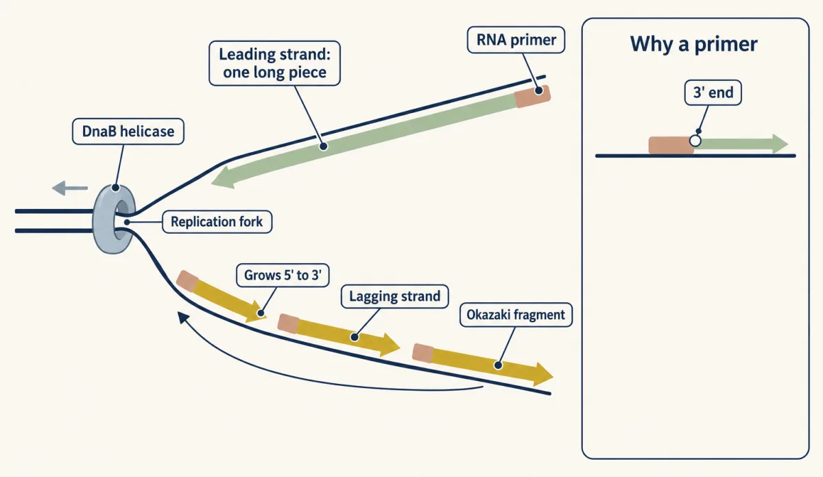
Copying happens at a Y-shaped replication fork. Every DNA polymerase extends only a free 3’-OH, whose hydroxyl attacks the incoming dNTP and releases pyrophosphate (see enzyme action). The replicative DNA polymerases also cannot start a chain: each needs a primer, a short paired stretch with a 3’ end to extend. The templates are antiparallel, so how can one fork extend 3’ ends on both? The second film answers.
Read the transcript of “Why One Strand Is Built in Pieces”
Film transcript: Why One Strand Is Built in Pieces
Each daughter molecule keeps one old strand and builds one new strand. But the two old strands point opposite ways. How can one fork build both new partners? Keep the first film's colour map. Purple marks parental templates, and cyan marks new DNA. The parental duplex opens into a moving Y-shaped replication fork. Each purple strand must guide a cyan partner, but the two templates run in opposite directions. Every DNA polymerase adds a nucleotide only to a 3' end. New DNA therefore grows 5' to 3'. A copying polymerase also needs that end paired to its template. Replicative polymerases cannot start a chain. Primase must first supply a short paired primer.
The fork moves right, exposing more purple template. Which cyan strand can grow with that fork while its polymerase extends a 3' end? On one template, the growing 3' end faces the fork. Polymerase follows the opening helix and lengthens one continuous leading strand. Its direction of synthesis stays 5' to 3'. On the other template, the usable 3' end faces away from the fork. Polymerase cannot reverse its chemistry. Primase starts near the fork, then polymerase extends back toward the previous piece. As more template opens, the cycle starts again. These pieces are Okazaki fragments. The pieces must become one strand. In E. coli, DNA polymerase I removes each RNA primer while replacing it with DNA.
DNA ligase seals the remaining nick. The finished lagging strand is continuous, although it was built discontinuously. Several proteins keep the fork moving. DnaB helicase unwinds the parental duplex. Single-strand binding protein holds the templates apart, and gyrase relieves overwinding ahead.
DnaG primase places primers. DNA polymerase III, held by its β clamp, performs most synthesis. Eukaryotes keep the same geometry with different parts and many origins. CMG is the helicase. Pol α-primase starts chains. The usual summary assigns most leading synthesis to Pol ε. It assigns most lagging synthesis to Pol δ on PCNA. Names change, but the constraint does not. Every fork needs a way to open DNA, start new chains, extend them, replace starter material, and seal the products. The text compares these jobs and the evidence behind them. Polymerases extend a 3' end, so one strand is built in pieces. Both strands still obey the same chemistry.
The enzyme never changes synthesis direction to solve the geometry. This constraint turns opposite templates into different building patterns.
From the film
- Polymerases extend a 3' end, so one strand is built in pieces.
- Both new strands grow 5' to 3', although they grow in opposite directions at a fork.
- Primase restarts the lagging strand as the fork exposes more template.
- Primer removal and ligase turn short fragments into one continuous strand.
Testing the pieces
The second film named the leading strand and the lagging strand’s Okazaki fragments, not the evidence for them.
In 1968 Reiji Okazaki, Tsuneko Okazaki, Kiwako Sakabe, Kazunori Sugimoto and Akio Sugino tested for pieces. They grew E. coli B at 20°C to slow replication, gave radioactive-thymidine pulses as short as five seconds, denatured the DNA and sized the labelled chains on alkaline sucrose gradients. After a five-second pulse most label was in a slow component of about 11S; with longer labelling it moved into long DNA. They suggested pieces of about 1,000 to 2,000 nucleotides, leaving their direction and joining for further proof. Eukaryotic Okazaki fragments are shorter, about 100 to 200 nucleotides.
The fork’s jobs, organism by organism
The table adds what the second film skipped: origin opening, helicase direction and primer removal.
| Job | E. coli (one origin, oriC, circular chromosome) | Eukaryotes (many origins, linear chromosomes) |
|---|---|---|
| Open the origin | DnaA binds oriC and opens a short AT-rich stretch | In G1, inactive MCM2-7 rings load at origins |
| Unwind | DnaB ring (loaded by DnaC, ATP-driven) moves 5’ to 3’ on the lagging-strand template; SSB coats single strands; gyrase, a topoisomerase, relieves overwinding | In S phase, Cdc45 and GINS join MCM as the active CMG helicase, which moves 3’ to 5’ on the leading-strand template |
| Prime | DnaG primase makes short RNA primers | Pol α-primase makes RNA and extends it briefly with DNA |
| Extend | Pol III holoenzyme; its β sliding clamp keeps it on DNA for thousands of nucleotides | In the usual summary, Pol ε most leading-strand and Pol δ most lagging-strand synthesis, both on the PCNA clamp |
| Remove primers | Pol I removes each primer with its 5’ to 3’ exonuclease while filling the gap | Pol δ displaces primer material as a 5’ flap; FEN1 cuts short flaps, Dna2 helps with longer ones, RNase H has a smaller part |
| Seal | DNA ligase | DNA ligase I |
Arthur Kornberg’s laboratory purified Pol I first, but a 1969 mutant with very little Pol I polymerase activity still grew; Pol III is the replicative polymerase.
Predict. The E. coli K-12 chromosome is about 4.6 million base pairs. Two forks each move at about 1,000 nucleotides per second. How long does one round take, and how can cells divide about every 20 minutes in rich medium?
Check your prediction
Each fork copies half: (4.6 × 10⁶ / 2) / 1,000 = 2,300 seconds, about 38 minutes. Fork speed rises only modestly with growth rate, far too little to close the gap, so new rounds begin at oriC before earlier rounds finish, and each division still receives a finished chromosome.
Three layers of accuracy
Polymerases first select bases: the active site closes well around a correct pair and poorly around a mispair, leaving roughly one error per 104 to 105 nucleotides. They then proofread: a 3’ to 5’ exonuclease removes a mispaired 3’ end. In E. coli Pol III that activity is in the ε subunit. Proofreading improves accuracy about 100-fold, and up to 1,000-fold depending on polymerase and mispair. Not every polymerase proofreads: eukaryotic Pol α lacks a functional proofreading exonuclease. Mismatch repair then fixes errors after the fork (DNA repair and recombination). Together they bring the E. coli rate to roughly one error per 109 to 1010 base pairs per round.
Predict. At that rate, about how often does copying a 4.6-million-base-pair chromosome add an error?
Check your prediction
About one copy in 200 to 2,000. These are orders of magnitude, not fixed constants; assay and sequence matter.
Linear chromosomes need finishing work
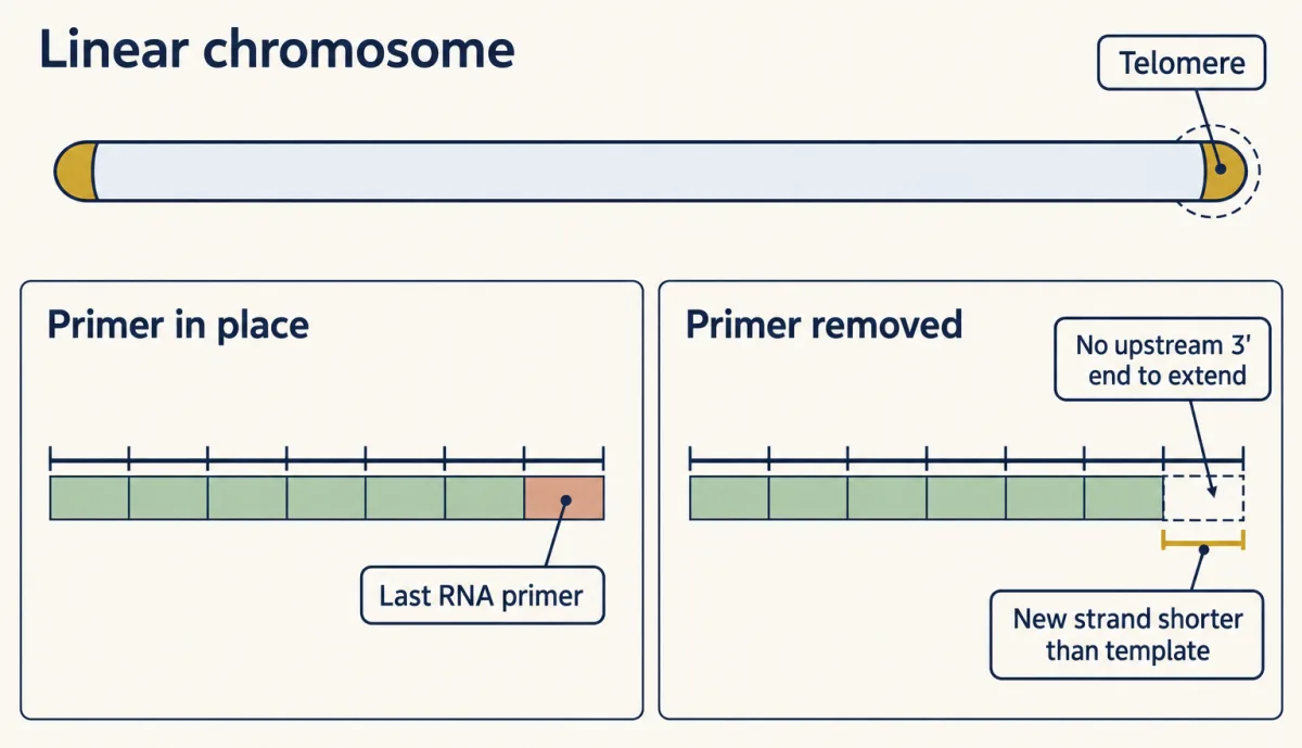
Predict. When an RNA primer is removed, why can a circular chromosome always fill the gap, while the gap left by the last primer at a linear tip may not be filled?
Check your prediction
A circle has DNA behind every gap, supplying a 3’ end for extension. At a linear tip, no upstream 3’ end may remain. This is the classical end-replication problem.
By the mid-1990s it was clear that real telomeres end in a single-stranded 3’ overhang of the G-rich strand, which leading-strand synthesis cannot regenerate and telomerase makes up; the lagging strand has its own finishing problem (Going deeper).
Telomeres are repeated sequences at chromosome ends: TTGGGG in the ciliate Tetrahymena and TTAGGG in humans. In 1985 Carol Greider and Elizabeth Blackburn found that Tetrahymena extracts added TTGGGG repeats to synthetic telomeric primers; heat and protease destroyed the activity. In 1989 they reported that the enzyme’s RNA contains CAACCCCAA, suggested as the template.
Telomerase is a reverse transcriptase (it makes DNA from an RNA template) that carries its own RNA and extends the G-rich 3’ strand. The C strand is filled in separately; in engineered human cells that required the CST complex with Pol α-primase. Most human somatic cells make little telomerase, so their telomeres shorten as they divide; germline cells and most cancer cells keep it active. Blackburn, Greider and Jack Szostak shared the 2009 Nobel Prize in Physiology or Medicine.
Design problem
E. coli are given a 5-second pulse of tritiated thymidine, and their DNA is denatured and sized. Right after the pulse, most label is in pieces of about 1,000 to 2,000 nucleotides. After a 60-second chase in unlabelled thymidine, the label is in long DNA. In a ligase-defective mutant, the short pieces persist after the chase. What do you conclude, and what control would you add?
Saved in this browser only.
One way to do it
Conclusion. At least some new DNA is made as short pieces later joined into long strands. The chase shows the same labelled molecules moving from short to long, a precursor becoming a product, and the mutant shows that joining needs ligase.
Controls. Long-labelled DNA handled the same way should stay long (not extraction breakage); repair mutants should still make the pieces (not repair patches). Hybridizing pulse-labelled pieces to separated strands of a region with known fork direction tests the semidiscontinuous prediction: pieces mostly from the lagging strand. Early data could not exclude pieces on both strands, and later E. coli work by Tye and colleagues in 1977 suggested that excising misincorporated uracil also makes short fragments, so a uracil-excision-deficient strain gives a cleaner test.
Going deeper
In a replisome rebuilt from purified budding-yeast proteins, Pol δ helped establish leading strands before handing off to Pol ε, so the usual division is dynamic.
Work published in 2024 added a second problem on the lagging strand: in budding-yeast replisomes rebuilt on linear DNA, no primers were laid on the overhang, and lagging-strand synthesis stopped in a zone more than 26 nucleotides from the template end. CST with Pol α-primase supplies the C-strand fill-in. This refines, rather than overturns, the classical last-primer model.
Your notes
Select any text in the lesson to highlight it or add a note to it. Your highlights and notes are saved in this browser and collected in your notebook.
This browser is not letting the site save anything (a private window or strict privacy settings), so highlights and notes will be lost when you leave the page.
How we measure it
Equilibrium density-gradient centrifugation in CsCl
DNA is spun for many hours in concentrated caesium chloride until a density gradient forms and each DNA species settles where the solution matches its buoyant density. DNA made with heavy nitrogen-15 bands apart from ordinary DNA, so the bands report how parental and new material are shared among molecules.
Pulse labelling and alkaline sucrose gradients
Cells are given radioactive thymidine for seconds, then the DNA is denatured in alkali and sized by sedimentation. The size of the newest chains shows whether DNA is made continuously or as short pieces that are joined later; a chase in unlabelled thymidine shows where the label goes next.
Cell-free primer-extension assay
A cell extract or purified enzyme is given a short synthetic primer and labelled nucleotides, and the products are sized on a gel. A ladder of added repeats, lost after heat or protease treatment, identified the telomerase activity in Tetrahymena.
Reconstitution with purified proteins
Replication is rebuilt in a tube from purified proteins and origin DNA, then single components are left out or added. In budding yeast this showed which polymerase and clamp each strand needs in that defined reaction.
Practice questions on this lesson (4) → MCAT-style, original, each with an explanation
Where this lesson fits
Before thisReview first
- DNA Is the Genetic MaterialThis course · Lesson 12
You need to know DNA carries heredity and how its labelled forms were tracked before following old and new strands here.
After thisStudy next
- DNA Repair and RecombinationThis course · Lesson 15
Mismatch repair, the third layer of accuracy, is worked out there along with other ways cells fix DNA.
- Mutation: Where Variation Comes FromThis course · Lesson 14
Replication errors that escape proofreading and repair become the mutations whose rates and origins are studied there.
- Recombinant DNA, Cloning and PCRThis course · Lesson 22
PCR copies DNA with primers and a polymerase extending 3' ends, the same rules used at the fork.
Elsewhere on the siteFilms, simulations and method films in other courses
- One Strand Is EnoughFilm · Biology
It shows how each strand of the double helix can serve as a template for rebuilding its partner.
- Copy a piece of DNASimulation · Biology
You can copy a piece of DNA there and watch base pairing build the new strand.
- How One Set Becomes TwoFilm · Biology
It places DNA copying in interphase before the copies are separated into two nuclei.
Suggested by an AI model from the lessons themselves, then checked. How we use AI
Finish line
Test yourself
9 questions
Practice and MCAT-style, with explained answers.
Next · Lesson 14
Mutation: Where Variation Comes From
Ten bacterial cultures meet the same virus: why does one leave 52 resistant colonies while five leave none?
Continue →