Cell Biology · Unit 2: Membranes and compartments
The Nuclear Envelope and Nuclear Transport
Rewritten on : new text, flashcards and film. Your notes and highlights stay, but the text around them may have moved.
If one pore carries traffic both ways, what decides which way each protein goes?
Pause when the reading reaches a film, and offer to play it. Saved on this device.
Watch the film now, or keep listening
Builds on:Protein Sorting into CompartmentsMembrane Organization and TopologyOrganelles as Distinct EnvironmentsWhy these?
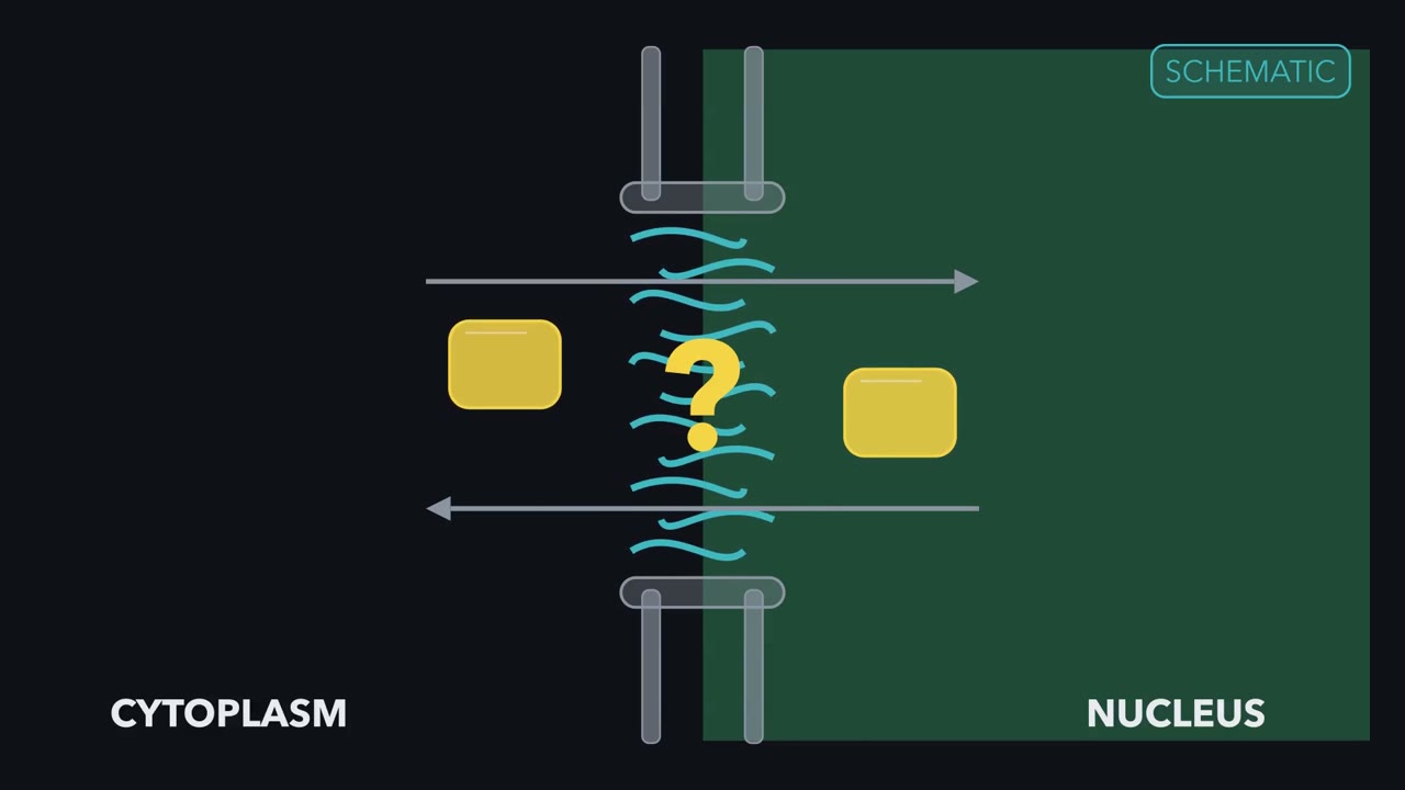 ▶ Watch One Pore, Two Directions · 3:48
Read the transcript of “One Pore, Two Directions”
Film transcript: One Pore, Two Directions
The nuclear pore is not a one-way door. So how can the same pore deliver one protein inward and another outward? Watch one pore between the cytoplasm and nucleus. Soluble cargo floats within either compartment instead of sitting in a membrane. Here is a first picture. For soluble protein cargo, the nuclear pore is a selective passage, not a one-way motor. Small grey particles slip between moving teal chains. Larger yellow cargo crosses slowly alone. Receptors engage the barrier for faster passage. Cargo carries labels that transport-receptor proteins can read. A blue NLS tag recruits blue importin α, which holds cargo.
Importin β joins it and engages the pore. An orange NES tag can recruit the purple export receptor CRM1. The difference between the two sides comes from Ran, a small switch protein. Ran is on with GTP and off with GDP. Green RanGTP is abundant in the nucleus.
Grey RanGDP dominates in the cytoplasm. This unequal distribution is the Ran gradient. In the simplest form, the import complex threads through the pore. It moves from cytoplasm to nucleus without a directional push. In the nucleus, green RanGTP binds importin β and separates it from importin α. Importin α then releases the blue-tagged cargo where RanGTP is high. All three receptor and cargo pieces are now distinct. Now predict. If RanGTP became equally common on both sides, could importin still unload only inside? Hold the two-sided picture in your mind.
No. Importin could still cross. But on either side, it might drop cargo or fail to pick it up. Net import would lose its one-sided rule. Restore the gradient, and cargo release again favors the nuclear side. Export reverses the same rule.
Nuclear green RanGTP helps purple CRM1 grip orange-tagged NES cargo. The complex crosses outward. In the cytoplasm, Ran-binding proteins first help the cargo come free. Hydrolysis then turns RanGTP into RanGDP. This frees CRM1 for another round. Nuclear RanGTP tells importins to unload and exportins to load. Blue importins carry NLS cargo in. Purple CRM1 carries many NES cargoes out. One asymmetry gives opposite receptors useful directions. In Ran-dependent protein transport, GTP hydrolysis maintains the gradient; it does not push cargo through the pore. In Ran-dependent transport, Ran mainly sets where cargo loads and unloads.
Pore proteins still help on each face, but the pore sets no direction of its own. Most messenger RNA export uses another receptor.
From the film
- For soluble protein cargo, the nuclear pore is a selective passage, not a one-way motor.
- In Ran-dependent protein transport, nuclear RanGTP tells importins to unload and exportins to load.
- Importins carry NLS cargo inward, while CRM1 carries many NES cargoes outward.
- In Ran-dependent transport, GTP hydrolysis maintains the gradient; it does not push cargo through the pore.
A protein sits mostly in the cytoplasm. Add one export-blocking drug, and within an hour it piles up in the nucleus. The simplest explanation is that it has been crossing the border all along. In an ordinary eukaryotic cell, soluble nuclear proteins are synthesized on cytosolic ribosomes, while messenger RNA and ribosomal subunits made inside must leave. How can one border sort traffic moving both ways?
The first film gave a first picture of Ran-dependent soluble-protein traffic; it leaves out the border itself, how much it leaks, and the evidence. The page maps the border and measures its leak, the second film follows the experiments, and the page ends on the drug puzzle.
The envelope, lamina and pore
To test a two-compartment transport model, first map the physical border and the spaces it separates.
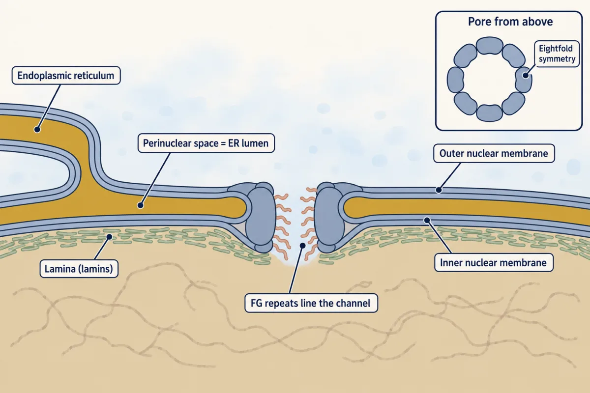
The nuclear envelope’s inner and outer lipid bilayers are described as about 20 to 40 nm apart in animal cells. The outer membrane is continuous with the endoplasmic reticulum, so the perinuclear space between them is ER lumen, the space enclosed by the ER membrane. The membranes meet around each pore’s water-filled channel.
Predict. A soluble tracer is placed in the perinuclear space. Without crossing a membrane, which space can it reach: the cytosol, nucleoplasm or ER lumen?
Check your prediction
The ER lumen. It and the perinuclear space are topologically equivalent: they are continuous without crossing a membrane. The pore instead joins cytosol to nucleoplasm. See membrane organization and topology.
Under the inner membrane lies the lamina, a lamin meshwork that gives the nucleus mechanical stability and bridges envelope and chromatin. Each pore has an eightfold-symmetric scaffold built from multiple copies of roughly 30 kinds of nucleoporin. Nucleoporins with disordered phenylalanine-glycine, or FG, repeats line the channel and form its selective barrier.
A soft barrier, measured
The first film used the simplest case; here is the fuller picture: passive passage slows continuously rather than stopping at one size. Textbooks once proposed a sharp 40 kDa limit. In 2016 Timney and colleagues bleached cytoplasmic fluorescence from different-sized reporters in live budding yeast and followed the excess nuclear signal relaxing, or returning, toward equilibrium. Model that excess as one exponential:
x(t) / x(0) = exp(-t / τ)
Their normalized time constants were τ = 15 s for 27 kDa GFP and τ = 413 s for a 67 kDa reporter. Assume these values apply to the cell and ignore their uncertainty.
- t / τ
- Excess left, exp(−t/τ)
- Half-time, τ × ln 2
In this model, the excess nuclear signal falls as exp(−t/τ), using the normalized τ values the lesson gives (Timney et al. 2016, yeast; uncertainty ignored). These are model curves, not measured traces; τ grew about 27-fold (413/15 = 27.5) for a 2.5-fold rise in mass (67/27 = 2.48), so the pore is a soft barrier, not a cutoff. Only these two reporters are given; the picture does not interpolate between them.
Predict. After one minute, will the larger reporter have relaxed by half? Estimate the fraction of excess signal left for each reporter before opening the calculation.
Check your prediction
No. For GFP, 60/15 = 4, so exp(-4) = 0.018: 1.8% remains. For the 67 kDa reporter, 60/413 = 0.145, so exp(-0.145) = 0.865: 86.5% remains. The larger reporter is far from halfway to equilibrium.
The half-time is t½ = τ × ln 2. It is 15 × 0.693 = 10.4 s for GFP and 413 × 0.693 = 286 s for the 67 kDa reporter. At 826 s, two time constants for the larger reporter, exp(-2) = 0.135, so only 13.5% remains. A 2.5-fold rise in mass (67/27 = 2.48) increased the time constant about 27-fold (413/15 = 27.5), yet did not stop exchange. Passive permeability falls steeply but continuously with size: this is a soft barrier, not a cutoff.
These values describe relaxation of excess signal in a yeast nucleus, not the time one molecule spends in one pore. The pore’s physical fine print comes in Going deeper.
A leak this slow means a large protein that must reach the nucleus quickly needs a label and a receptor. The second film picks up here: how experiments found those labels and their readers, and what each one proved.
Read the transcript of “Move It, Mutate It, Block It”
Film transcript: Move It, Mutate It, Block It
How did anyone find a nuclear address, or prove what it does? Chapter one showed receptors reading labels, with Ran controlling where cargo loads and unloads. Start with nucleoplasmin, a frog oocyte protein of five copies, each with a tail. In 1982, Dingwall, Sharnick and Laskey clipped the tails away.
Bare cores injected into the cytoplasm stayed out. A larger pentamer keeping one tail went in. One attached tail was sufficient for final accumulation. More tails changed the rate, not how much eventually reached the nucleus.
One more injection put bare cores straight into the nucleus. They remained there. Why that injection matters is a question the page asks you. Which route does cargo take? In 1984, Feldherr and colleagues injected gold grains coated with nucleoplasmin. The coated grains spanned about 5 to 20 nm. They entered the nucleus.
Electron microscopy located transport at the pores. Now predict. Coat the same gold with an inert polymer instead of nucleoplasmin. Where will most of the grains end up? Mostly outside the nucleus. Gold coated with nucleoplasmin lacking its transport part was also largely kept out. So the coat, not the gold, decided entry. Next, a label you could move.
Kalderon, Smith and colleagues traced the address of SV40 large T antigen to a short stretch. The normally cytoplasmic reporters were beta β-galactosidase and pyruvate kinase. Attached to either one, the stretch caused nuclear accumulation. Next, change it. Swapping lysine 128 for threonine abolished nuclear accumulation. Moving, mutating, and blocking are complementary tests. Each result supports only a limited claim. By 1995, importin α was shown to recognize the signal. Importin β was shown to dock at the pore. Leucine-rich export signals still had no known reader. In 1997, Fornerod, Mattaj and colleagues tested CRM1. Extra CRM1 stimulated export of Rev.
It also stimulated export of U snRNAs. Leptomycin B inhibited both export pathways. CRM1, RanGTP and an export signal formed a complex in vitro. All three results pointed at CRM1. Together, they made a stronger case than any one result alone. The same year, Izaurralde, Görlich and colleagues collapsed the Ran gradient in oocyte nuclei. They injected RanGAP1, RanBP1, or a defective Ran mutant. The mutant could not stably hold GTP. All three treatments depleted nuclear RanGTP. Major import and export pathways stopped.
Export was more readily inhibited than import. None of these results proved everything. Move it, mutate it, block its reader: each supports one claim. The page's drug puzzle stays unresolved.
From the film
- In frog oocytes, bare nucleoplasmin cores injected into the cytoplasm stayed out, while a larger pentamer with one tail went in.
- Nucleoplasmin-coated gold entered through pores; inert-coated gold was largely kept out, so the coat decided entry.
- The SV40 large-T signal made two cytoplasmic proteins nuclear, and one lysine change abolished that accumulation.
- Extra CRM1, leptomycin B and an NES–RanGTP binding test converged on CRM1 as an export receptor.
- Moving, mutating and blocking are complementary tests; each result supports only a limited claim.
Signals: what an experiment can prove
The second film traced how the labels were found; here is what they are.
A classical nuclear localization signal (NLS) is a basic amino-acid sequence recognized by importin α. It can have one basic cluster, such as SV40 large T antigen’s Pro-Lys-Lys-Lys-Arg-Lys-Val, or two clusters separated by a spacer, as in nucleoplasmin. Unlike many ER signal sequences in protein sorting, it is ordinarily not cut off. Its effect depends on protein context.
In the classical pathway, importin α binds the NLS, importin β binds importin α, and importin β engages FG repeats in the pore. A nuclear export signal (NES) recognized by CRM1, also called exportin 1, is a pattern of spaced hydrophobic residues, often leucines.
Entry is not retention
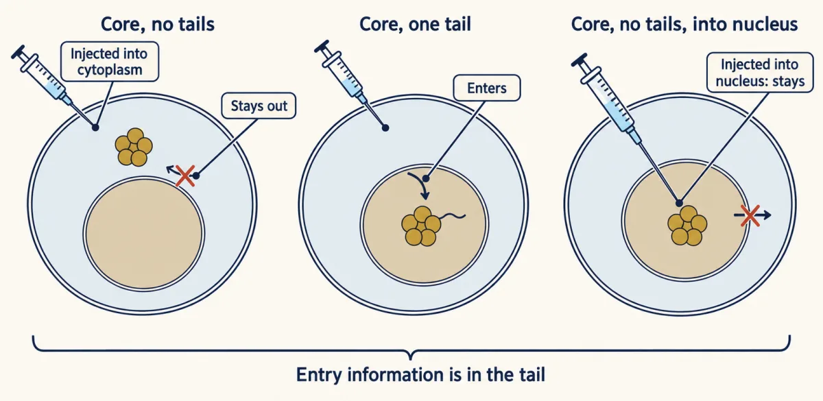
In 1982 Dingwall, Sharnick and Laskey clipped the tails from pentameric nucleoplasmin while leaving its core. Tail-less cores injected into Xenopus oocyte cytoplasm stayed out; a larger one-tail pentamer entered. Tail-less cores injected into the nucleus stayed there. The second film leaves the inference from that third injection to you.
Predict. Why is the nuclear injection essential? What conclusion would be unsafe without it?
Check your prediction
It shows that the tail is needed for entry, not simply for retention. Without the nuclear injection, a cytoplasmic core might have entered and leaked back out. The larger one-tail pentamer entering also argues against size alone deciding passage.
Route and signal
In 1984 Feldherr, Kallenbach and Schultz injected nucleoplasmin-coated gold particles, about 5 to 20 nm across including the coat. Electron microscopy placed transported gold at pores. Transport-deficient nucleoplasmin or inert-polymer coatings were largely excluded, showing that entry depended on a transport-competent coat, not gold itself.
That same year Kalderon, Roberts, Richardson and Smith transferred the SV40 large-T signal region to β-galactosidase and pyruvate kinase; both reporters became nuclear. Changing lysine 128 to threonine abolished accumulation. Earlier that year Lanford and Butel had found a cytoplasmic T-antigen mutant with lysine 128 changed to asparagine.
Predict. A transferred signal makes a cytoplasmic reporter nuclear, while a one-residue mutant does not. What has this shown, and what remains unproved?
Check your prediction
The sequence is sufficient for nuclear accumulation in those reporters, and that residue matters. The transfer test does not by itself show that the sequence is necessary in its original protein; for T antigen, deletions and Lanford and Butel’s lysine-128 mutant supplied that evidence. Nor does transfer show that importin α binds the sequence directly or that every protein context exposes it equally.
Crossing is not direction
The second film showed that blocking Ran stops traffic; it did not separate passage through the pore from directional accumulation over many transport rounds. The next experiment makes that separation.
Ran is a small GTPase that works like the switches in GTPase switches: on with GTP and off with GDP. In an in vitro experiment, transportin, another import receptor, carried cargo across pores without Ran or nucleotide. Nuclear RanGTP, but not its hydrolysis, was needed to release cargo and return the receptor; hydrolysis appeared necessary to restore import competence for further rounds. Receptor-assisted crossing is best described as facilitated diffusion: a receptor speeds diffusive passage through the pore rather than delivering a power stroke. Repeated directional accumulation still requires energy to maintain and reset the Ran system.
The receptors read the Ran gradient. Importins load cargo where RanGTP is low, in the cytoplasm, and release it when nuclear RanGTP binds. Exportins such as CRM1 bind cargo only together with RanGTP in the nucleus and release it in the cytoplasm, after Ran-binding proteins and GTP hydrolysis take the complex apart.
The evidence accumulated in steps. Adam and Gerace purified import-stimulating nuclear localization signal (NLS) binders in 1991; Moore and Blobel showed import needs Ran in 1993; and Görlich, Laskey and colleagues assigned NLS recognition to importin α and pore docking to importin β in 1995. In 1997 Izaurralde, Mattaj, Görlich and colleagues collapsed Ran asymmetry in oocyte nuclei with RanGAP1, RanBP1 or a Ran mutant unable to hold GTP. Major import and export pathways stopped, export more readily than import.
The same year Fornerod, Ohno, Yoshida and Mattaj found three converging results in oocytes and in vitro: extra CRM1 stimulated export of Rev and U snRNAs; leptomycin B inhibited that export; and CRM1 bound nuclear export signal (NES) cargo cooperatively with RanGTP.
This rule is scoped to Ran-dependent transport. Most messenger RNA leaves through NXF1, also called TAP, largely independently of Ran; spliced mRNA export proceeded without Ran in a Xenopus oocyte assay. RNA preparation before export belongs to RNA processing. Cells can also regulate import, for example by masking or exposing an NLS, to switch transcription factors on or off; see signaling to the nucleus.
Predict and design: the drug puzzle
Both films now meet in one perturbation: the loading-and-unloading rule, and the habit of claiming only what a result shows.
Leptomycin B inhibits CRM1-dependent export. Protein Z is cytoplasmic at steady state in cultured cells, but within an hour of adding the drug it is nuclear.
Predict. What is the simplest interpretation, and what does nuclear accumulation fail to prove?
One way to do it
Z must enter the nucleus fast enough to accumulate within an hour once export is blocked. The simplest interpretation is that Z shuttles: it enters through its own NLS, rides on a partner with one or, if small enough, diffuses; it leaves through CRM1 using its own NES or a partner’s. Export normally wins at steady state. The drug result alone proves neither that Z has its own NLS nor that it binds CRM1 directly.
Test the model in five ways:
- NES necessity. Mutate key hydrophobic residues in a candidate leucine-rich signal. Z should become nuclear without drug. Check expression, folding and partner binding, because the mutation could disrupt more than export.
- NLS necessity. Mutate a candidate basic cluster. If it is Z’s main route in, Z should stay cytoplasmic even with leptomycin B. Remaining entry could reflect another signal, a partner or passive diffusion.
- Signal sufficiency. Attach each candidate signal to a large inert reporter, such as a multi-GFP fusion that diffuses only slowly. The NLS fusion should become nuclear. Adding the NES should shift it toward the cytoplasm, and leptomycin B should reverse that shift.
- Direct shuttling. Fuse cells so two nuclei share one cytoplasm, block new protein synthesis and ask whether Z moves from a labelled nucleus to an unlabelled one. Alternatively, photoactivate nuclear Z and watch it leave.
- Controls. Check cell health, include a known CRM1 cargo, and, if available, rescue with a leptomycin-resistant CRM1 mutant.
Going deeper
These details give the receptor-recycling machinery and the pore’s structure.
The receptor-recycling machinery
RCC1, Ran’s exchange factor, sits on chromatin and loads GTP onto Ran, making RanGTP in the nucleus. Cytoplasmic RanGAP1, helped by RanBP1, stimulates hydrolysis and keeps cytoplasmic RanGTP low. NTF2 returns RanGDP to the nucleus. During classical import, nuclear RanGTP separates importin β from importin α; Nup50 helps α release cargo. Importin β returns with RanGTP, while CAS returns α with RanGTP. During export, RanBP1 helps dismantle cytoplasmic CRM1-cargo-RanGTP before RanGAP-stimulated hydrolysis frees the receptor.
The pore is not one fixed mesh
Passive entry of inert probes into digitonin-permeabilized HeLa cells, whose plasma membranes had been selectively opened with detergent, supported a model with a dominant FG-domain mesh radius of about 2.6 nm and a small fraction of wider channels. That is a model-dependent description, not a literal universal hole size. Whether FG domains behave more like a cohesive gel or a brush of waving chains remains debated. Nor is the scaffold rigid: fission-yeast pores constricted reversibly during energy depletion and hypertonic stress. In permeabilized HeLa cells, single NLS-carrying molecules interacted with pores for about 10 ms on average.
Lamina fine print
Lamins belong to the intermediate-filament family. In cryo-electron tomograms of extracted mouse fibroblasts lacking vimentin, a cytoplasmic intermediate-filament protein, A- and B-type lamins formed tetrameric filaments only about 3.5 nm thick. Mutations in LMNA, which encodes A-type lamins, cause laminopathies. A mechanically weaker nucleus is one proposed mechanism, and why one gene produces such varied diseases remains argued. Budding yeast has no lamins.
Your notes
Select any text in the lesson to highlight it or add a note to it. Your highlights and notes are saved in this browser and collected in your notebook.
This browser is not letting the site save anything (a private window or strict privacy settings), so highlights and notes will be lost when you leave the page.
How we measure it
Compartment-specific microinjection
A protein is injected into either the cytoplasm or the nucleus of a large cell such as a Xenopus oocyte, and its location is read later. Comparing the two injection sites separates a failure to get in from a failure to stay in.
Gold-particle electron microscopy
Colloidal gold coated with a protein is injected, the cells are fixed and sectioned, and the dense grains are located by electron microscopy. Where the grains sit shows the route a cargo takes, and changing the coating while keeping particle size fixed shows what the route selects for.
Signal-transfer reporter fusions
A candidate signal is attached to a protein that normally stays in one compartment, with an unfused reporter and a point-mutant signal as controls. If the fusion moves and the mutant does not, the sequence is sufficient in that reporter.
FRAP-based nuclear exchange assay
In living cells expressing a fluorescent reporter, the reporter's fluorescence in one compartment is bleached with light and the return to equilibrium is followed over time. Fitting the time course gives an exchange time constant, which can be compared across reporter sizes, mutants or species.
Practice questions on this lesson (3) → MCAT-style, original, each with an explanation
Where this lesson fits
Before thisReview first
- Protein Sorting into CompartmentsThis course · Lesson 7
Signal sequences and receptors that send proteins to compartments are the idea the nuclear signals here build on.
- Membrane Organization and TopologyThis course · Lesson 4
The envelope's two bilayers and the outer membrane's continuity with the ER rest on how membranes and their faces are organized.
- Organelles as Distinct EnvironmentsThis course · Lesson 6
You need to know how compartments keep different contents separate to see why a nucleus needs selective transport.
After thisStudy next
- Nuclear Architecture and Membraneless CompartmentsThis course · Lesson 10
It takes the nucleus further, asking how it organizes compartments inside without membranes after this lesson's border.
- Protein Quality Control and Organelle TurnoverThis course · Lesson 9
Shuttling and turnover of proteins by receptors and tags extends this lesson's idea that traffic is read from signals.
- Cytoskeletal Polymers and DynamicsThis course · Lesson 14
Another GTPase-driven polymer system whose GTP cycle sets direction, reusing this lesson's switch logic.
Suggested by an AI model from the lessons themselves, then checked. How we use AI
Finish line
Test yourself
8 questions
Practice and MCAT-style, with explained answers.
Next · Lesson 9
Protein Quality Control and Organelle Turnover
When material piles up in a cell, is more arriving or less leaving, and how can you tell?
Continue →