Cell Biology · Skim version
Cyclin-CDK Control of the Cell Cycle
Read the full lesson → Skim the whole course →
The guiding idea
From yeast to humans, one kinase family drives every cell cycle, and it works only while a partner protein is present. Why would a cell destroy that partner every time it divides?
Every eukaryotic cell cycle runs on one conserved engine: a cyclin-dependent kinase (CDK) switched on by a cyclin partner.
From the film: One Rise, One Fall
- In the sea-urchin egg record, cyclin is made steadily and destroyed at every division, making a sawtooth; a CDK is inactive alone and needs a bound cyclin to switch on.
- Wee1 holds loaded CDK1-cyclin B pairs off through G2, then Cdc25 releases them, so activity snaps on all at once.
- CDK activity rises and falls once per cycle; the rise carries the cell into mitosis.
- The APC/C destroys securin and cyclin B, so the sisters separate and activity falls; a destroyed protein must be made again, so the fall runs one way.
- Origins are licensed only when activity is low and fire only when it is high, so the genome is copied once per cycle.
From the film: A Snapshot of the Cycle
- Because the genome is copied once per cycle, a diploid cell holds 2C of DNA in G1 and 4C in G2 and mitosis, with S-phase cells in between.
- Cells stained with propidium iodide and read one at a time in a flow cytometer give a histogram: a 2C peak, a 4C peak and S smeared between.
- DNA content alone cannot tell G0 from G1, or mitosis from G2.
- A short EdU pulse marks cells in S; histone H3 phosphorylation begins in late G2, spreads over condensing mitotic chromosomes and fades during anaphase.
- No single stain marks G0 cleanly.
-
The record: a sawtooth and a snap
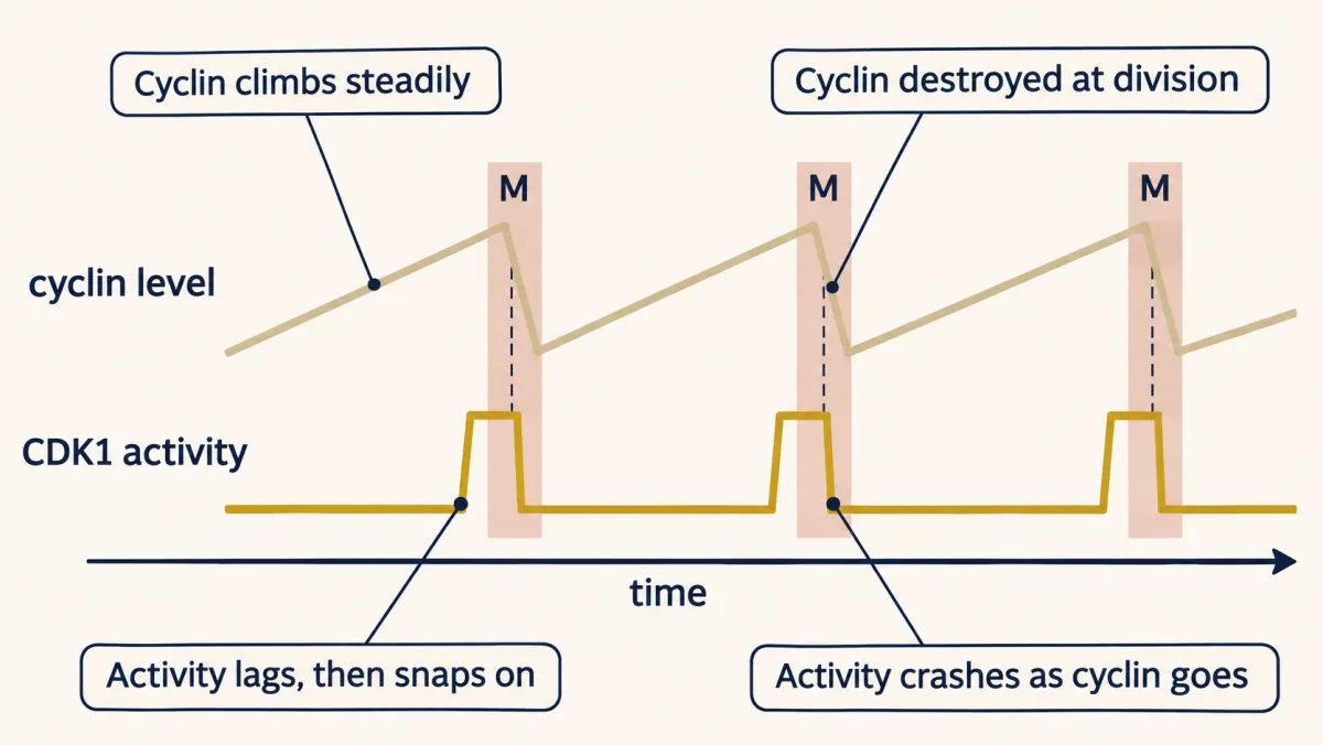
Cyclin climbs steadily and is destroyed at each division, but the kinase it switches on stays low, then snaps on just before mitosis and crashes as cyclin goes. A schematic, not data: the exact waveform depends on organism and stage. AI-generated illustration -
The engine: a kinase and a partner that comes and goes
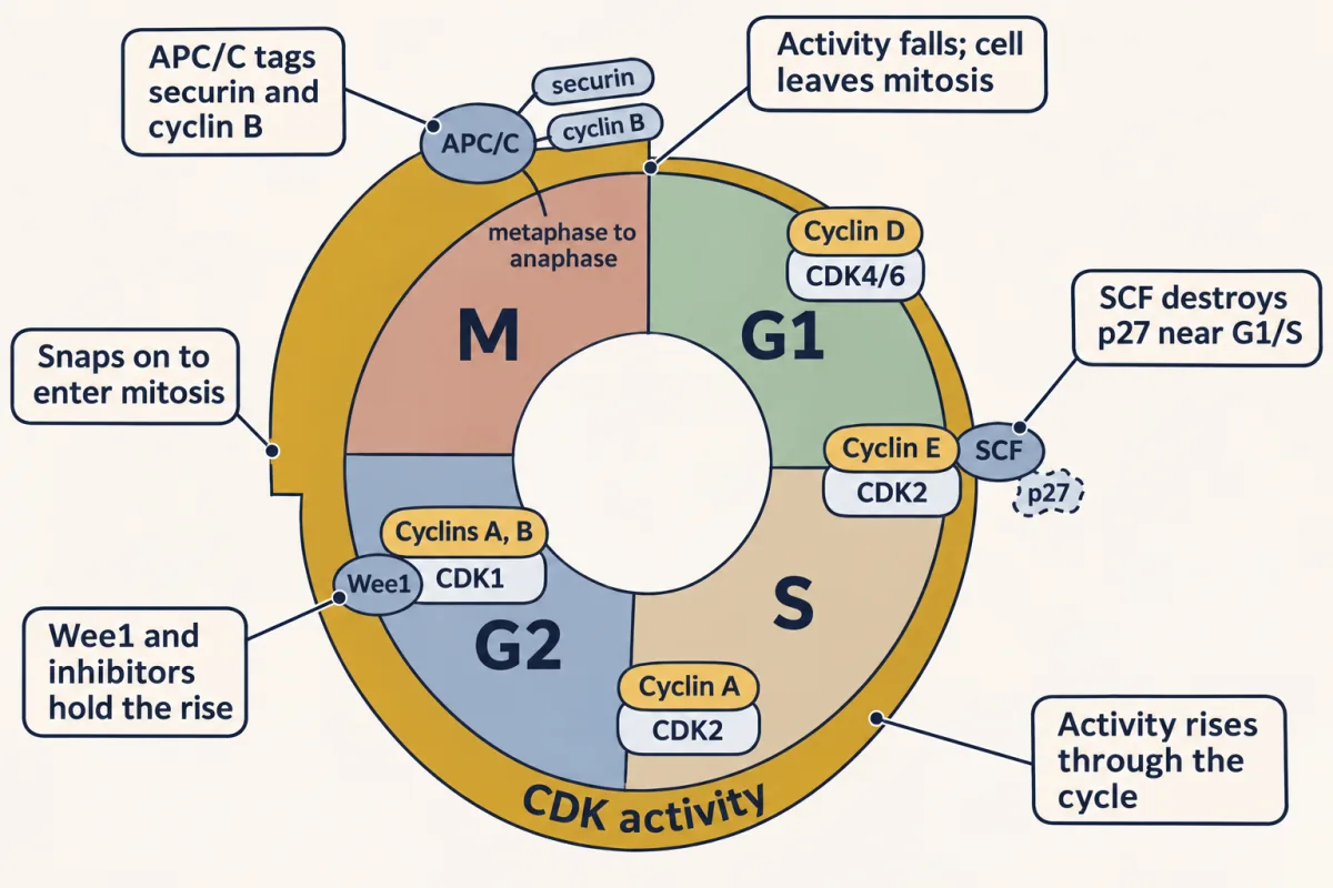
CDK activity rises and falls once per cycle: successive cyclin-CDK pairs carry the rise, brakes such as Wee1 and the inhibitor p27 (destroyed by the SCF around G1/S) hold it back, it can snap on to enter mitosis, and the APC/C ends it by tagging securin and cyclin B. A schematic of mammalian cells; the pairings are the usual division of labour, not a strict rule. AI-generated illustration -
Holding the rise back, and letting it go
-
The fall: destruction makes it one way
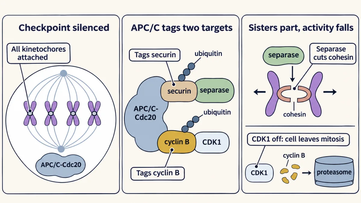
Once the spindle checkpoint is silenced, the APC/C with Cdc20 tags two targets: securin, so separase cuts cohesin and the sisters separate, and cyclin B, so CDK1 activity falls and the cell leaves mitosis. A destroyed protein has to be made again, which helps make the step run one way. AI-generated illustration -
Copy once: low activity licenses, high activity fires
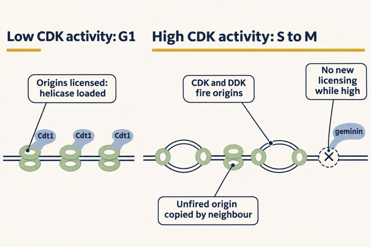
Licensing needs low CDK activity and firing needs high activity, so an origin can fire at most once per cycle; new licences wait until activity falls again, normally at the exit from mitosis. AI-generated illustration -
How the engine was found: frogs, yeast and sea urchins
-
Measuring the states
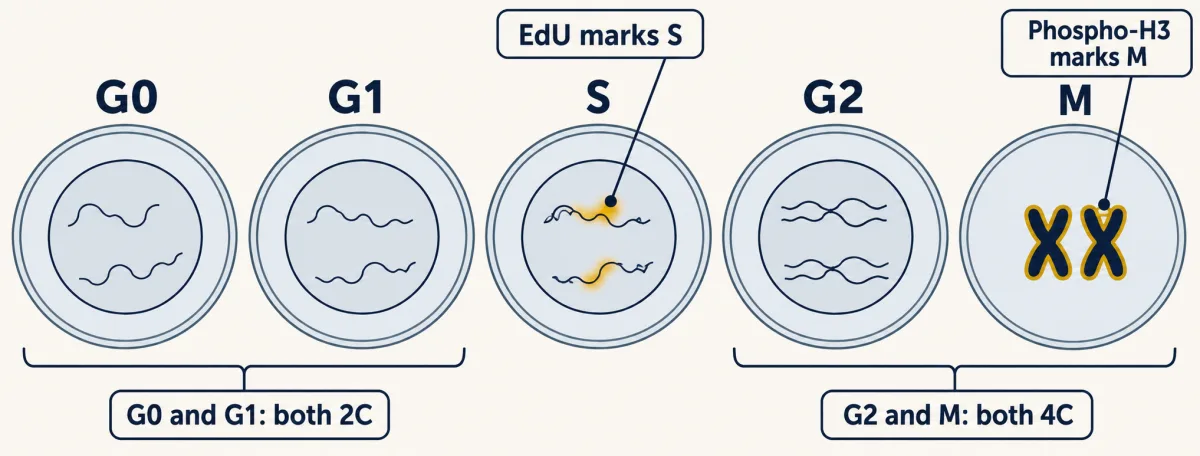
DNA content alone cannot separate G0 from G1 or mitosis from G2. A short EdU pulse marks cells in S, and phosphorylated histone H3 marks condensed mitotic chromosomes. AI-generated illustration -
Going deeper
Where this lesson fits
Before thisReview first
- Experiments That Distinguish MechanismsThis course · Lesson 3
The cell-fusion, extract and temperature-sensitive mutant experiments here use the reasoning for telling mechanisms apart taught there.
- Protein Quality Control and Organelle TurnoverThis course · Lesson 9
Regulated destruction of proteins by ubiquitin tagging and the proteasome is used here to reset the cycle, so you need that idea.
- The Nuclear Envelope and Nuclear TransportThis course · Lesson 8
Cyclin B-CDK1 and licensing factors move in and out of the nucleus, which you need to understand regulated nuclear access.
After thisStudy next
- Checkpoints and Irreversible TransitionsThis course · Lesson 18
It takes the Wee1, Cdc25, p21 and APC/C levers from here and asks how checkpoints make transitions irreversible.
- Mitosis and Chromosome SegregationThis course · Lesson 19
It uses the CDK1 rise, and the APC/C destruction of securin and cyclin B, to explain chromosome segregation.
- Cytokinesis and Organelle InheritanceThis course · Lesson 20
It builds on the fall of CDK activity at mitotic exit, which triggers furrow formation and organelle partitioning.
Elsewhere on the siteFilms, simulations and method films in other courses
- How One Set Becomes TwoFilm · Biology
It shows the copy-then-separate sequence of chromosomes that the cyclin-CDK engine times.
- Mitosis stepper and phase counterSimulation · Biology
Its phase counter shows how the proportion of cells in each phase relates to the cycle.
- Pulse-chase labellingMethod film
Following a cohort labelled in a short window is the logic behind the EdU pulse used to mark S-phase cells.
Suggested by an AI model from the lessons themselves, then checked. How we use AI