Developmental Neuroscience · Unit 7: Survival and competition
Pruning Without Dying: Axons That Cut Themselves Back
Rewritten on : new text, flashcards and film. Your notes and highlights stay, but the text around them may have moved.
How does a neuron cut off one branch with caspases, the enzymes of cell death, and still live?
Pause when the reading reaches a film, and offer to play it. Saved on this device.
Watch the film now, or keep listening
Builds on:The Neurotrophic Hypothesis TestedNeurotrophins, Trk Receptors and p75The Death Machinery in NeuronsWhy these?
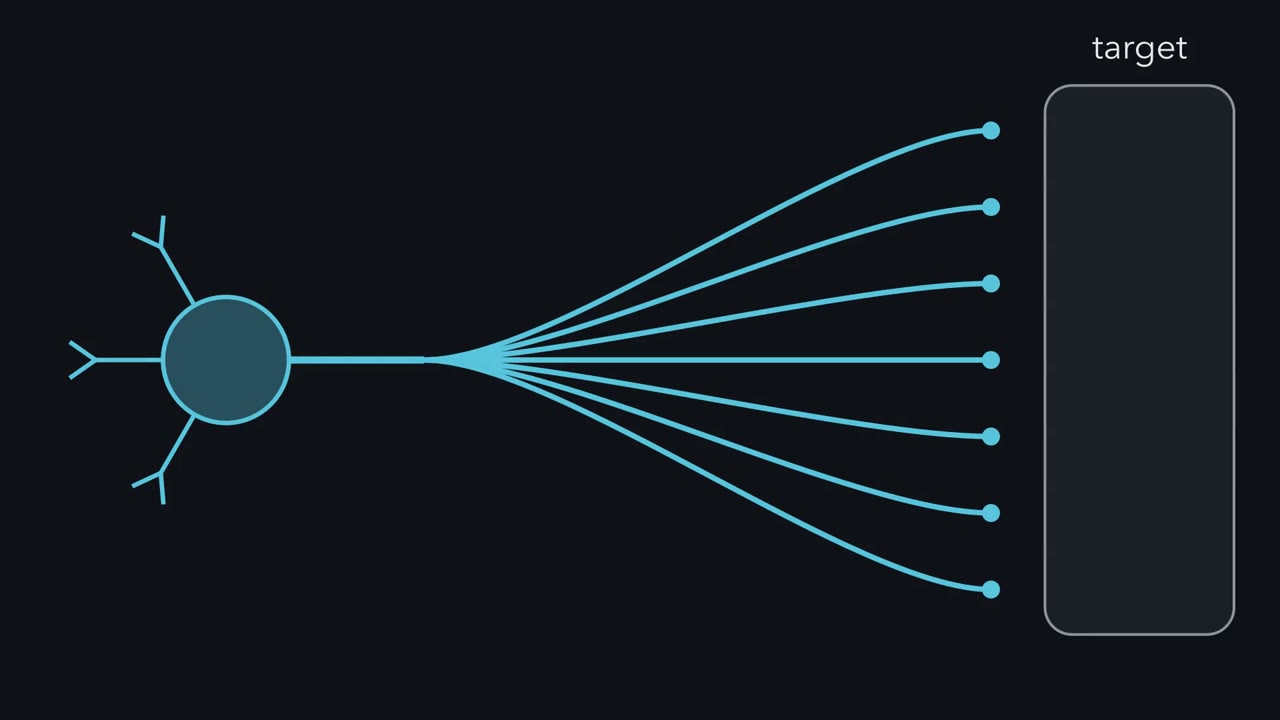 ▶ Watch Where Support Is Lost · 2:16
Read the transcript of “Where Support Is Lost”
Film transcript: Where Support Is Lost
A young neuron grows more branches than it keeps. Who decides which branch goes? Pruning removes an axon branch, or a dendrite, from a neuron that survives. Development first builds a rough draft, then carves it. Think of a sculptor who piles up too much clay. Sympathetic neurons need NGF, a growth factor supplied by their target. Now grow them in a dish divided into compartments. The cell bodies sit in one compartment. Their branches cross into side compartments. The fluids stay apart, so each compartment can have NGF, or not. Remove NGF from one side compartment only. Which branches degenerate?
Only the branches in that compartment. Branches of the same neuron next door, still in NGF, stay. Now remove NGF from every compartment. What happens to the neuron? The neuron dies, and its axons go with it. Here is the one idea for this lesson. Where support is lost decides what is lost. Lose support at the axons while the cell body keeps it, and the axons degenerate while the neuron lives. Lose it everywhere, and the neuron dies. In the animal, target NGF normally reaches the cell body through signals sent back along the axons. So pruning and death follow the same rule. But they share enzymes, and are wired differently. Both use caspases, the enzymes of cell death.
So how can one branch use them, and the neuron live? This rule covers branches lost after support is lost. Some branches go on a fixed program instead. Those come later. So to predict what is lost, first ask where the support was lost.
From the film
- Where support is lost decides what is lost.
- In NGF-dependent sympathetic neurons in culture, axons that lose NGF while the cell body keeps it degenerate and the neuron lives.
- A neuron that loses NGF everywhere dies, and its axons go with it.
- Pruning and death share caspases but are wired differently.
Grow sympathetic neurons in a dish divided into compartments, so that one neuron’s branches reach into separate compartments. Take nerve growth factor (NGF) away from one compartment, and the branches there degenerate. Branches of the same neuron next door, still in NGF, stay.
That experiment, from Campenot’s compartmented cultures (1977, 1982), is pruning in miniature. Development builds more connections than the adult circuit keeps, and pruning is how neurons cut the excess back. This lesson asks three questions. What chooses the branch that goes? How is it removed? And how does a neuron cut off one branch with caspases, the enzymes of cell death, and still live?
Lose support here, lose the branch here
NGF, nerve growth factor, is a trophic factor: a growth factor, supplied by the target, that a neuron needs to survive. Developing sympathetic neurons depend on it, and the lesson on the neurotrophic hypothesis shows how strictly. A compartmented culture lets you give or withhold NGF at one part of a neuron while the rest stays in it. The cell bodies sit in one compartment, and their neurites, the growing branches, cross into side compartments whose fluids stay apart.
Predict. Remove NGF from one side compartment only. Which branches degenerate? Then remove NGF from every compartment. What happens to the neuron?
Check your prediction
Only the branches in the deprived compartment degenerate; branches of the same neuron still in NGF survive. That does not mean every part of a neurite must be bathed in NGF: Campenot found that the cell bodies and the near ends of the neurites could do without it, as long as the far ends reached NGF. Remove NGF from every compartment and the neurons themselves die, as the experiments below show.
The one idea to carry through this lesson. Where support is lost decides what is lost. During a neuron’s window of dependence on NGF, in culture, take the trophic signal away from its axons, or some of them, while the cell body still has enough support, and the deprived axons degenerate while the neuron lives. Take it away from the whole neuron, and the neuron dies. Pruning and death follow the same rule, but run on caspase routes that are wired differently, as the sections below show.
What the rule covers.
- Which neurons. NGF-dependent sympathetic neurons, and NGF-dependent sensory neurons, during their window of dependence. The experiments in this lesson were done in sympathetic neurons.
- Which pruning. Pruning by local degeneration after a branch loses trophic support, including the losing branch in a competition. It does not cover stereotyped pruning, where a developmental program picks the branch.
Building, then sculpting
Development can be divided into two kinds of events. Progressive events build a rough draft of the nervous system: neurons are born, migrate, form layers, and send out axons that find their targets. Regressive events then sculpt the circuits: whole neurons die, synapses are removed, and axons and dendrites are pruned. Think of a sculptor who first piles up more clay than the figure needs, then carves.
Pruning has a narrow meaning here: removal of an axon branch or a dendrite from a neuron that survives. That separates it from the neuron death of the lesson on death machinery in neurons, where the whole cell goes. The word covers two physical processes. In retraction, a branch pulls back toward the cell body. Much of its material returns to the cell, though some retracting branches also shed small pieces that glia engulf. In local degeneration, the branch breaks into pieces in place, and other cells engulf the pieces. A good description of any pruning event says which one happened.
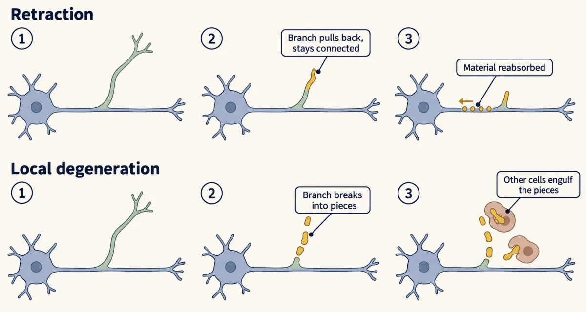
Pruning has two broad triggers in the animal. In competitive pruning the outcome depends on competition between axons, for example for trophic support, or through differences in activity. In stereotyped pruning the branch to be removed is set by a genetic program, so the same branch goes in every animal. The stereotyped cases come at the end, as the contrast; the last of them is pruned by retraction.
One neuron, two ways to lose an axon
Read the transcript of “Same Knives, Two Routes”
Film transcript: Same Knives, Two Routes
A neuron's cell body and its axon share one cytoplasm. Both contain caspases. So how can caspases run in one and not the other? Caspases are proteases that dismantle a cell during apoptosis, a built-in program of cell death. To find out, you must deprive one place and not the other. In 2013, Corey Cusack, Mohanish Deshmukh and colleagues grew mouse sympathetic neurons in a microfluidic chamber. The cell bodies sat in one compartment. Their axons grew through narrow grooves into a second. The fluids stayed apart. Local deprivation removes NGF from the axon compartment only. Global deprivation removes it from both. In normal neurons, what happens in each experiment?
Local deprivation: the axons degenerate, and the cell bodies live. That is pruning, in a dish. Global deprivation: the neurons die, and their axons go with them. That is developmental cell death, in a dish. Now fill in a table, one genotype per row. Does axon breakdown need caspase-6 in both experiments? Delete it, and predict each one.
Without caspase-6, locally deprived axons were protected. After global deprivation, the axons still degenerated as the neurons died. So the two experiments have different needs. A second mutant shows whether they are two programs.
Now the other end of the pathway. During apoptosis, the protein Apaf-1 builds the apoptosome. The apoptosome switches on Caspase nine then switches on caspase-3. Local pruning needed caspase-9, caspase-3 and caspase-6. Does it need Apaf-1 too?
No. Without Apaf-1, locally deprived axons still degenerated, and the cell bodies lived. But after global deprivation, these neurons resisted death. Death of the whole neuron needed Apaf-1. So in this study, caspase-6 mattered only for the local route. Apaf-1 mattered only for the global route. Each route needs something the other does not. They share enzymes, but they are wired differently. Caspases become active in the deprived axon. What keeps their activity low in the cell body? The same study found two things that keep caspases off in the cell body. One is XIAP, which blocks active caspase-9 and caspase-3. The other is the proteasome. Neurons lacking XIAP built up active caspase-3 in their cell bodies.
Same enzymes, two wirings, and an inhibitor guarding the part that must live.
From the film
- After local NGF deprivation in mouse sympathetic neurons, axons degenerate and the cell bodies live; after global deprivation the neurons die.
- In this study, caspase-6 was needed for local axon pruning, and Apaf-1 was needed for death of the whole neuron.
- The two routes share enzymes such as caspase-9 and caspase-3 but are wired differently.
- XIAP and the proteasome keep caspases off in the cell body, guarding the part that must live.
Caspases are proteases that dismantle a cell during apoptosis, a built-in program of cell death; the Cell Signaling course covers how they are activated in the lesson on death receptors, Bcl-2 proteins and caspases. Initiator caspases, such as caspase-9, start a cascade that switches on executioner caspases, such as caspase-3 and caspase-6, which do the cutting. A sympathetic neuron’s cell body and its axon share one cytoplasm, and both contain caspases. To ask how the neuron uses them in one place and not the other, you need to deprive one place and not the other.
In 2013 Corey Cusack, Mohanish Deshmukh and colleagues grew mouse sympathetic neurons in a microfluidic chamber, a modern compartmented culture: cell bodies on one side, axons grown through narrow grooves into the other, fluids kept apart. Both sides started with nerve growth factor (NGF). That setup allows two experiments:
- Local deprivation: remove NGF from the axon compartment only.
- Global deprivation: remove NGF from both compartments.
In normal neurons, after local deprivation, the deprived axons degenerated and the cell bodies, still bathed in NGF, lived. That is pruning in a dish. After global deprivation, the neurons died, and their axons went with them. That is developmental cell death in a dish.
Both outcomes involve caspases. Are they one program run at two strengths, or two programs? Mutant neurons answer that.
Predict. Delete caspase-6. Do the axons still break down after local deprivation? After global deprivation?
Check your prediction
Caspase-6 mattered in only one. Without it, locally deprived axons were spared. After global deprivation, deleting caspase-6 no longer saved the axons: they degenerated as the neurons died, since axon breakdown during whole-cell death rides on apoptosis. On its own, that does not rule out one program at two strengths, since a stronger signal might simply not need caspase-6. The second mutant gives much stronger evidence.
The second mutant tests the other end of the pathway. During apoptosis the usual activator of caspase-9 is the apoptosome, a complex built by the protein Apaf-1; caspase-9 then switches on caspase-3. Local pruning needed caspase-9, caspase-3 and caspase-6.
Predict. Does local pruning also need Apaf-1? Predict both experiments in neurons lacking Apaf-1.
Check your prediction
| Neurons | NGF removed from axons only | NGF removed everywhere |
|---|---|---|
| Normal | axons degenerate; cell bodies live | neurons die, axons too |
| Lacking caspase-6 | axons protected | neurons die, axons with them |
| Lacking Apaf-1 | axons still degenerate; cell bodies live | neurons resist death |
In this study, local pruning did not need Apaf-1, and death of the whole neuron did. Each route needs something the other does not, which points to two differently wired routes that share enzymes, rather than one program at two strengths.
What about the axons of Apaf-1-null neurons after global deprivation? Cusack and colleagues summarize Apaf-1 as required only for axon degeneration during apoptosis, which implies those axons are spared too: an inference from their model rather than a result described here.
Guarding the part that must live
During local deprivation, caspases become active in the axon. What keeps caspase activity low in the cell body? The same study found that X-linked inhibitor of apoptosis protein (XIAP), a protein that binds and blocks active caspase-9 and caspase-3, and the proteasome kept caspases switched off in the cell bodies. The proteasome is the cell’s machine for destroying proteins tagged with ubiquitin, covered in the lesson on ubiquitin and proteolysis. After local deprivation, neurons lacking XIAP built up active caspase-3 in their cell bodies. An inhibitor guards the part of the cell that must live.
Fly sensory neurons that prune their dendrites at metamorphosis suggest a similar idea. In 2006 Darren Williams, James Truman and colleagues used a genetically encoded reporter to find caspase activity. It appeared only in the dendrites being removed. Suppressing caspases kept the dendrites; activating caspases throughout the neuron killed it.
In the same year Chay Kuo, working with Lily Jan and Yuh-Nung Jan, found evidence on how the caspase is let loose. The ubiquitin-conjugating enzyme UbcD1 helps send DIAP1, an inhibitor that holds the fly caspase Dronc in check, to the proteasome. Their model: lose the minder and the caspase can run. Where that removal happens, and how caspase activity stays in the dendrites and out of the cell body, is not fully worked out. In the mouse, by contrast, XIAP was shown to protect the cell body.
Winners and losers: who loses a contest
Read the transcript of “Who Loses, and When It Commits”
Film transcript: Who Loses, and When It Commits
In a dish, the experimenter decides where support is lost. In the animal, who decides? Many sympathetic neurons send branches toward targets that make NGF. The branches compete. When NGF is limited, a branch that wins enough is stabilized. One left with too little is weakened and removed. Two receptors matter. TrkA is the NGF receptor. p75 binds all neurotrophins, including BDNF. One axon has strong TrkA signalling, the other weak. BDNF is present. Which axon goes?
The weak one. On axons with weak TrkA signalling, BDNF acting through p75 caused degeneration. Axons with strong TrkA signalling were protected. And the winning neurons made the BDNF. So a winner does more than take the NGF. It also sends a signal that removes the loser.
The neurotrophic hypothesis lesson showed the same winner-removes-loser idea for whole neurons. Here it works on axons. So how does an axon come apart after NGF is withdrawn? Not all at once. First, the axon waits through a long latent phase. It looks nearly normal. Late in latency, swellings called spheroids appear. Calcium gathers in them. Then the spheroids rupture.
Then the axon fragments. That is the catastrophic phase. Two receptors split the work. p75 is needed for spheroids to form. DR6 is needed for the catastrophic degeneration that medium from deprived axons speeds up. The Deppmann laboratory's model is that spheroids gate the step from waiting to catastrophe. It is not yet a settled mechanism. Where support is lost decides what is lost. In a contest, the winner helps decide too.
From the film
- Sympathetic axons compete for limited target-derived NGF, and a branch left with too little is weakened and removed.
- On axons with weak TrkA signalling, BDNF acting through p75 caused degeneration, and the winning neurons made the BDNF.
- A deprived axon passes through a latent phase, then spheroids, then catastrophic fragmentation.
- The laboratory's model is that spheroids gate the commitment to catastrophe, but it is not a settled mechanism.
In the dish, the experimenter decides where support is lost. In the animal, target-derived nerve growth factor (NGF) normally reaches a sympathetic neuron at its axon terminals and signals back to the cell body. So the whole neuron dies when too little of that signal reaches the cell body overall, not after some fixed number of branches lose it. Which branches lose support depends on competition for limited target-derived NGF. As the lesson on competition for targets shows, a branch that wins enough support is stabilized, and one left with too little is weakened and removed.
What removes the loser? In 2008 Singh, David Kaplan, Freda Miller and colleagues studied sympathetic neurons in compartmented cultures and in mice. Two receptors matter. TrkA is the NGF receptor tyrosine kinase. p75 is a receptor of the TNF-receptor family that binds all neurotrophins, including brain-derived neurotrophic factor (BDNF).
Predict. Two axons compete, one with strong and one with weak TrkA signalling. BDNF is present. Which axon degenerates, and through which receptor?
Check your prediction
The weak one, through p75. On axons with weak TrkA signalling, BDNF acting through p75 caused degeneration, while axons with strong TrkA signalling were protected. And the winning neurons made the BDNF. In one model, winning axons release BDNF in an activity-dependent way. A winner therefore does more than take the NGF. It also sends a signal that removes the loser, and its own TrkA signalling makes it resistant to that signal.
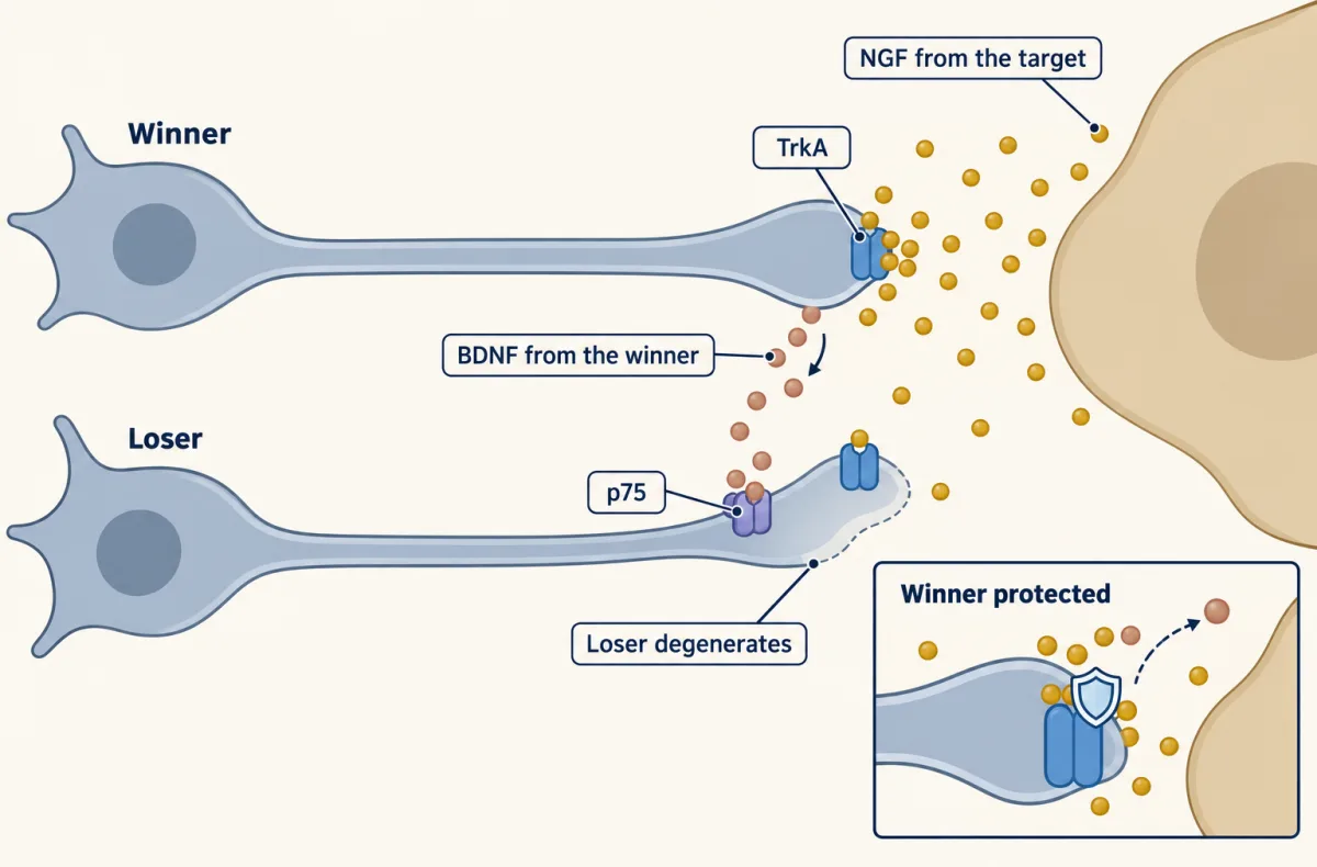
This is the axon-scale version of a whole-neuron model in the lesson on the neurotrophic hypothesis, whose rule is make too many, then let the target keep what it can feed. There, neurons with strong NGF signalling release BDNF and neurotrophin-4 (NT-4), which act through p75 on weak neighbours and push them toward death (Deppmann and colleagues, 2008). Singh and colleagues’ 2008 work is the evidence for axons: the same winner-removes-loser idea at the smaller scale. The pairing of TrkA with p75 is the subject of the lesson on Trk and p75 receptors.
When a deprived axon commits
A deprived axon does not come apart at once. Following axons deprived of nerve growth factor (NGF) over time, Yong, Gamage and colleagues in the Deppmann laboratory (J Neurosci 2019) divided the process into three phases.
- Latent phase. A long period in which the axon looks nearly normal.
- Transition. Late in latency, swellings called axonal spheroids appear.
- Catastrophic phase. The axon fragments.
Live imaging showed calcium rising in axons before catastrophic degeneration and gathering in spheroids; calcium signalling is covered in the lesson on calcium signaling. Spheroids, but not the rest of the axon, exposed phosphatidylserine, a membrane lipid that phagocytes recognize. Then they ruptured, letting in dextran tracers, sugar chains that cannot cross an intact membrane, of 3 and 10 kilodaltons but not of 70; the calcium signal was lost as the dextran entered. Medium from axons already deprived of NGF, called conditioned medium, sped fresh deprived axons into catastrophe, so deprived axons release something that does this.
Two members of the TNF-receptor family, a receptor family that includes the death receptors, split the work. p75 was needed for spheroids to form, and DR6 was not. DR6, not p75, was needed for the catastrophic degeneration that conditioned medium speeds up. p75 acted through Rho, a small GTPase that reorganizes actin, covered in the lesson on integrins and Rho GTPases; blocking Rho or actin dynamics prevented spheroids.
Are spheroids just a sign that the axon is failing, or a step that sets the timing? The laboratory’s model is that spheroids gate the transition from latency to catastrophic degeneration: they set when the axon commits. The evidence here is one rescue: forcing Rho on in p75-null axons restored both spheroids and degeneration. That fits a gate but does not prove one; the gate is the authors’ interpretation, not a settled mechanism.
Development and injury differ. For sympathetic axons deprived of NGF, caspases are required for developmental degeneration but not for degeneration after injury. After a nerve is cut, the detached part of the axon undergoes Wallerian degeneration, which uses a partly different program; the lesson on injury and regeneration covers it.
When a program, not support, picks the branch
So far, lost support picked the branch. In stereotyped pruning a developmental program does: in the examples below a hormone or a repulsive cue triggers the pruning, and no loss of trophic support is needed to explain it.
Start with the fly. When a fruit fly turns from larva into adult, each gamma neuron in its mushroom body, a learning centre of the fly brain, loses both of its larval axon branches within about eighteen hours, and the neuron survives. Later in metamorphosis the same neuron grows a new adult branch.
Liqun Luo’s group used MARCM, a genetic method that labels one or a few neurons in an otherwise unlabelled brain and can make only those neurons mutant. In 2000 Tzumin Lee, Luo and colleagues showed that a gamma neuron needs the steroid receptor EcR-B1, working with its partner USP (Ultraspiracle), inside itself to remodel. The ecdysone receptor is a nuclear receptor, a ligand-activated transcription factor of the kind covered in the lesson on nuclear receptors. The hormone 20-hydroxyecdysone rises at metamorphosis, and within the mushroom body EcR-B1 is made in the gamma neurons. The hormone gives the timing; the receptor helps decide which neurons respond.
Predict. Gamma neuron branches break into pieces in place, like a locally deprived mouse axon, which needs caspases to do it. Would blocking the fly’s main apoptosis activators, or expressing the caspase inhibitor p35, stop this pruning?
Check your prediction
No. In fly brains fixed at two-hour intervals over the first eighteen hours of metamorphosis (Watts, Hoopfer and Luo, 2003), the branches blebbed, then appeared as separate fragments, then were gone, while the main axon shaft stayed continuous. Separated pieces are what local degeneration predicts, since a retracting branch would stay connected to the cell. In the same study, blocking the fly’s main apoptosis activators or expressing p35 did not stop this axon pruning, so the fly mushroom body does not appear to rely on the usual apoptosis machinery. What it did need was the ubiquitin-proteasome system: blocking ubiquitin tagging, or the proteasome, kept the larval branches in place. The requirement was inside the pruning neuron.
Mammals use guidance cues as well. In the mouse hippocampus, the axons of dentate granule cells, the mossy fibres, form a main bundle and a short infrapyramidal tract. After birth the infrapyramidal tract is cut back. In 2003 Anil Bagri, Marc Tessier-Lavigne and colleagues found that in mice lacking neuropilin-2 or Plexin-A3, parts of the receptor for the repulsive cue Sema3F, the tract stayed long, and later work showed that Sema3F itself is required. Cultured hippocampal neurons retracted axon branches when exposed to semaphorins, and later electron microscopy found the tract’s branches removed without breaking up or being engulfed by glia: pruning by retraction. Sema3F, best known for repelling growing axons, here acts later to trim the infrapyramidal tract; the lesson on semaphorins covers these cues and their receptors. In this tract, then, a repulsive cue is required for the pruning, and nothing in the evidence points to lost trophic support as the trigger.
Design problem
You have a two-compartment chamber and sympathetic neurons from wild-type mice and from Apaf-1-null mice. In one set of chambers you remove nerve growth factor (NGF) from the axon compartment only. In another you remove NGF from the whole dish. Predict, for each genotype, which axons degenerate and which neurons die.
Saved in this browser only.
One way to do it
Local deprivation: in both genotypes the deprived axons degenerate and the cell bodies survive, because the cell bodies still have NGF and pruning uses caspase-9, -3 and -6 without Apaf-1. Global deprivation: wild-type neurons die, with their axons, through the apoptosome. Apaf-1-null neurons resist that death, and Cusack and colleagues’ model predicts their axons are spared too. The same caspases are reached by two routes: the whole-cell route needs Apaf-1 to build the apoptosome and activate caspase-9; the local route does not.
Going deeper
Bax in both routes
Bax is a Bcl-2 family protein that, in apoptosis, triggers the release of cytochrome c from mitochondria. In Cusack and colleagues’ chambers, axons lacking Bax were protected after local deprivation, and earlier work had shown that apoptosis needs Bax. So both routes need Bax as well as caspase-9 and caspase-3. How Bax acts in the local route, which does not need Apaf-1, is not settled.
Latent phases differ with the kind of deprivation
Yong, Gamage and colleagues found that local and global nerve growth factor (NGF) deprivation give different latent phases, so how the axon was deprived changes how long it waits before catastrophe.
Spheroids after injury
A later study from the laboratory (Yong and colleagues, Sci Rep 2020) found that, after injury, adding NAD+ or expressing WldS suppresses Rho-dependent spheroid formation.
Wallerian degeneration, WldS, SARM1 and DR6 after injury
Lunn and colleagues described the WldS mouse, whose cut axons survive far longer, in 1989; Coleman’s group later identified the gene. WldS and loss of SARM1 protect strongly against injury-induced degeneration, WldS did not block the developmental axon pruning Hoopfer and colleagues tested, and whether SARM1 contributes to degeneration after trophic deprivation is still debated. Gamage and colleagues (Curr Biol 2017) found that DR6 promotes degeneration after injury (a 2026 study with two DR6 knockout lines found no such effect), which raises a question: are there convergent mechanisms between pathological and developmental regression?
Your notes
Select any text in the lesson to highlight it or add a note to it. Your highlights and notes are saved in this browser and collected in your notebook.
This browser is not letting the site save anything (a private window or strict privacy settings), so highlights and notes will be lost when you leave the page.
How we measure it
MARCM mosaic labelling
Mitotic recombination in a fly makes a few cells lose a repressor, so only those cells express a marker such as membrane GFP, and they can be made homozygous mutant at the same time. One labelled neuron can then be followed, and a gene's requirement inside that neuron tested, in an otherwise normal brain.
Compartmented and microfluidic cultures
Neurons are plated so their cell bodies sit in one compartment and their axons grow through narrow channels into another, with the fluids kept apart. Changing the medium on one side only, for example removing NGF from the axons, separates what happens to the axon from what happens to the cell.
Live imaging of calcium and membrane integrity
Axons loaded with a calcium dye are filmed after deprivation, along with Annexin V, which binds exposed phosphatidylserine, and labelled dextrans of different sizes. Rapid, diffuse filling of an axon with dextran marks a break in its membrane (dotted uptake can be endocytosis), and the timing of each signal orders the events of degeneration.
Knockout mice
The knockout mice used here lack both working copies of one gene in every cell. Comparing neurons from knockout and wild-type mice, here for p75, DR6, caspase-6 and Apaf-1, asks whether that gene is needed for a given step.
Practice questions on this lesson (3) → MCAT-style, original, each with an explanation
Where this lesson fits
Before thisReview first
- The Neurotrophic Hypothesis TestedThis course · Lesson 29
You need the idea that neurons compete for limited target-derived NGF to follow why a branch is weakened and removed.
- Neurotrophins, Trk Receptors and p75This course · Lesson 30
The loser-removal step uses p75 acting against weak TrkA signalling, so you need how these receptors work.
- The Death Machinery in NeuronsThis course · Lesson 31
Pruning is set against whole-neuron death, so you need the caspase and apoptosome routes from there.
After thisStudy next
- Synapse Elimination: One Muscle Fiber, One AxonThis course · Lesson 37
Removal of excess axon inputs by competition, here at a muscle fiber, continues the pruning-by-competition idea.
- Microglia and Complement Prune SynapsesThis course · Lesson 41
It shows how other cells engulf and remove synapses and pieces of neurons during pruning.
Elsewhere on the siteFilms, simulations and method films in other courses
- Sealed or TornFilm · Cell Biology
It shows how a cell's own caspases dismantle it, the apoptosis routes that axon pruning reuses locally.
- Cell death assaysMethod film
The lesson's caspase-dependent axon degeneration versus neuron death is read with annexin V, TUNEL and similar assays.
Suggested by an AI model from the lessons themselves, then checked. How we use AI
Finish line
Test yourself
8 questions
Practice and MCAT-style, with explained answers.
Next · Lesson 35
Building the Neuromuscular Junction: Agrin, Lrp4 and MuSK
A mouse without the muscle kinase MuSK dies at birth: its motor axons reach the muscle, but no junction forms.
Continue →