Developmental Neuroscience · Unit 7: Survival and competition
The Neurotrophic Hypothesis Tested
Rewritten on : new text, figures, flashcards and film. Your notes and highlights stay, but the text around them may have moved.
Neutralize one protein in a newborn mouse and its sympathetic ganglia waste away. Neurons compete for a limiting amount of target-derived factor, and the amount limits how many live. Does NGF decide how many neurons there are?
Pause when the reading reaches a film, and offer to play it. Saved on this device.
Watch the film now, or keep listening
 ▶ Watch Too Small, or Lost? · 2:56
Read the transcript of “Too Small, or Lost?”
Film transcript: Too Small, or Lost?
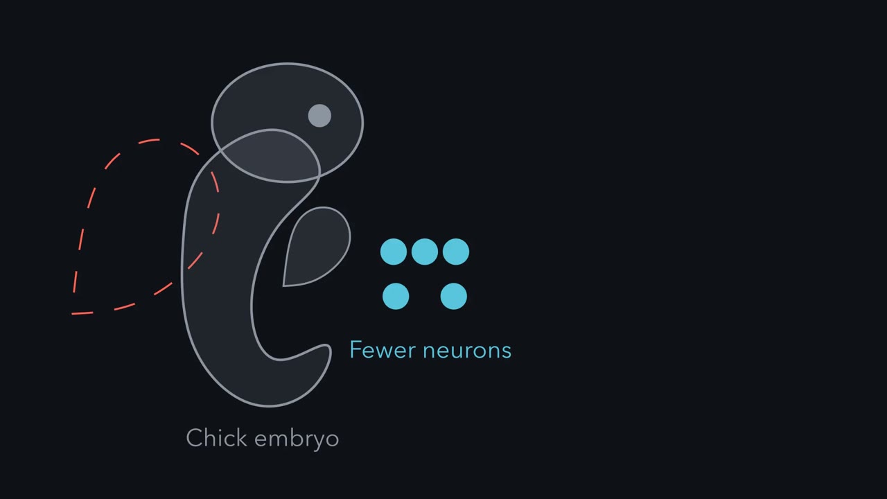
Remove one wing bud from a chick embryo. The neurons serving that limb come out fewer. Were they never made? Or were they made, and then lost? First, two words. A target is the tissue an axon grows to and connects with, such as a limb muscle or a gland. A ganglion is a cluster of neuron cell bodies outside the brain and spinal cord. Spinal ganglia sit beside the cord and hold sensory neurons. Sympathetic ganglia hold neurons of the sympathetic nervous system. In 1934, Viktor Hamburger removed one wing bud from chick embryos about three days old. On the operated side, the neurons serving that limb came out reduced.
Hamburger first read this as the limb controlling how many cells grew in the spinal cord and its ganglia. But one count at the end hides a choice. A small ganglion has two possible histories. Predict. What are the two histories? And what would you measure to tell them apart?
Fewer neurons might have been made. Or the normal number might have been made, and then lost. Both give a small ganglion at the end. To separate them, you count at a series of stages. You also mark the cells that are dividing, and the cells that are dying. In 1949, Hamburger and Rita Levi-Montalcini did exactly that, in chick spinal ganglia. They followed the ganglia stage by stage, after removing or adding limb tissue. Many neurons formed and grew fibres before the deficit appeared. Degenerating cells became conspicuous later. The neurons had been made. Then they were lost.
So the limb seemed to keep neurons alive. It was not only calling them into existence. Three conclusions follow. First, neurons are made in excess, and then lost. Second, whether they are kept depends on the target. Third, the target must supply something neurons need. If it supplies too little for all of them, they must in effect compete. Neither the something nor the competition had been shown. They were inferences. The neurotrophic hypothesis would have to earn them, one test at a time. Here is the one idea to carry through. Make too many, then let the target keep what it can feed. The test case is nerve growth factor, NGF. Acting as a survival factor, it decides how many neurons made stay alive.
It can never raise that number above the number made. So a small ganglion is a question about history. Count across time before you decide.
From the film
- Many neurons are made and then lost, so a small ganglion is a question about history: count across time before you decide.
- Whether neurons are kept depends on the target, which must supply something they need.
- Make too many, then let the target keep what it can feed; as a survival factor, NGF cannot raise survivors above the number made.
Inject a newborn mouse with an antiserum against one protein, and its sympathetic ganglia waste away. Give it extra of the same protein, and they swell.
The protein is nerve growth factor, NGF, and the neurotrophic hypothesis explains its power: neurons are made in excess, the targets they reach supply a limiting amount of a survival factor, and the neurons compete for it. This lesson follows the tests of that idea one at a time and ends on a limit: what extra NGF can and cannot do to the number of neurons.
Too small, or lost?
The idea began with chick embryos and a puzzle about size. Two words first. A target is the tissue an axon grows to and connects with, such as a limb muscle or a gland. A ganglion is a cluster of neuron cell bodies outside the brain and spinal cord; the spinal ganglia beside the cord hold sensory neurons, and the sympathetic ganglia hold neurons of the sympathetic nervous system, the fight-or-flight division that speeds the heart and readies the body for action.
In 1934 Viktor Hamburger removed one wing bud from chick embryos about three days old and left the other side as the comparison. The neurons serving that limb came out reduced on the operated side. He first read this as the limb controlling how many cells grew in the spinal cord and its ganglia.
Predict. A spinal ganglion on the operated side ends up smaller. Name two different histories that could have produced it, and say what you would measure to tell them apart.
Check your prediction
Fewer neurons might have been made, or the normal number made and then lost. A single final count cannot separate the two, because both give a small ganglion. You need counts at a series of stages, with dividing and dying cells marked.
In 1949 Hamburger and Rita Levi-Montalcini did that, following chick spinal ganglia stage by stage after removing or adding limb tissue. Many neurons formed and grew fibres before the deficit appeared, and degenerating cells became conspicuous later. The neurons had been made, and then were lost.
Three conclusions follow from this early work.
- Neurons are made in excess and then lost.
- Whether they are kept depends on the target.
- So the target must supply something neurons need, and if it supplies too little for all of them, they must in effect compete for it.
Neither the “something” nor the competition had been shown. Those were inferences, and the hypothesis would have to earn them. How many neurons die, and when, is the subject of the lesson on naturally occurring cell death.
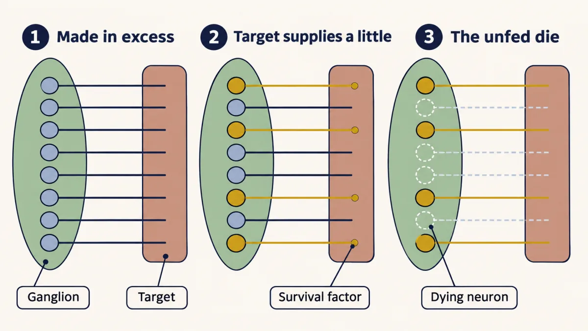
The one idea to carry through this lesson. Make too many, then let the target keep what it can feed. Acting as a survival factor, nerve growth factor (NGF) decides how many of the neurons that were made stay alive. It can never raise that number above the number made.
What the rule covers.
- Which neurons. NGF-dependent populations during their death windows: sympathetic neurons, and the sensory neurons that carry TrkA, the receptor NGF binds, mostly small neurons that sense pain and temperature. Limb motor neurons and some other populations fall outside it, and older animals are much less sensitive.
- Which effect. Only NGF’s survival effect. NGF also affects division, growth and branching.
- A ceiling, not a count. The rule says how many can survive at most, not how many do.
The hypothesis and what it predicts
Read the transcript of “A Limiting Supply”
Film transcript: A Limiting Supply
If neurons are made in excess and the target decides who stays, what exactly would the target be doing? The neurotrophic hypothesis puts it in one line. Neurons compete for a limiting amount of neurotrophic factor for survival. The targets supply it. Axons that reach these tissues take it up. Neurons that get too little die by apoptosis, a built-in program of cell death. Stated this way, the hypothesis makes predictions you can check one at a time. Remove the target, and more neurons die. Hamburger's wing-bud removal, read across time, fits. Add target, and fewer die. Add factor, and fewer die. Remove factor, and more die. And most of the death should happen around the time axons reach the target and begin to compete.
That is a checklist. The rest of this lesson tests the first four items, one at a time. In 1976, Margaret Hollyday and Viktor Hamburger tested the second item. They grafted an extra leg onto chick embryos, beside the normal one. Predict. With more target, what should happen to the lumbar motor neurons, before the normal loss and after it?
At day 6, before the normal loss, there were no more motor neurons than usual. Later, more motor neurons remained, in embryos whose grafts moved vigorously. They proposed a word for it. Hypo-thanasia: not extra birth, but reduced death. One caution. That result came from motor neurons, and they do not depend on NGF. So the graft supports a limiting target resource. It says nothing about which molecule. Hollyday and Hamburger left open whether the scarce resource was synaptic space or a trophic agent. Hold on to the shape of the idea. Make too many, then let the target keep what it can feed. The next tests need a molecule. That molecule is nerve growth factor, NGF.
Takeaway: the hypothesis predicts reduced death, not extra birth. When counts change, tests must ask which happened.
From the film
- Neurons compete for a limiting amount of neurotrophic factor from their targets, and neurons that get too little die by apoptosis.
- The hypothesis predicts reduced death, not extra birth, so when counts change the tests must ask which happened.
- An extra-leg graft in chick embryos supports a limiting target resource, but motor neurons do not depend on NGF, so it does not say which molecule.
Stated formally: neurons compete for a limiting amount of neurotrophic factor, which their targets supply and which axons reaching those targets take up, and neurons that get too little die by apoptosis, a built-in program of cell death. The hypothesis makes five predictions you can check one at a time; the rest of the lesson tests the first four.
- Remove the target, and more neurons die. Hamburger’s wing-bud removal, read across time in 1949, fits.
- Add target, and fewer die. Tested next.
- Add factor, and fewer die. Tested in Add it, take it away.
- Remove factor, and more die. Tested by antibody and then by genetics.
- Most of the death should happen around the time axons reach the target and begin to compete.
In 1976 Margaret Hollyday and Hamburger ran the add-target test. They grafted an extra leg onto chick embryos beside the normal one. Lumbar motor neuron numbers were not raised at day 6, before the normal loss, but more remained later in embryos whose grafts moved vigorously. They proposed a word for it, hypothanasia: not extra birth but reduced death.
The caution: these were motor neurons, which do not depend on nerve growth factor (NGF). The result supports a limiting target resource but says nothing about which molecule; Hollyday and Hamburger left open whether it was synaptic space or a trophic agent.
Add it, take it away
Read the transcript of “Add It, Take It Away”
Film transcript: Add It, Take It Away
How do you show that a protein keeps neurons alive? You add it, and you take it away. Each test shows something different. A mouse tumour grafted into a chick embryo drew in sensory and sympathetic fibres. Stanley Cohen purified the activity as a protein. The question here is what that protein does in a normal animal. Two words do the work. A factor is sufficient if it is enough, by itself, to produce an effect. It is necessary if the effect does not happen without it. Each experiment tests one of these, and no more. Sufficiency first. In 1960, Rita Levi-Montalcini and Barbara Booker gave the purified protein to young mammals. Their sympathetic ganglia were greatly enlarged.
Injected into chick embryos, the factor makes sensory and sympathetic ganglia overgrow. A bigger ganglion does not say whether neurons were saved or born. That needs counts, which come later. Now necessity. Cohen raised an antiserum, serum containing antibodies against one molecule. It blocked NGF's activity in culture. Levi-Montalcini and Booker injected it into newborn mice, with rats and other mammals for comparison. The sympathetic ganglia were destroyed. The procedure became known as immunosympathectomy.
Predict. Give the same antiserum to older animals. Does it destroy their sympathetic ganglia too? It has much less effect. Dependence on NGF is tied to a developmental window. And the window differs between populations. NGF dependent sensory neurons are hit hardest by earlier exposure.
But an antiserum carries a doubt. It might kill neurons by binding something on them other than NGF. So how would you remove NGF without using an antibody at all? Takeaway: adding NGF is enough to enlarge the ganglia. Taking it away shows it is necessary, if the antibody is clean. Settling that doubt took genetics.
From the film
- Adding NGF is enough to enlarge ganglia, but a larger ganglion does not say whether neurons were saved or born.
- An antiserum against NGF destroyed sympathetic ganglia in newborn mice, which shows necessity only if the antibody hit nothing else.
- Dependence on NGF is tied to a developmental window, and the window differs between populations.
You met the discovery in the lesson on finding a signal: a mouse tumour grafted into a chick embryo drew in sensory and sympathetic fibres, an activity it released could do the same in a culture dish, and Stanley Cohen purified that activity as a protein. The question here is what the protein does in a normal animal.
Two words do most of the work from here on. A factor is sufficient if it is enough, by itself, to produce an effect. It is necessary if the effect does not happen without it. Each experiment below tests one of these and no more.
Sufficiency. In 1960 Levi-Montalcini and Barbara Booker gave the purified salivary-gland protein to young mammals and reported greatly enlarged sympathetic ganglia. Purified factor injected into chick embryos likewise makes sensory and sympathetic ganglia overgrow. A larger ganglion does not yet say whether neurons were saved or born.
Necessity, by antibody. Cohen raised an antiserum, serum containing antibodies against one molecule, that blocked NGF’s activity in culture. In companion papers that year, Levi-Montalcini and Booker injected it into newborn mice, with rats and other mammals for comparison. The sympathetic ganglia were destroyed. The procedure became known as immunosympathectomy.
The same antiserum has much less effect on the sympathetic ganglia of older animals, so dependence on nerve growth factor (NGF) is tied to a developmental window, and the window differs between populations. NGF-dependent sensory neurons are hit hardest by earlier exposure: in 1980 Johnson and colleagues destroyed rat dorsal root ganglion neurons with maternal antibody to NGF that reached the fetuses in utero.
An antiserum carries a doubt. It might kill neurons by binding something on them other than NGF. How would you remove NGF without an antibody?
Delete it, and watch the time course
Read the transcript of “Made, Then Lost”
Film transcript: Made, Then Lost
A mouse lacking the NGF receptor is born with too few sympathetic neurons. Were they never made, or made and then lost? A knockout mouse has a gene disrupted, so it makes no working protein. No antibody is involved. In 1994, Crowley and colleagues reported mice lacking NGF. Smeyne, Klein, Barbacid and colleagues reported mice lacking a working TrkA, the receptor for NGF. Both mutants lose sympathetic neurons and many sensory neurons. The antibody doubt is settled. The sensory losses fall on neurons that express TrkA, mostly small neurons that sense pain and temperature. But a count at birth still cannot say whether the neurons were ever made.
In 1996, Anne Fagan, Story Landis, Barbacid and colleagues counted one ganglion at a series of ages. It was the superior cervical ganglion, the sympathetic ganglion at the top of the neck. Here are the axes. Neuron number runs up the side. Embryonic and postnatal age runs along the bottom. Predict. If TrkA is needed for survival but not for production, what shape does the mutant curve take?
Loss of TrkA did not affect neurogenesis, neuronal markers, or initial axon growth. In this schematic, the two counts match at embryonic day 13.5, soon after the ganglion forms. By embryonic day 15.5, the mutant ganglion already has fewer neurons. The gap keeps growing.
The ganglion formed, and then lost its neurons. The neurons were produced. What failed was survival. This time course is the signature evidence that the loss is a failure of survival, not production. Counts of dividing and dying cells showed why. More neurons were dying, and division was normal. How does NGF hold off that death? Neurons that get too little die by a death that needs the protein Bax. Mice lacking Bax keep sympathetic neurons that would otherwise die. In culture, they survive when NGF is withdrawn. So NGF works by holding back a Bax dependent death program. Takeaway: without TrkA, sympathetic neurons are made and then lost. NGF acts as a survival factor.
From the film
- Mice lacking NGF or TrkA lose sympathetic neurons and many sensory neurons, which settles the antibody doubt.
- In TrkA-null mice the counts matched early and the deficit appeared later, so the neurons were produced and then lost.
- NGF works by holding back a Bax-dependent death program.
A knockout mouse has a gene disrupted so that it makes no working protein, and it settles the antibody doubt. In 1994 Crowley and colleagues reported mice lacking nerve growth factor (NGF), and Richard Smeyne, Ruediger Klein, Mariano Barbacid and colleagues reported mice lacking a functional TrkA. TrkA is a receptor tyrosine kinase (the lesson on receptor tyrosine kinases), and its family has its own lesson, the lesson on Trk and p75 receptors. Both mutants lose sympathetic neurons and many sensory neurons.
A ganglion counted only at birth in a TrkA-null mouse looks depleted, which could mean too few precursors, failed differentiation or excess death. The question from the chick is back: were its neurons never made, or made and lost?
Predict. In 1996 Anne Fagan, Story Landis, Smeyne, Barbacid and colleagues counted the superior cervical ganglion, the sympathetic ganglion at the top of the neck, in TrkA-null mice at a series of embryonic and postnatal ages. Sketch neuron number against age for normal and TrkA-null mice. What shape do you expect if TrkA is needed for survival but not for production?
Check your prediction
Loss of TrkA did not affect neurogenesis (the birth of neurons from dividing precursors), neuronal markers or initial axon growth, but the receptor was absolutely necessary for the survival of sympathetic neurons after embryonic day 15.5. The counts were alike at embryonic day 13.5, soon after the ganglion forms. By embryonic day 15.5 the mutant ganglion already had about 15 percent fewer cells, and the gap kept growing. The neurons were produced; what failed was survival.
That time course is the signature evidence for a failure of survival. Fagan’s team also counted dividing cells and pyknotic (condensed, dying) nuclei: the shortfall came from more dying cells, not fewer dividing ones.
How does NGF keep a neuron alive? Neurons that get too little NGF die by a death that needs the pro-apoptotic protein Bax. Mice lacking Bax keep sympathetic neurons that would otherwise die, and in culture, sympathetic neurons lacking Bax survive when NGF is withdrawn. So NGF works by holding back a Bax-dependent death program. That story is told in the lesson on death machinery in neurons.
What extra NGF can do
Read the transcript of “The Ceiling, and the Sharing Puzzle”
Film transcript: The Ceiling, and the Sharing Puzzle
If NGF keeps neurons alive, can extra NGF make a ganglion bigger without limit? Give a chick embryo extra NGF every day, through the period when its neurons normally die. Predict. No change? More survivors? New ganglia in the wrong places? Fibres invading every tissue?
More of the neurons survive than normal. Survivors also grow larger and sprout more fibres. But no neuron changes its type. In 1981, Hamburger, Brunso-Bechtold and Yip counted chick dorsal root ganglia across the death period. NGF reduced the normal death.
How do you know they were saved, not born? Count neurons and dying nuclei at several stages. Mark dividing cells. Then check that the neurons carry markers of their usual type. Very early in the chick, extra NGF can also increase division in the primary sympathetic ganglia. That happens before sympathetic axons reach their targets. The extra division is NGF acting as something other than a survival factor. Now the limit. A survival factor cannot save a neuron that was never made. Take a ganglion that has made 1,000 neurons, with none added. Each needs one unit a day from a shared supply. With 500 units a day, the target limits survivors. With 3,000, the number made limits them.
In this ideal model, survivors are at most the smaller of the neurons made and how many the supply can support. It is a budget bound, not a dose law. Extra factor raises the second term. Acting as a survival factor, nothing raises the first. Now a puzzle. Spread those 500 units evenly over all 1,000 neurons. Predict. Every neuron gets half of what it needs. How many survive?
It depends. Every neuron is equally short, so all are at risk at once. Each death frees factor for the rest. But a neuron already committed to dying cannot use it. So the count depends on timing. Slow commitment and quick sharing leave close to 500. Fast commitment leaves far fewer, perhaps none. Equal sharing alone does not say how many survive.
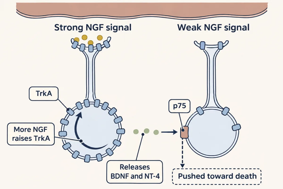
When supply is short, matching it means the factor ends up with some neurons, not spread over all. Slow, random loss can get there. Competition that picks winners early is another route. In mouse sympathetic neurons, evidence supports a model of active competition. NGF raises its own receptor, TrkA, in the neurons that receive it. A neuron that gets a little more can take up still more. Strong signallers release two related factors, BDNF and NT-4. They act through another receptor, p75. They push neighbours with weak NGF signalling toward death. Winners are amplified, and losers are punished. One caution. The ceiling holds only for the population measured.
Limb motor neurons do not depend on NGF. Takeaway: extra NGF saves neurons, but never more than were made. How many survive depends on how the factor is shared.
From the film
- Extra NGF given during the death period saves neurons that would have died, but never more than were made.
- In the ideal model, survivors are at most the smaller of the neurons made and the number the supply can support.
- How many survive when supply is short depends on how the factor is shared, and in mouse sympathetic neurons evidence supports active competition.
- The ceiling holds only for the population measured, and limb motor neurons do not depend on NGF.
Every test so far says one thing: Nerve growth factor (NGF) keeps neurons alive. Now turn that around and ask what follows from it.
Predict. Give a chick embryo extra NGF every day through the period when its sympathetic and sensory neurons normally die. Which happens: no change; more of the neurons survive than normal; new sympathetic ganglia appear in the wrong places; or sympathetic fibres invade every tissue? And is there a limit to how large the ganglia can get?
Check your prediction
More of the neurons survive. Given during the death period, NGF enlarges the ganglia mainly because more of the neurons that were made escape the normal death. The survivors also grow larger and sprout more fibres, so the “invade” option is not entirely wrong, but survival support is not expected to change neuron type. Extra survival support should preserve neurons in the existing ganglia; by itself it does not specify new ganglion locations. Ectopic ganglia would need evidence of migration, aggregation or survival at ectopic sites, not necessarily extra births. In 1981 Hamburger, Brunso-Bechtold and Yip counted chick dorsal root ganglia across the death period and found that NGF reduced the normal death.
There is a limit: a survival factor can save a neuron that would have died, not one that was never made.
How would you know the extra neurons were saved, not born? A larger ganglion at one late age cannot tell you. Three checks can.
- Count across the window. Count neurons, and pyknotic nuclei, in the same ganglia, treated and untreated, at several stages: soon after the ganglion forms, during the death period, and after it. If NGF acts on survival, the counts match early and diverge only during the death period, and the treated ganglia contain fewer pyknotic nuclei.
- Mark division and follow the progeny. A short pulse of a proliferation marker such as BrdU or tritiated thymidine, counted soon after, shows how many precursors are copying DNA, not how many neurons are produced. Combine such proliferation and cell-cycle measurements with pulse-chase birthdating or lineage tracing across the whole interval to test whether cumulative neuron production changed.
- Check identity and lineage. Confirm the expected type with several markers, such as tyrosine hydroxylase, an enzyme sympathetic neurons need to make noradrenaline, and follow the original cohort by lineage tracing or pulse-chase birthdating. Markers alone cannot exclude a fate change, because converted cells would carry the same markers in the same place.
The checks matter in the chick. Very early, before sympathetic axons reach their targets, extra NGF can also increase division in the primary sympathetic ganglia, the first sympathetic clusters to form, and it also reduces the low level of death there. The extra division is NGF acting as something other than a survival factor, and these checks catch it.
The ceiling
Put the logic in one line, as an ideal model:
neurons surviving ≤ the smaller of (neurons made) and (supply per day ÷ the least each survivor needs per day)
The model takes one group of N neurons already made, with none added, and a shared supply of F units per day, the target’s plus any you add, from which every neuron needs at least q units per day. S survivors take at least S × q, which cannot exceed F, so S ≤ the smaller of N and F/q, rounded down. It is a bound for this model, not a measured law linking an NGF dose to neuron number. Work it for a ganglion that has made 1,000 neurons, each needing one unit a day.
| Supply per day | Neurons made | Survivors, at most | What limits them |
|---|---|---|---|
| 500 units | 1,000 | 500 | the target |
| 1,000 units | 1,000 | 1,000 | both |
| 3,000 units | 1,000 | 1,000 | the number made |
In the middle row the two limits are equal, so either one alone would stop survivors at 1,000.
Extra factor raises the second term. Acting as a survival factor, nothing raises the first. Once every neuron that was made is supported, more factor adds no survivors. When the three checks together support a survival effect, the larger ganglia are made of neurons that would have died, and their number stays at or below the number made. Real ganglia can fall short of the ceiling. Neurons differ in how much they need, so survival rises gradually with dose. Some die for reasons NGF cannot fix, so even plenty of NGF can leave the count below the number made. And if winners take more than they need, fewer survive.
neurons surviving = the smaller of (neurons made) and (neurons the target's factor can support). Extra factor raises the second term; nothing about a survival factor raises the first. The numbers are relative units: this is a sketch of the logic, for a population and its own factor such as sympathetic neurons and NGF.
A puzzle hidden in the ceiling
The first row states only a bound. To reach it, all 500 units would have to go to 500 neurons, enough for each, with none wasted. Suppose instead the target spread them evenly over all 1,000.
Predict. Every neuron gets half of what it needs. How many survive?
Check your prediction
It depends on timing, and the answer could be none. If survival needs a full unit, every neuron is equally short and at risk at once. Then:
- Some neurons commit to dying.
- Each death frees factor for the rest, because the continuing supply can be redirected to the neurons still taking it up, but a neuron that has already committed cannot use it.
- If neurons commit slowly and freed factor is shared out quickly, close to 500 can survive. If many commit before it is shared out again, far fewer are left, perhaps none.
Equal sharing alone does not say how many survive.
So the “at most” is doing real work: when supply is short, survivors reach it only if the factor ends up with some neurons rather than spread over all, through slow, random loss with quick re-sharing or through competition that picks winners early.
In mouse sympathetic neurons, evidence supports a model in which competition does this actively. NGF raises the level of its own receptor, TrkA, in the neurons that receive it (shown in mouse and rat sympathetic neurons), so a neuron that gets a little more can take up still more. Neurons with strong NGF signalling also release two related factors, brain-derived neurotrophic factor (BDNF) and neurotrophin-4 (NT-4). These act through a second receptor, p75, on neighbours whose NGF signalling is weak, and push them toward death (Deppmann and colleagues, 2008). In this model, competition is more than a shortage: winners are amplified and losers are punished.
The ceiling holds only for the population you measured, with its own factor. One found for sympathetic neurons and NGF does not transfer to limb motor neurons, which do not depend on NGF: in chick embryos several other factors each reduce part of their normal death, and extra NGF should leave it roughly unchanged.
Permissive for survival, instructive for wiring
Read the transcript of “Permissive or Instructive”
Film transcript: Permissive or Instructive
NGF keeps neurons alive. Does it also choose where their axons go? A permissive signal allows an outcome that something else chooses. An instructive signal chooses the outcome itself. Fuel lets a car go anywhere. The driver picks where. Fuel is permissive. The driver is instructive. Removing a signal and restoring its normal pattern cannot tell the two apart. It only shows the signal is needed. Instead, keep the signal present, but change where or when it is delivered. Then ask whether the pattern still forms. If the pattern stays put, something else reads position, and the signal was permissive. If the pattern follows the delivery, it is instructive.
In 1989, Edwards and colleagues directed NGF to the pancreatic islets of otherwise normal mice. Islets are small clusters of hormone-making cells scattered through the pancreas. Predict. Where do the sympathetic fibres go?
Innervation of the islets rose sharply. The surrounding tissue gained no matching rise. The pattern followed the delivery. In 1993, Hoyle and colleagues ran the converse test. Their mice had sympathetic neurons that made NGF themselves. Now NGF was made by the neurons too, not only by the target. Axons still reached their tissues. But inside them, they branched sparsely. Making NGF in the islets of these mice restored islet innervation. So, in the sympathetic targets tested, NGF is permissive for reaching a target. It is instructive for how densely, and where, axons innervate within it. Here the fuel analogy breaks. One more test. In mice lacking both NGF and Bax, sympathetic neurons survive.
With death blocked, any defect left is not about survival. They fail to innervate many of their targets. Put the lesson together. The target is needed. Adding NGF saves neurons. Removing it, by antibody or by gene, kills them. The TrkA mutant shows the neurons were made, then lost. Acting as a survival factor, NGF cannot raise survivors above the number made. NGF keeps already specified neurons alive, and types differ in that need. Where it is supplied helps decide which connections stay. How some neurons come to win is the subject of the lesson on competition for targets. Takeaway: for survival, NGF is permissive. Within a target, it also instructs where axons branch.
From the film
- For survival, NGF is permissive: it keeps alive neurons that were already specified as sympathetic or sensory.
- To tell permissive from instructive, keep the signal present but change where or when it is delivered, and ask whether the pattern follows.
- In the targets examined, NGF also instructs how densely and where axons branch within a target.
Survival is not the whole story. Nerve growth factor (NGF) passes the tests of necessity and sufficiency for survival, but neither test says whether NGF chooses anything. A permissive signal allows an outcome that something else chooses. An instructive signal chooses the outcome itself. Fuel lets a car go anywhere; the driver picks where. Fuel is permissive, the driver instructive. The analogy breaks in one place, as you will see: within a target, NGF also acts like the driver.
Removing a signal and restoring its normal pattern cannot separate the two; it shows only that the signal is needed. What separates them is changing where or when the signal is delivered while keeping it present. Supply it evenly everywhere, or at an arbitrary new place or time, and ask whether the pattern still forms. If it does, something else is reading position, and the signal was permissive. If the pattern follows the delivery, the signal is instructive.
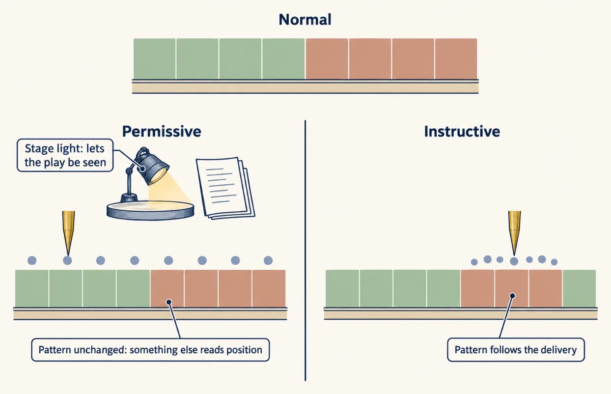
Predict. In 1989 Edwards and colleagues directed NGF to the pancreatic islets of otherwise normal mice. Islets are small clusters of hormone-making cells scattered through the pancreas. If NGF only permits survival, where should sympathetic fibres go? If it instructs where they branch, where should they go?
Check your prediction
Sympathetic innervation of the islets rose sharply, with no matching rise in the tissue around them. The pattern followed the delivery, so here NGF was instructive.
Hoyle and colleagues in 1993 ran the converse test. They made mice whose sympathetic neurons made NGF themselves, so the target no longer held the only supply. Axons still reached their tissues, so reaching them did not need the target to be the only source. But they branched sparsely inside them, and making NGF in the pancreatic islets of these mice restored islet innervation. In the targets examined, the results fit a model in which NGF is permissive for reaching a target and instructive for how densely, and where, axons innervate within it. The experiments altered NGF sources; they did not show that every guidance-relevant gradient was absent.
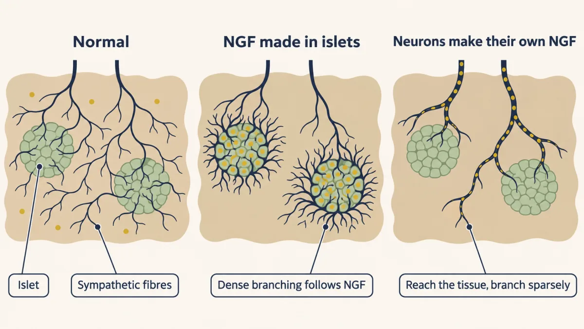
For survival, NGF behaves as permissive: it keeps alive neurons that were already specified as sympathetic or sensory. For other effects, such as axon growth, branching and the rise in TrkA described above, it is not merely permissive. Blocking death gives a cleaner test. In mice lacking both NGF and Bax, sympathetic neurons survive without NGF, so any remaining defect is not about survival. They fail to innervate many of their targets.
Putting it together
Four of the five predictions from the start of the lesson now have tests, each of its own kind. The fifth, the timing of death, needs counts of when axons arrive and when neurons die in the same population; see Going deeper.
| Experiment | Animal and age | Observed result | What it supports | What it does not establish |
|---|---|---|---|---|
| Remove wing bud | chick embryo | fewer neurons later; they had formed first (1949) | implicates the target in keeping neurons alive | which molecule |
| Graft extra leg | chick embryo | no more motor neurons at day 6; more later in embryos whose grafts moved vigorously | extra target can reduce death | which limiting resource; motor neurons are not NGF-dependent |
| Inject NGF | young mammals, chick embryos | larger ganglia | NGF is enough to enlarge the ganglia | whether neurons were saved, born or just grew larger |
| Extra NGF, counted across the death period | chick embryo | normal death reduced (1981) | the extra neurons were saved | anything above the number made |
| NGF antiserum | newborn mice and rats | sympathetic ganglia destroyed | necessity | that the antibody hit only NGF |
| NGF antiserum, later | older animals | much smaller effect | a window of dependence | — |
| NGF-null, TrkA-null | mouse | sympathetic and TrkA sensory neurons lost | genetic necessity; settles the antibody doubt | made versus lost |
| TrkA-null, counted over time | mouse superior cervical ganglion | normal neurogenesis and count at embryonic day 13.5; deficit by 15.5 that grows, with more dying cells | survival failed, not production | — |
| Bax-null | mouse | sympathetic neurons kept | the death runs through Bax | what else NGF does |
| NGF and Bax both deleted | mouse | neurons survive; many targets not innervated | NGF is needed for more than survival | — |
| NGF moved (made in islets, or by the neurons themselves) | mouse | more fibres in islets that make NGF; sparse branching inside tissues when neurons make their own | instructive for density within a target | that this holds in every target |
For NGF-dependent neurons the guiding idea holds up: make too many, then let the target keep what it can feed. Survival and place are linked, though. Nerve growth factor (NGF) does not create a sympathetic or sensory lineage, but because dependence on it differs among types, its availability can influence which types survive. Where it is supplied helps decide which connections persist, and within a target where axons branch.
The ceiling also left a question open: when supply is short, which neurons win, and how? That is the subject of the lesson on competition for targets.
Design problem
Factor X keeps cultured enteric neurons, the neurons of the gut wall, alive. Design the series of experiments in living animals that would show X is a target-derived neurotrophic factor for them, and say what each experiment can and cannot prove.
Saved in this browser only.
One way to do it
Walk the evidence table, with X in place of NGF, but first check the premise.
- Is there a death period? Define which enteric neurons you mean, and measure when they are born, migrate, mature and die. Do not assume an NGF-like death window. If normal death is rare, adding X may not raise survival because endogenous support already suffices; that does not rule out a survival requirement, which removal tests (steps 2 and 3). X may also act on precursors; test that separately.
- Sufficiency. If there is a death period, give extra X then. Count neurons and dying cells across ages, with pulse-chase birthdating and lineage markers. More neurons, fewer dying cells, unchanged production and stable identity support a survival effect. This cannot show that the animal needs X.
- Necessity by antibody. Neutralize X during defined developmental intervals, even without a normal death window, and measure induced death, production, migration and differentiation. More death means the neurons need something the antibody removes, but the antibody might bind something else.
- Necessity by genetics. Delete the gene for X, or for its receptor, conditionally to time the loss. Loss of the same neurons removes the antibody doubt.
- Made versus lost. Count the mutant ganglia at a series of ages. Normal production followed by later loss, with more dying cells and no neurons leaving, means survival failed, not production.
- Target-derived. Show that the target tissue makes and releases X. Delete X only in that tissue, leaving the tissue intact, and check that the neurons die and that local X rescues them. Removing or enlarging the tissue supports the idea but changes many things at once.
- Mechanism and the ceiling. Find which death program these neurons use, block it, and ask whether they then survive without X. Follow one birthdated group: extra X should never leave more of it alive than were born.
Going deeper
Where NGF is made
On the list of NGF-producing targets are the eye, salivary gland, pineal gland, blood vessels, spleen, heart and fat.
Which neurons depend on NGF
Large neurons that sense muscle stretch depend on a different neurotrophin. Basal forebrain cholinergic neurons, which respond to nerve growth factor (NGF) in culture, still develop in NGF-null mice. Not every neuron that can respond to a factor needs it to exist.
How a distant target reaches the cell body
The factor acts at the axon ending, and a signal returns to the cell body to support survival.
When the superior cervical ganglion starts to depend
In the mouse superior cervical ganglion some neurons may come to depend on neurotrophins a little before target innervation is complete.
A fly example of permissive versus instructive
In 1985 Kathryn Anderson and colleagues used this logic in fly eggs. Embryos from Toll mutant mothers are dorsalized: they lack ventral, belly-side structures. When wild-type cytoplasm was injected into them, the ventralmost part of the rescued pattern formed at the injection site, wherever the needle went in. The pattern followed the delivery, which is the instructive result.
Your notes
Select any text in the lesson to highlight it or add a note to it. Your highlights and notes are saved in this browser and collected in your notebook.
This browser is not letting the site save anything (a private window or strict privacy settings), so highlights and notes will be lost when you leave the page.
How we measure it
Antibody neutralization (immunosympathectomy)
An antiserum against a purified factor is injected into an animal to remove the factor's activity. If a population dies, it needed something the antiserum removed. The caveat is specificity: an antiserum might also bind something else.
Gene knockout in the mouse
A knockout is a mouse in which a gene has been disrupted so that it makes no working protein. Deleting the gene for NGF or for its receptor TrkA tests necessity without relying on an antibody, though the gene is missing from every cell for the whole of life.
Neuron counts across the death window
Neurons are counted in serial sections at a series of ages before, during and after the normal period of cell death, together with dividing cells and pyknotic (condensed, dying) nuclei. The time course shows whether a population was never made or was made and lost.
Birthdating with a proliferation marker
A labelled nucleotide such as tritiated thymidine or BrdU is taken up only by cells copying their DNA, so it marks the cells that divide while it is present. A short pulse counted soon after measures how many cells are copying DNA, not how many neurons are produced; following the labelled cells until they become neurons (pulse-chase birthdating) shows whether a factor changed how many neurons were born.
Practice questions on this lesson (3) → MCAT-style, original, each with an explanation
Finish line
Test yourself
8 questions
Practice and MCAT-style, with explained answers.
Next · Lesson 30
Neurotrophins, Trk Receptors and p75
Many developing neurons live only if their target supplies enough of the right neurotrophin. How does the neuron read that factor, and how can the same factor sometimes kill?
Continue →← Lesson 28 · Too Many Neurons: Naturally Occurring Cell Death