Developmental Neuroscience · Unit 6: Axons find their way
Semaphorins and Repulsion
Rewritten on : new text, flashcards and film. Your notes and highlights stay, but the text around them may have moved.
Pain fibres and muscle afferents enter the spinal cord side by side. One kind stops in the dorsal horn; the other runs on to the motor neurons. What sorts them?
Pause when the reading reaches a film, and offer to play it. Saved on this device.
Watch the film now, or keep listening
Builds on:The Growth Cone: The Tip That Steers the AxonPioneers, Guideposts and the Logic of Guidance CuesNetrin and DCC: One Cue, Two AnswersWhy these?
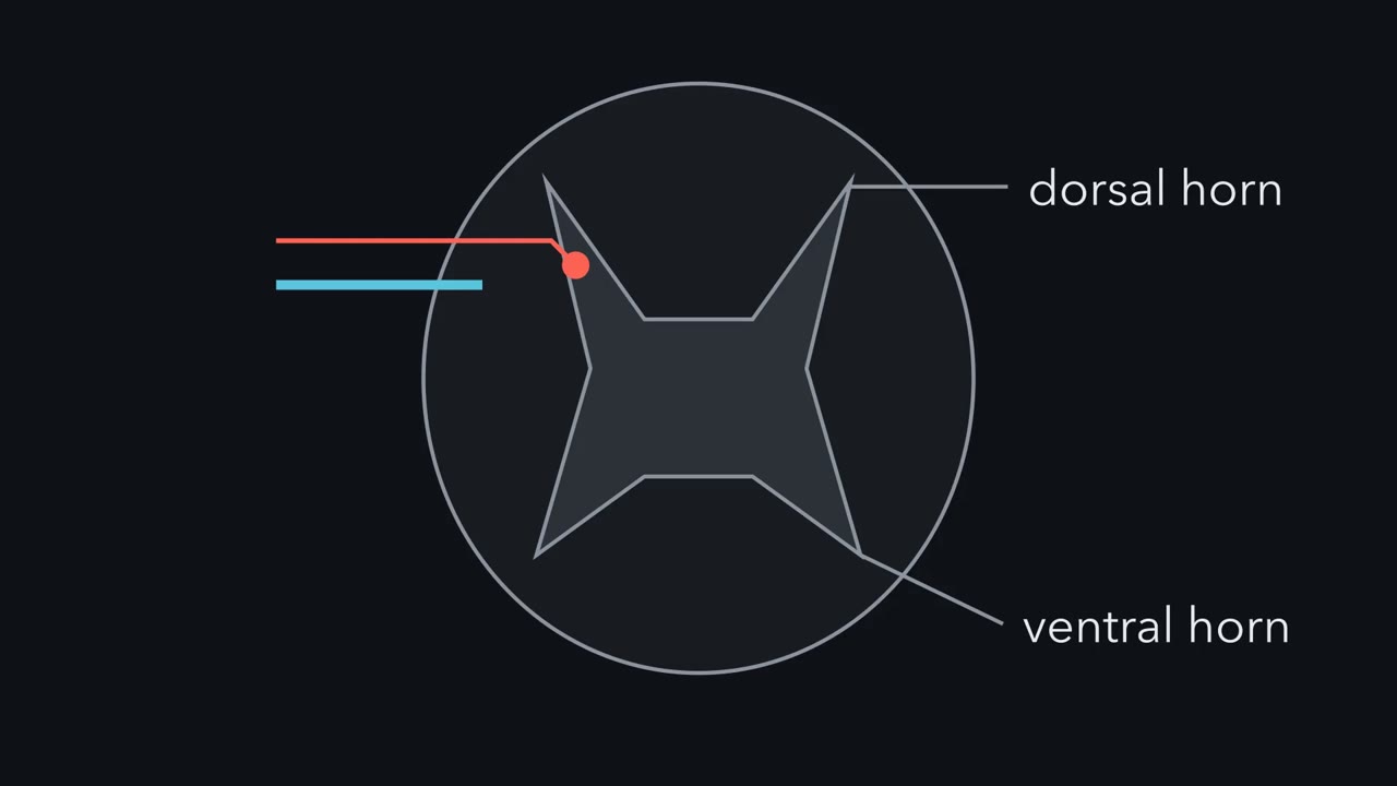 ▶ Watch The Cue Marks the Place · 2:47
Read the transcript of “The Cue Marks the Place”
Film transcript: The Cue Marks the Place
Two kinds of sensory axon enter the spinal cord side by side. One kind stops in the dorsal horn. The other runs on to the motor neurons. What sorts them? The ones that stop are small pain and temperature fibres. They depend on nerve growth factor The ones that go on are large muscle stretch fibres. They depend on neurotrophin-3 (NT-3).
The ventral cord was known to release something that inhibits sensory axon growth. A map needs walls as well as roads. To find a wall, you need an assay. Raper and Kapfhammer grew chick sensory neurons in a dish. Then they added membranes from embryonic brain.
Within minutes, many of the spread growth cones folded into thin tips. That is collapse. The fraction that collapsed was the readout. Remove the membranes, and the effect reverses. Would any membrane do this? And what kind of molecule is doing it?
Membranes from liver or fibroblasts did not cause collapse. So not just any membrane would do. Heat and trypsin destroyed the activity. So it was a protein. Raper's group purified it from chick brain and named it collapsin. Today it is called Sema3A, a semaphorin.
Then came a first clue. Growth cones from the retina did not respond to it. Here is the idea to carry through. The cue marks the place. The growth cone decides what it means. Does a growth cone answer the cue at all? First, it needs the receptor.
Which way does it answer? That depends on its internal state. If it is repelled, does it turn, or collapse? In typical assays, a gradient from one side turns it away. A high dose all round can fold it up, where the preparation supports collapse. Layout, dose, exposure and the cell all matter.
The rule has limits. Which cue is made, where, and how much still matter. And in the living cord, other cues share the job. Back to the two axons. One wall, two answers. The takeaway: the same cue stops one axon, and barely affects the other.
From the film
- Sema3A, made in the ventral cord, repels NGF-dependent pain fibres but at the stage tested barely affects NT-3-dependent muscle afferents.
- The collapse assay led to collapsin, now Sema3A, a protein that folds up growth cones, though retinal growth cones did not respond to it.
- The cue marks the place and the growth cone decides what it means, which explains selectivity but not the whole map.
Two kinds of sensory axon enter the spinal cord side by side. Pain and temperature fibres stop in the dorsal horn. Muscle afferents run on into the ventral horn to reach the motor neurons. Both enter at the same place. What sorts them?
A map needs walls as well as roads. An axon on its way to a target passes many places where it must not go: the wrong side of the spinal cord, the wrong layer, the wrong nerve. Cues whose message is “Don’t go there” are repulsive cues, and the semaphorins are one of the largest and best-studied families of them.
The puzzle in the spinal cord
Sensory neurons in a dorsal root ganglion are of several kinds. Two of them set the puzzle.
| Pain and temperature neurons | Muscle stretch neurons | |
|---|---|---|
| Cell size | small | large |
| Neurotrophin they depend on | NGF | NT-3 |
| Its receptor on the neuron | TrkA | TrkC |
| Where the axon ends in the cord | dorsal horn | ventral horn, on motor neurons |
The ventral cord was known to release something that inhibits sensory axon growth. That raises three questions.
- Why does one tissue keep out one class of axon and admit another?
- How does a growth cone hear a secreted cue when the protein that binds it has almost no tail inside the cell?
- Why can the same cue repel one part of a neuron and attract another?
The one idea to carry through this lesson. The cue marks the place; the growth cone decides what it means. In the spinal cord, Sema3A is made where pain fibres must not go.
- Does a growth cone answer at all? It needs the receptor: neuropilin to catch the cue, plexin to signal. (At the stages tested, pain fibres answer strongly and muscle afferents weakly; the chick retinal growth cones in the first collapsin assay did not collapse.)
- Which way does it answer? That depends on its internal state, such as its cyclic GMP level. (A cortical axon is repelled; the same neuron’s dendrite is attracted.)
- If it is repelled, does it turn or collapse? In typical assays, a gradient from one side turns it away, and a high dose all round can fold it up where the preparation supports collapse. Layout, dose, exposure time and the cell all shape which you see. (A pipette versus a bath.)
What the rule covers. Secreted class 3 semaphorins, mainly Sema3A, acting through neuropilin-plexin receptors on vertebrate growth cones: sensory axons from dorsal root ganglia in explants and in the spinal cord, Xenopus spinal neurons in turning assays, and cortical pyramidal neurons. Three limits matter.
- “Decides” does not mean the cue carries no information: which semaphorin is made, where and how much all matter, and membrane semaphorins bind plexins directly, with no neuropilin.
- The receiver’s state is not fixed: muscle afferents appear to lose their sensitivity as they grow into the ventral cord.
- In the living cord Sema3A shares the job with other cues, so the rule explains selectivity, not the whole map.
A growth cone that folds up
You met the growth cone as both sense organ and motor: a fan of actin-filled filopodia and lamellipodia at the axon’s tip. It turns toward the side where actin builds and away from the side where actin falls apart. The guidance cues lesson sorted cues into four kinds: contact attraction, contact repulsion, long-range attraction and long-range repulsion. Semaphorins work in both repulsive modes. Some are secreted and repel from a distance; others sit in cell membranes and repel on contact. Repellents fence off wrong territories, so an axon growing between them is channelled along its path, though other cues tell it where to stop.
A repellent needs an assay. In 1990 Jonathan Raper and Josef Kapfhammer, at the University of Pennsylvania, grew chick dorsal root ganglion neurons on laminin and added a suspension of embryonic brain membranes. Within minutes the growth cones collapsed, folding from a spread fan into a thin tip. The effect reversed when the membranes were gone.
Predict. Is this something any membrane would do, and what kind of molecule is doing it? Name two controls that would tell you.
Check your prediction
Membranes from other tissues are the first control: membranes from liver or fibroblasts did not cause collapse, so collapse was not a response to any membrane at all. Treatments that destroy proteins are the second: the activity was destroyed by heat and by trypsin, so it was a protein. Counting the fraction of collapsed growth cones gave a fast, graded readout that could follow the activity through each step of purification.
Collapsin, the first vertebrate semaphorin
In 1993 Yuling Luo, David Raible and Raper purified the protein from chick brain, a glycoprotein of about 100 kilodaltons, and named it collapsin. Retinal growth cones did not respond to it: the cue was present, but those growth cones did not hear it.
Collapsin’s first half resembled fasciclin IV, a guidance protein cloned the year before in the grasshopper by Alex Kolodkin, Corey Goodman and colleagues. So a membrane protein in an insect had a secreted relative in a bird. Later in 1993 Kolodkin, David Matthes and Goodman named the whole family the semaphorins, after the semaphore, a way of signalling with flags. Collapsin is now Sema3A, the semaphorin this lesson follows. A naming key: collapsin (chick) = Semaphorin III (the older vertebrate name) = Sema3A; fasciclin IV (grasshopper) = Semaphorin I. The vertebrate classes differ in how the protein is held:
| Vertebrate class | How it is held |
|---|---|
| Class 3, including Sema3A | secreted |
| Classes 4 to 6 | span the membrane |
| Class 7 | held by a lipid anchor |
Semaphorins also work far outside the nervous system, in immune cells, heart, blood vessels and bone.
One tissue, two axons
Read the transcript of “One Tissue, Two Axons, One Receptor”
Film transcript: One Tissue, Two Axons, One Receptor
Sema3A is made in the ventral cord. Pain fibres stay out, and muscle afferents go in. Is the difference in the cue, or in the axons? Here is the test. Set a dorsal root ganglion in a collagen gel, beside a piece of ventral cord. Grow it once with nerve growth factor and once with neurotrophin-3 (NT-3). The medium largely decides which neurons grow. Where will the axons grow in each dish?
With NGF, the pain fibres grow. They grow poorly on the side facing the cord. The ring of axons, the halo, is lopsided. With NT-3, the muscle afferents grow. Their halo is nearly round. Same tissue, two answers. Messersmith, Kolodkin and colleagues found this pattern in rat explants. Cells secreting Sema3A did the same.
So how does a growth cone hear Sema3A? Two groups found the receptor independently. It was neuropilin. Neuropilin binds Sema3A tightly. But its tail inside the cell is short, with nothing like an enzyme. Where is the wire? Two more groups pointed to the plexins. These membrane proteins have a large part inside the cell. In engineered cells, neuropilin alone binds the cue, but the cell does not respond. With plexin as well, binding is tighter. Now the cells shrink and round up.
Other work tested neurons, which keep their other receptor parts. There, a neuropilin with no inner tail still let growth cones collapse. Now give sensory neurons extra plexin with its inner part cut off. They still have their own normal plexin. What happens?
The response is blocked. The broken plexin interferes with the cell's own working receptors. One likely idea: it joins neuropilin and the cue in complexes that cannot signal. A mutant that blocks the normal protein this way is called a dominant negative. So neuropilin catches, and plexin signals.
Inside, the plexin switches off a small GTPase of the Ras family. That protein helps integrins grip the surface. Other plexin pathways act on actin and microtubules. Together, the growth cone loses grip and actin. In a growth cone that Sema3A repels, where this happens shapes what you see. Typically, a gradient from one side turns it away, and a strong dose all round can fold the fan up, in preparations that support collapse.
The takeaway: for Sema3A, the working receptor is neuropilin and plexin together. No pair, no answer in these experiments.
From the film
- In explants, the ventral cord repels axons grown in NGF but has little effect on axons grown in NT-3.
- Neuropilin catches Sema3A and plexin signals, and in these experiments no neuropilin-plexin pair means no response.
- A repelled growth cone typically turns away from a gradient on one side, and a high dose all round can make it collapse where the preparation supports collapse.
Back to the spinal cord, where Sema3A is made in the ventral cord. The test is an explant co-culture: an explant, a piece of living tissue, is set in a collagen gel beside a candidate source, and its axons grow out in a ring, the halo. Measure the axons on the side facing the source and on the far side. A near-to-far ratio near 1 means outgrowth is symmetric. A ratio well below 1 means the near side grew less, as a diffusible repellent or a local growth inhibitor would cause. Lengths compared with control cells, and turning assays, tell those apart.
Predict. You culture a dorsal root ganglion in a collagen gel next to a piece of ventral spinal cord, once in medium with nerve growth factor (NGF) and once in medium with neurotrophin-3 (NT-3). The neurotrophin in the medium largely decides which neurons survive and extend axons. Predict the outgrowth in each case.
Check your prediction
In NGF, the axons come mostly from small TrkA neurons, the pain and temperature fibres. Expect them to grow poorly on the side facing the ventral cord and well on the far side: an asymmetric halo, the mark of a diffusible repellent. In NT-3, the axons come mostly from large TrkC neurons, the muscle afferents. Expect a nearly symmetric halo.
That is the result. In 1995 Elizabeth Messersmith, Kolodkin and colleagues, with explants of embryonic rat tissue, showed that the ventral cord’s activity repels axons grown in NGF but has little effect on axons grown in NT-3. Cells secreting Semaphorin III did the same, and the protein is made in the ventral cord. The first MCAT question below puts made-up numbers on this kind of result.
So one tissue treats two kinds of axon differently. At this stage the NGF-dependent class responds to Sema3A, through a neuropilin-plexin receptor, and the NT-3-dependent class mostly does not, so only the first is kept out. To test the idea, add an antibody against neuropilin-1 or against Sema3A: the NGF halo should become more symmetric.
The NT-3 result is a snapshot at one stage, and the answer can change with time. In chick, Raper’s group found that antibodies against collapsin neutralized the ventral cord’s repellent. Their data also suggest that muscle afferents lose their sensitivity as they grow into the ventral cord.
Be clear about what the explants do and do not show. A ventral cord explant tests everything the cord releases. Cells secreting Sema3A test Sema3A alone. Neither shows whether Sema3A is the only cue doing this job in an animal, and it probably is not: mice lacking Sema3A have clearly disorganized peripheral nerves, and some sensory axons enter spinal cord regions they normally avoid, so other cues probably share the work in the living cord.
Neuropilin catches, plexin signals
Who receives Sema3A? Two groups found the answer independently, in back-to-back papers in the August 1997 issue of Cell: Zhigang He and Marc Tessier-Lavigne by expression cloning (putting pools of cDNA into cells and finding which pool lets them bind the tagged cue), and Alex Kolodkin, David Ginty and colleagues. The protein was neuropilin. He and Tessier-Lavigne also showed that antibodies to neuropilin block Semaphorin III repulsion of sensory axons.
Neuropilin presented a puzzle. It bound Sema3A tightly, but its tail inside the cell was only about forty amino acids long, with nothing that looked like an enzyme. A receptor has two jobs: catch the signal and pass it on. Neuropilin plainly does the first. Where was the wire for the second?
In 1999 two groups, Takahashi, Strittmatter and colleagues, and Tamagnone, Comoglio and colleagues, pointed to the plexins, a large family of membrane proteins with a substantial part inside the cell. The decisive tests added and removed the two parts. Read each table one row at a time and predict the last column before you look across. First, engineered cells that normally ignore Sema3A:
| Engineered cells carry | Does Sema3A bind? | Does the cell respond? |
|---|---|---|
| Neither protein | no | no |
| Neuropilin-1 alone | yes | no |
| Plexin-A1 alone | little or none | no |
| Neuropilin-1 and Plexin-A1 together | yes, more tightly | yes: the cells shrink and round up |
Then neurons, which already have their own receptor parts and change one of them:
| Neurons carry | Does Sema3A bind? | Does the cell respond? |
|---|---|---|
| A neuropilin lacking its tail inside the cell (other work) | yes | yes: growth cones still collapse |
| Extra Plexin-A1 lacking its part inside the cell (sensory neurons) | expected, through the neurons’ own neuropilin | no: the response is blocked |
Predict. The last row is the odd one. The neurons still make their own normal plexin. Why would adding a broken plexin stop them responding?
Check your prediction
The broken plexin interferes with the cell’s own working receptor complexes. One likely explanation is that it joins neuropilin and Sema3A in complexes that cannot pass the signal on; the blocking experiment alone does not settle the exact mechanism. A mutant that blocks the normal protein in this way is called a dominant negative. Taken with the row above it, the result splits the jobs: neuropilin’s tail is not needed, and the plexin’s inner part is. Neuropilin catches; plexin signals.
That is the first part of the lesson’s rule: in these experiments, no neuropilin-plexin pair, no response to Sema3A. Having the pair is necessary; it does not by itself guarantee a strong response. Some plexins bind membrane semaphorins directly, while most class 3 semaphorins reach plexins through neuropilin.
What does the plexin’s inner part do? It contains a domain that acts as a GAP, a protein that speeds GTP hydrolysis and so switches a small GTPase off (see GTPase switches and Ras to MAPK). The target is a Ras-family GTPase, R-Ras or Rap depending on the report, and both help hold integrin adhesions to the substrate, so switching either off lowers adhesion. Plexin signalling also engages Rho-family GTPases and effectors such as MICAL and CRMP2 that act on actin and microtubules. The net result is local loss of actin and adhesion, the cytoskeletal response described in integrins and Rho GTPases.
Turn or collapse
Predict. Take a growth cone that Sema3A repels. In one dish, a micropipette releases Sema3A to one side of it. In another, Sema3A is added evenly to the whole dish, as in the collapse assay. What does each growth cone do?
Check your prediction
On one side, Sema3A typically loosens that side, and the growth cone turns away. All round, a high enough dose can loosen every side, and in sensory growth cones the fan folds up: collapse. These are typical outcomes in assays that support both, not a switch every growth cone must show; dose, exposure time and the preparation matter as well as layout.
Worked problem: what a gradient gives the receptors. Use simple binding, fraction occupied = c / (c + Kd), with illustrative Kd values: 0.2 nM for the neuropilin-plexin pair and 1.0 nM for neuropilin alone (the second MCAT question uses the same values). Let the concentration fall by 10% of its mean from the near edge of the growth cone to the far edge, an illustrative figure of the order estimated for pipette turning assays.
Step 1: a gradient centred near Kd. Near side 0.21 nM, far side 0.19 nM. Occupancy is 0.21 / 0.41 = 0.512 near and 0.19 / 0.39 = 0.487 far. The near side is about 2.5 percentage points more occupied: a signal on one side.
Step 2: the same 10% gradient at 50 nM. Near side 52.5 nM, far side 47.5 nM. Occupancy is 0.9962 near and 0.9958 far. The difference is 0.04 percentage points, about sixty times smaller. Almost every receptor is occupied, so even a gradient looks nearly uniform.
Step 3: a bath. With the same concentration on every side and identical receptors, there is no near-to-far difference in occupancy at any concentration, so no directional signal.
Step 4: what the receptor sets. For a small percentage gradient like this one, the near-to-far difference is greatest when the mean concentration is close to Kd. With neuropilin alone, the difference at 0.2 nM is only about 1.4 percentage points. Affinity sets the concentrations over which occupancy differs most, but neuropilin alone cannot signal, so only the pair can act on a difference.
The sketch describes receptor occupancy, not behaviour: it cannot predict a turn or a collapse, and it ignores amplification, adaptation and averaging over time. Collapsin was active at roughly 10 picomolar in the original assay, and these illustrative Kd values cannot say how many receptors were occupied there. How sensitive a growth cone is depends on binding and on how occupied receptors are coupled to a response.
Same cue, opposite answers
Read the transcript of “Same Cue, Opposite Answers”
Film transcript: Same Cue, Opposite Answers
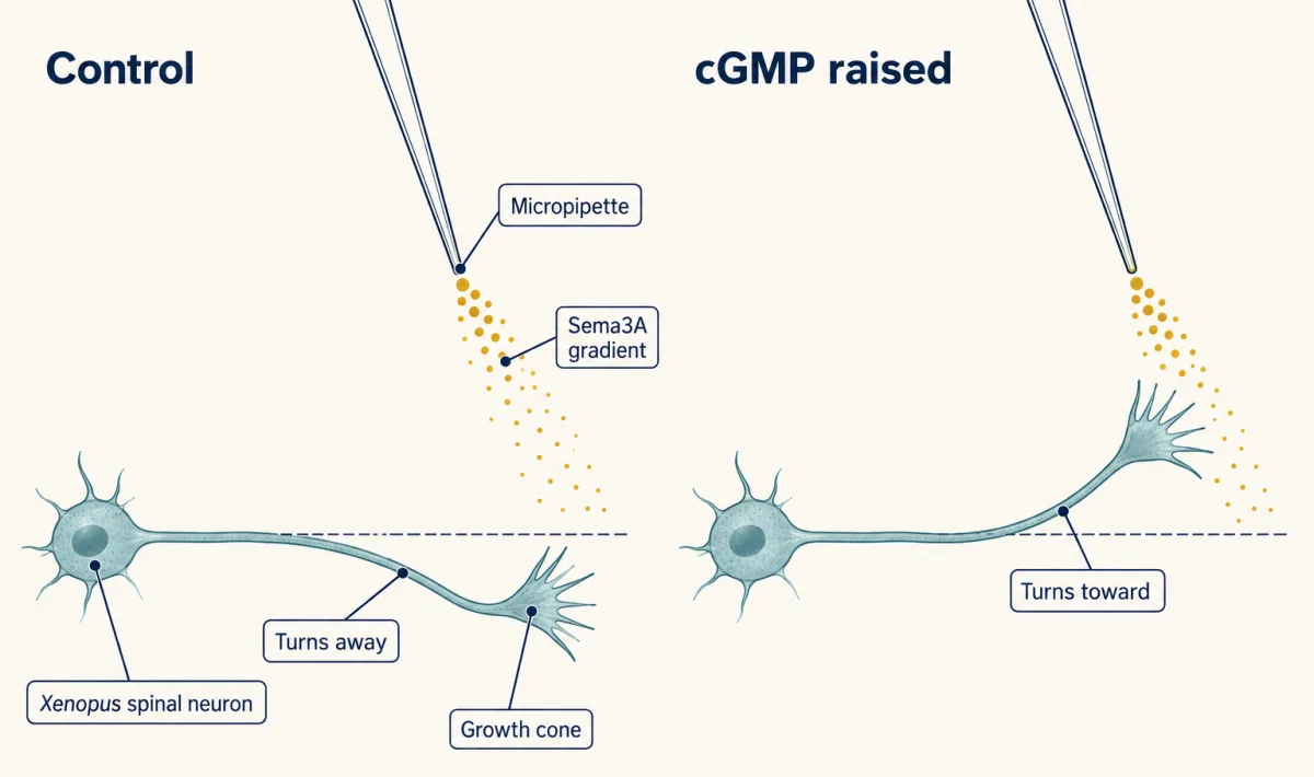
Is repulsion a fixed property of Sema3A? Or can the growth cone change the answer? Song, Poo and colleagues grew single frog spinal neurons in a dish. A fine pipette set up a small gradient of Sema3A beside one growth cone. The growth cone turned away. Now raise cyclic GMP inside the growth cone. Same pipette, same gradient. What happens?
The growth cone turned toward the Sema3A. The gradient did not change. The reader did. Cyclic GMP is a second messenger made by guanylate cyclases. Here, its level sets the sign of the answer. A similar switch, with cyclic AMP, governs netrin. Does a living neuron use this switch? Look at the developing cortex. Sema3A is high near the pial surface, the outer surface of the brain. It repels the axons of cortical neurons downward. But the apical dendrite of the same neuron grows up, toward it. Why the difference? Soluble guanylate cyclase, an enzyme that makes cyclic GMP, sits mainly in the developing dendrite. It is needed for that attraction.
One cue, one neuron, two answers, set by where the enzyme is. Now replay the rule. The cue marks the place. The growth cone decides what it means. The pain fibre and the muscle afferent differ in how strongly they answer the cue. The axon and the dendrite differ in which way they answer. For a repelled growth cone, a pipette gradient typically gives a turn. A strong dose in a bath can give a collapse, where the preparation supports it. The takeaway: semaphorins keep axons out of wrong places. But only axons that can hear them, and only in the sense their state allows.
From the film
- Raising cyclic GMP turned Xenopus growth cones from repelled by a Sema3A gradient to attracted to it.
- In developing cortex, one neuron's axon is repelled by Sema3A while its apical dendrite is attracted, and soluble guanylate cyclase sits mainly in the dendrite.
- Pain fibres and muscle afferents differ in how strongly they answer the cue, while axon and dendrite differ in which way.
Is repulsion a fixed property of Sema3A? In 1998 Hongjun Song, Mu-ming Poo and colleagues put single Xenopus spinal neurons in culture and set up a small Sema3A gradient with a micropipette, a growth-cone turning assay. The growth cones turned away.
Predict. Now raise cyclic GMP inside the growth cone and repeat with the same gradient. Does the growth cone turn away more sharply, ignore the gradient, or do something else?
Check your prediction
It turns toward Sema3A: with the cyclic GMP pathway activated, the same growth cones were attracted. The experimenters reapplied the same gradient conditions, repositioning the pipette relative to the growth cone’s new direction for each test of the same neuron, so the change is in how the growth cone reads the cue.
The senses and cGMP lesson introduced cGMP as a second messenger made by guanylate cyclases. Here its level sets the sign of the answer; a similar switch with cAMP governs netrin, covered in netrin and DCC.
Does a living neuron use this? In the developing cortex, Sema3A is high near the pial surface, the brain’s outer surface. Franck Polleux and Anirvan Ghosh, with colleagues, had shown that it repels cortical axons downward. In 2000 Polleux, T. Morrow and Ghosh showed it also attracts the apical dendrite of the same pyramidal neurons upward. Soluble guanylate cyclase, an enzyme that makes cGMP, sits mainly in the developing apical dendrite and is needed for that attraction: one cue, one neuron, two answers.
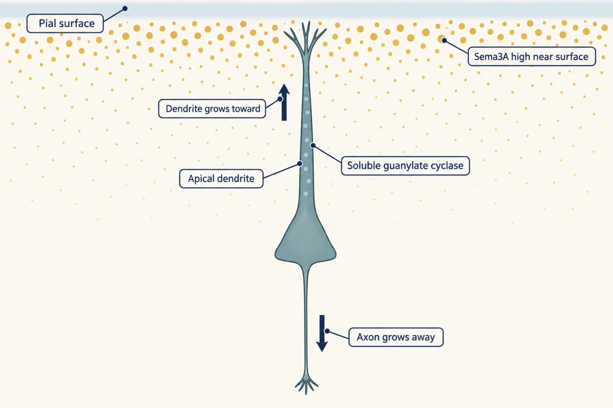
So from the spinal cord to the cortex, the cue marks the place and the growth cone decides what it means: the pain fibre and the muscle afferent differ in how strongly they answer, the axon and the dendrite in which way.
Where next: the same family keeps working after axons arrive. It prunes axon branches, covered in axon pruning, and limits dendritic spines, covered in CNS synapses.
Design problem
In the explant experiment, axons grown in neurotrophin-3 (NT-3) barely respond to Sema3A. Design experiments to show whether they ignore it for lack of a receptor or for another reason, and not simply because they never meet it.
Saved in this browser only.
One way to do it
First make sure they meet it. Set NT-3 explants beside cells secreting Sema3A, with control cells as the comparison, over a series of distances or doses. NGF-supported explants in the same set-up confirm that the source is active, though not the exact gradient at each NT-3 growth cone. Measure absolute outgrowth and collapse against matched controls as well as near-to-far asymmetry, and use turning assays for direction: a symmetric halo alone cannot tell weak signalling from growth inhibited equally on both sides (600 to 150 micrometres on both sides still gives a ratio of 1) or from too little spatial stimulation.
Next, ask what they carry. Compare how much neuropilin-1 and A-class plexin the NT-3-supported and NGF-supported growth cones have, and where it sits, at the same age. One possibility is that the NT-3 class lacks part of the pair or has less of it. The result could also show that both carry it, which would point instead to a difference in signalling state.
Then test the answer. If a part is missing or low, express it in the NT-3-supported neurons, with controls for the expression itself, and repeat the explant test. If the receptor is the reason, they should now respond: an asymmetric halo, with outgrowth and turning measured as above. As a check on the other side, a dominant-negative plexin in NGF-supported neurons should abolish their response by the same measures.
Two caveats. Sensitivity may change with age, so test at more than one. And the neurotrophin in the medium chooses which neurons grow, and may also change how they respond (see Going deeper), so compare the two classes in the same medium where you can.
Going deeper
Fasciclin IV and the semaphorin family, in detail
Fasciclin IV marks bands of epithelial cells in the grasshopper limb bud, and one band lies where the growth cones of the Ti1 pioneer neurons make a sharp turn. Embryos cultured in an antibody against fasciclin IV built that pathway abnormally. The 1993 paper naming the family described related genes in flies, in humans and in two viruses, all sharing one large region now called the sema domain.
Two pairs of papers
Neuropilin was first described in the frog nervous system by the laboratory of Hajime Fujisawa. The plexin papers of 1999, by Takahashi, Strittmatter and colleagues and by Tamagnone, Comoglio and colleagues, also appeared together in one issue of Cell.
Which GTPase does plexin switch off?
In the first case studied, Plexin-B1, which is the receptor for a membrane semaphorin rather than for Sema3A, the reported target is R-Ras. Later work with purified plexins points instead to its relative Rap.
How the receptor is switched on
Structures of Sema3A with neuropilin-1 and Plexin-A2 show neuropilin bridging the semaphorin dimer and the plexin (see the structures on this page). Signalling proceeds through plexin dimerization or rearrangement, which relieves autoinhibition of a split GAP domain. There are exceptions to the neuropilin route: Sema3E binds Plexin-D1 without neuropilin.
What the explant contrast mixes together
Beyond choosing which neurons grow, the neurotrophin a sensory neuron sees has been reported to change how sensitive its growth cones are to Sema3A. If so, the nerve growth factor (NGF) and neurotrophin-3 (NT-3) explant contrast mixes which neurons grow with how they respond. An open question is how much of the selectivity seen in the dish is set by receptor presence, by the choice of plexin and neuropilin co-receptors, and by internal state such as cGMP, and how these change as afferents mature.
Your notes
Select any text in the lesson to highlight it or add a note to it. Your highlights and notes are saved in this browser and collected in your notebook.
This browser is not letting the site save anything (a private window or strict privacy settings), so highlights and notes will be lost when you leave the page.
How we measure it
Growth-cone collapse assay
Cultured neurons with spread growth cones are exposed to a membrane extract or a purified protein for a set time, then fixed, and the fraction of growth cones that have folded into thin tips is counted. Because it is fast and graded, it can follow an activity through each step of purification.
Explant co-culture in collagen
A piece of embryonic tissue, such as a dorsal root ganglion, is set in a three-dimensional collagen gel a short distance from another tissue or from cells secreting a candidate cue. Axons that grow less on the near side than the far side are read as repelled; axons that grow more on the near side, as attracted. Turning assays help separate steering from simple effects on growth.
Expression cloning and ligand binding
The candidate ligand is fused to an enzyme tag such as alkaline phosphatase and applied to cells transfected with pools of cDNA. Cells that turn color carry a protein that binds the ligand, and repeated subdivision of the positive pool isolates the gene.
Growth-cone turning assay
A micropipette releases puffs of a cue to one side of a single growth cone of a cultured neuron, setting up a small gradient. The angle the axon turns, toward or away, is measured, and drugs or receptor mutants show what the turn depends on.
Practice questions on this lesson (3) → MCAT-style, original, each with an explanation
Where this lesson fits
Before thisReview first
- The Growth Cone: The Tip That Steers the AxonThis course · Lesson 3
You need how the growth cone steers by building and losing actin to follow why repulsion makes it fold up.
- Pioneers, Guideposts and the Logic of Guidance CuesThis course · Lesson 22
The four kinds of cue, including contact and long-range repulsion, are used here without being re-taught.
- Netrin and DCC: One Cue, Two AnswersThis course · Lesson 23
It introduces the idea that a cue's sign depends on the growth cone's internal state, which is applied here with cGMP.
After thisStudy next
- Topographic Maps: Sperry's Eye and Eph GradientsThis course · Lesson 26
It uses repulsive cues acting on growth cones with different receptor levels to build an ordered map.
- Pruning Without Dying: Axons That Cut Themselves BackThis course · Lesson 34
Semaphorin signalling here goes on to prune axon branches after they have arrived.
- Synapses in the Brain: Adhesion, Scaffolds and AstrocytesThis course · Lesson 36
It takes the same semaphorin family on to limiting dendritic spines and shaping synapses.
Suggested by an AI model from the lessons themselves, then checked. How we use AI
Finish line
Test yourself
9 questions
Practice and MCAT-style, with explained answers.
Next · Lesson 26
Topographic Maps: Sperry's Eye and Eph Gradients
Turn a newt's eye upside down, let the nerve regrow, and it turns its head down toward a lure held above it. Why does it not learn?
Continue →← Lesson 24 · Crossing the Midline Once: Slit, Robo and Comm