Developmental Neuroscience · Unit 11: Rebuilding, evolving, and going wrong
Injury and Regeneration: Why the Brain Does Not Regrow
Rewritten on : new text, flashcards and film. Your notes and highlights stay, but the text around them may have moved.
A cut nerve in your arm can grow back. A cut tract in your spinal cord mostly cannot. Is the fault in the neurons or in what surrounds them?
Pause when the reading reaches a film, and offer to play it. Saved on this device.
Watch the film now, or keep listening
Builds on:The Growth Cone: The Tip That Steers the AxonPruning Without Dying: Axons That Cut Themselves BackMyelination: Wrapping the Axon So the Signal JumpsWhy these?
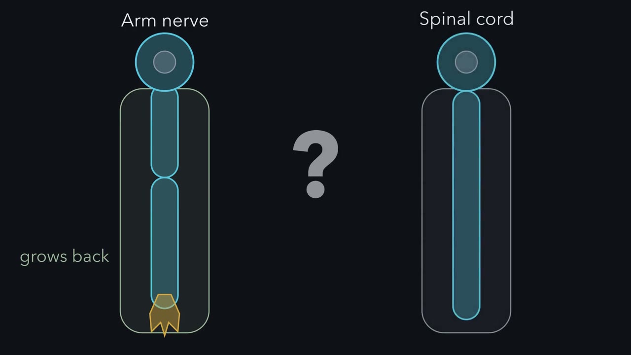 ▶ Watch One Cut, Two Fates · 2:07
Read the transcript of “One Cut, Two Fates”
Film transcript: One Cut, Two Fates
Cut a nerve in your arm, and its axons can grow back. Injure your spinal cord, and they mostly do not. Why? A cut splits each axon in two, and the two pieces have different fates. The piece cut off from its cell body breaks down, in a limb nerve and in the spinal cord alike. The attached piece can grow a new tip, depending on the neuron and on what surrounds it. In a mouse nerve, the cut-off piece looks normal for a day and a half to two days. Then it falls apart. Its debris is cleared quickly in a nerve, and slowly in the brain and cord. Meanwhile, after a delay, the attached piece may regrow. In people that is often about a millimetre a day.
Or it stalls, as many central axons do. A sensory neuron in a spinal ganglion offers a test. It has one nucleus and two branches. Injure each branch, in separate experiments. Which one regrows? The branch in the nerve regrows. The branch in the cord mostly does not. Same kind of neuron, different outcome. Is it the tissue, or what each injury tells the cell body?
Test the second. Cut the nerve branch first, and the cord branch regrows better later. This is the conditioning lesion. So the neuron's own state matters, and the tissue may too. So here is the one idea. One cut, two fates. The cut-off piece destroys itself, by a program it carries. The attached piece tends to regrow where the neuron's drive to grow outweighs the resistance of the tissue around it.
So ask two things of any cut. What does the cut-off piece do? And at the attached piece, which side wins?
From the film
- A cut splits an axon into two pieces with different fates.
- The piece cut off from its cell body destroys itself by a program it carries.
- The attached piece tends to regrow where the neuron's drive to grow outweighs the resistance of the surrounding tissue.
Cut a nerve in your arm and its axons can grow back. Injure your spinal cord and they mostly do not, which is why such an injury usually leaves a lasting loss of movement or feeling. Is the fault in the neurons, or in what surrounds them?
The axon is the long process that carries a neuron’s output; the cell body holds the nucleus and makes most of the cell’s proteins.
One cut, two pieces
A cut splits each axon in two. The distal piece, beyond the cut, is separated from its cell body. The proximal piece is still joined to it. The distal piece breaks down, in a limb nerve and in the spinal cord alike. The proximal piece may grow a new tip.
These responses overlap, and they run on different clocks.
| When | What happens |
|---|---|
| The first day and a half to two days (mouse sciatic nerve) | the distal piece looks normal (the latent phase) |
| Then | the distal piece falls to pieces |
| Days to weeks in a peripheral nerve; far longer, and incompletely, in the brain and spinal cord | the debris is cleared |
| After a delay, overlapping the clearing, for weeks to months or longer | the proximal piece may regrow, often at around a millimetre a day in humans (the rate depends on the nerve and the injury), or it stalls, as many central axons do |
So the lesson has two questions. Why does the distal piece die? And when can the proximal piece regrow?
The one idea to carry through this lesson. One cut, two fates. The piece cut off from its cell body destroys itself by a program it carries. The piece still attached tends to regrow where the neuron’s drive to grow outweighs the resistance of the tissue around it.
What the rule covers. It is written for mature mammalian axons broken by a cut or a severe crush, with the cell body surviving. It is not about the death of whole neurons, or about developmental pruning in general (see the neurotrophic hypothesis). The first half also holds in flies, and it runs in peripheral nerve and in the brain and spinal cord alike, though the timing differs. The second half is a rule of thumb: drive and resistance are not measured quantities. Fish regrow many central axons; in the rule’s terms, the balance there favours growth.
The cut-off piece destroys itself
Read the transcript of “The Stump That Kills Itself”
Film transcript: The Stump That Kills Itself
Why does a cut-off axon fall apart? Is it starving, or following orders? In 1850, Augustus Waller saw cut frog nerve fibres break up beyond the cut. He read it as a matter of nutrition. If it is starving, only replacing what it lacks should save it. If it is a program, deleting one part should stop it. What mouse would decide it?
One missing a part of the program, whose cut axons stay whole. But the first clue came by accident. In the slow mouse, cut axons last two weeks or more. Its mutation makes an odd fusion with an enzyme that makes NAD+. Remember that enzyme. So it might replace what the stump lacks. That alone cannot prove a program. Then came the deciding mouse. Mice lacking an ordinary gene, Sarm1, kept cut axons intact for a long time. The stump carries a program.
Why is a long axon so exposed? Picture a metre-long axon, 10 micrometres wide, holding about a thousand times the volume of a cell body 50 micrometres wide. It depends on deliveries from that body. The key delivery is NMNAT2, the least stable enzyme of its family. It turns NMN, nicotinamide mononucleotide, into NAD+.
The slow mouse's fusion protein is a longer-lived NMNAT. It keeps that job going after a cut. After a cut, it stops arriving. What happens to NMN, and to NAD+? NMN piles up, and NAD+ plus falls. Their ratio climbs as the enzyme fails. SARM1 senses that ratio. It is a ring of eight, held off while NAD+ Plus is bound. In the leading model, NMN competes for that site. Its binding shifts the outer domains and frees the TIR domains. The freed TIR domains come together into an enzyme that cleaves NAD+. In this model, each loss may push the ratio higher, and the slow drift becomes a crash.
Energy fails, calcium rises, and calpains, proteases switched on by calcium, cut the cytoskeleton. Mice lacking NMNAT2 from the start die around birth. Their growing axons stall. Remove Sarm1 too. What happens? Their axons grow, and the mice survive. Losing NMNAT2 works by tripping SARM1.
And it is not apoptosis. The stump degenerates even when caspases are blocked. So the cut-off piece is not simply starving. It carries a switch that waits for the deliveries to stop.
From the film
- The cut-off stump is not simply starving, because deleting one gene, Sarm1, keeps cut axons intact for a long time.
- NMNAT2 is a short-lived enzyme that must keep arriving from the cell body, and after a cut its loss lets SARM1 destroy NAD+.
- Energy then fails, calcium rises and calpains cut the cytoskeleton, and this is not apoptosis.
In most mice, cut sciatic nerve axons fragment within two days. In one strain they stay intact for two weeks or more.
In a paper published in 1850, the physiologist Augustus Waller described cutting nerves that serve the frog’s tongue and following the fibres under the microscope. Over the following days the fibres beyond the cut broke up, while the parts still joined to the brain or spinal cord stayed normal. Breakdown of the distal stump is still called Wallerian degeneration. Waller read it as a matter of nutrition: a fibre depends on a centre for its upkeep. That is true, and it suggests a passive death, the way a severed branch withers. Starve the stump and it fails.
Design problem
After a nerve is cut, is the degeneration of the distal stump passive starvation, because it has lost its supply from the cell body, or an active program of self-destruction? Design a genetic test, and say what result would decide it.
Saved in this browser only.
One way to do it
Starvation and a program make different predictions. If the stump is starving, no change to a single gene of the neuron should save it for long, short of replacing what it lacks. A program has parts, and removing a part should stop it.
So make mouse lines that each lack one candidate gene, cut the sciatic nerve, and examine the distal stump at one, three, seven and fourteen days, by electron microscopy and by testing whether the nerve still conducts. If deleting a single gene keeps cut distal axons intact for weeks, while normal axons fragment in a day or two, the stump was not simply starving. It was executing a program, and the gene you deleted drives it. To show that the gene acts in the neuron and not in the cells around it, cut the axons of mutant neurons in culture, away from glia and immune cells, or delete the gene only in neurons.
The slow mouse, and a knockout that copies it
The first such mouse came by accident. In 1989 a substrain of C57BL mice was described in which cut sciatic nerve axons degenerated very slowly. It became known as Wlds, for slow Wallerian degeneration. In 2001 Michael Coleman’s group showed that the mutation fuses the start of a ubiquitination factor, Ube4b, to NMNAT1, an enzyme that makes NAD+ from nicotinamide mononucleotide (NMN). Keep that enzyme in mind: it is the clue to the whole program. Adding the fusion gene to normal mice protected cut axons, and more of it protected them more.
Then came a loss-of-function result of the kind the design problem asks for. In 2012 Marc Freeman’s laboratory and its collaborators reported that flies lacking a protein called dSarm keep severed axons for weeks, and that mice lacking its counterpart, Sarm1, keep cut axons intact for a long time, in the nerve and in culture. Deleting one ordinary gene gave protection like that of the odd fusion. The stump is not simply starving. Losing its supply sets off a program it carries, and the program dismantles it.
The program: a switch that waits for the deliveries to stop
Start with the problem the axon has. In an adult human, the axon of a motor neuron that moves a toe runs roughly a metre from the spinal cord. An axon that long and about 10 micrometres across can hold hundreds of times the volume of its cell body (about 1,200 times for a body 50 micrometres across, about 150 times for one 100 micrometres across). Many of its essential proteins are made in the cell body and carried down, though axons make some locally. The far end depends on a steady flow of deliveries, and the program is how an axon responds when that flow stops.
The current model, built by several laboratories, has four steps.
- A delivery that must keep coming. Nicotinamide mononucleotide adenylyltransferase 2 (NMNAT2) is the NMNAT isoform that works in axons. Like every NMNAT, it joins NMN to the adenylyl (AMP) part of ATP to make NAD+, a coenzyme the axon needs to make ATP. In 2010 Jonathan Gilley and Coleman showed that NMNAT2 is the least stable NMNAT isoform and must be replenished constantly by transport from the cell body (long-distance signaling). After a cut the deliveries stop, and NMNAT2 in the stump falls below a threshold before the axon breaks up. Blocking the proteasome, the cell’s protein-disposal machine, slows its loss (ubiquitin and proteolysis).
- A gauge that reads the gap. As NMNAT2 disappears, its substrate, NMN, piles up and its product, NAD+, starts to fall. So a rising ratio of NMN to NAD+ signals that NMNAT2 is failing, though NMN supply and NAD+ use also move it. SARM1 senses that ratio. It waits in the axon as a ring of eight, held shut by its ARM domain, the sensor part, while NAD+ is bound there. In the leading model, NMN competes for the same site, and a rise in NMN relative to NAD+ switches SARM1 on.
- An executioner. The TIR domain of SARM1, its enzyme part, cleaves NAD+. In 2015 the laboratories of Aaron DiAntonio and Jeffrey Milbrandt showed that forcing SARM1 TIR domains together is enough to destroy axonal NAD+ and trigger degeneration, and in 2017 that the TIR domain itself cleaves NAD+. Each NAD+ it destroys pushes the NMN-to-NAD+ ratio higher, so in this model the switch may feed itself, a proposed positive feedback. The slow drift becomes a crash: NAD+ falls fast, and ATP falls after it.
- Collapse. As energy fails, calcium rises inside the axon. Calpains, proteases switched on by calcium, cut the cytoskeleton, and the axon fragments, often after swelling into beads called spheroids.
Think of a dead-man’s switch, the kind that stops a train when the driver’s hand leaves the handle. The cell body is the hand, and every delivery of NMNAT2 says “still attached.” When the deliveries stop, the switch trips. The Wlds protein works because it is a longer-lived source of NMNAT activity that keeps the handle pressed after the cut. Deleting Sarm1 removes what the switch sets off. The analogy breaks in one place: a real switch trips at once, while this gauge drifts for hours, and how long the latent phase lasts in a living nerve depends on more than how fast NMNAT2 disappears.
Rule to keep: a cut axon loses NMNAT2, NMN builds up, SARM1 switches on and cleaves NAD+, and the axon falls apart; Wlds works upstream of SARM1 and loss of Sarm1 removes the executioner, so either holds the stump for weeks. This is a sketch of the logic in relative units, not a measurement, and it shows the leading model, not the last word.
Predict. Use the four steps to say which cut axons survive: (a) Wlds; (b) Sarm1-null; (c) Sarm1-null neurons in culture, given back a SARM1 whose TIR domain cannot cleave NAD+. One more case reaches back into development. Mice lacking NMNAT2 from the start have axons that stall as they grow, and the mice die around birth. (d) What happens, without any cut, in mice lacking both NMNAT2 and Sarm1?
Check your prediction
| Genotype | Cut? | What happens | What it places in the sequence |
|---|---|---|---|
| Wild type | yes | intact for a day and a half to two days in mouse nerve, then fragments | the latent phase, then the crash |
| Wlds | yes | intact for two weeks or more | a longer-lived NMNAT acts upstream |
| Sarm1-null | yes | intact for a long time | SARM1 is required |
| Sarm1-null plus SARM1 that cannot cleave NAD+ | yes, in culture | keeps its NAD+ and stays intact | SARM1’s NAD+-cleaving activity is what kills |
| NMNAT2-deficient and Sarm1-null | no | axons grow and the mice survive | NMNAT2 loss acts through SARM1 |
The last row is the decisive one (Gilley and colleagues, 2013 and 2015). Removing the switch rescues axons that lack the delivery, so losing the delivery works by tripping the switch. These are developing axons, outside the rule’s mature-axon scope. In established, uncut axons of cultured neurons, depleting NMNAT2 is also enough to make them degenerate (Gilley and Coleman, 2010).
This is not apoptosis, the death program of a whole cell (see Bcl-2 and caspases). Injury-induced degeneration of the stump proceeds when caspases are blocked or missing, while the developmental pruning of axons deprived of trophic factor needs them. The program also runs locally, in a stump with no nucleus. Stress kinases such as DLK and c-Jun N-terminal kinase (JNK) take part after injury (stress signals), and where they fit in the sequence is still being worked out.
Clearing the way
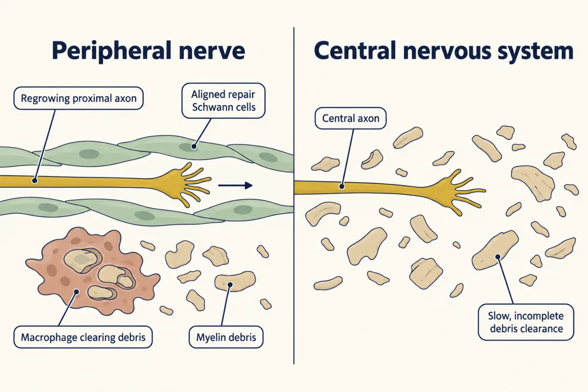
As the distal piece falls apart, its debris is cleared. Clearing helps a new axon use the path, though regrowth need not wait for it to finish. Here peripheral nerve and the central nervous system part company.
In a peripheral nerve, Schwann cells, the glia of peripheral nerve, become repair cells (a switch run by the transcription factor c-Jun). They do three jobs: they help break down myelin, they recruit macrophages from the blood to clear the rest, and they line up into tubes that guide regrowing axons back toward their targets. In the central nervous system, myelin debris is cleared far more slowly and incompletely.
The attached piece: drive against resistance
Read the transcript of “Drive Against Resistance”
Film transcript: Drive Against Resistance
The attached piece can regrow. So why does it stall in the brain and spinal cord? In 1981, Samuel David and Albert Aguayo changed mainly the tissue. In adult rats, they bridged the medulla to the spinal cord with peripheral nerve. If adult central neurons cannot grow long axons at all, nothing grows. If the tissue stops them, what should you see?
Central axons grew about 30 millimetres along the grafts. Back in central tissue, they soon stopped. Now keep the central tissue, and change mainly the neuron. In 2008, Zhigang He's laboratory deleted PTEN from retinal ganglion cells in mice. PTEN is a brake on cell growth. Then they crushed the optic nerve, which is central tissue. Will the axons cross?
Some do. They come from a minority of the neurons, but far more than normal. So it is not a gate. Each experiment got growth while changing mainly one side. It is a balance. Picture neurons spread out by drive, and a line set by the tissue. Only neurons past the line regrow. Lower the line, or push the neurons up, and some get through. Here the line sits in the thin tail. So doing both passes more than the two gains added. With another spread, the gains might only add. This is a model, not data. Myelin proteins such as Nogo collapse growth cones. Yet mice lacking three of them barely regrow more. Why?
One way to see it: most neurons may sit far below the line. Lowering it a little moves few across. Other barriers remain, too. The scar resists, but also helps. In mice, removing its astrocyte border reduced stimulated regrowth. Change the scar, do not remove it. And regrowth is not recovery. At a steady millimetre a day, a metre takes about a thousand days, nearly three years. Over that long a time, Schwann cells lose their repair state, and distant targets are hard to reach.
Blocking SARM1 may help failing axons that are still attached. A fully severed stump cannot rejoin its cell body on its own. For regrowth, no single fix is enough. None yet restores function reliably in people. One cut, two fates. A program for the cut-off piece, and a balance of drive against resistance for the attached one.
From the film
- Central axons grew about 30 millimetres along peripheral nerve grafts but stopped in central tissue, so the tissue resists regrowth.
- Deleting PTEN in mouse retinal ganglion cells let some axons cross a crushed optic nerve, so the neuron's own drive matters too.
- Regrowth is a balance, no single fix is enough, and regrowth is not the same as recovery.
The proximal stump forms a growth cone, the motile tip that leads a growing axon.
Nature has already run one comparison. A sensory neuron in a spinal (dorsal root) ganglion has one cell body, one nucleus and two branches. One branch runs out in a peripheral nerve to the skin or muscle. The other enters the spinal cord. Injure the peripheral branch and it regrows; injure the central branch inside the spinal cord and it mostly does not. Same neuron, same nucleus, different outcomes. The difference could lie in the tissue around each branch, or in what each injury tells the cell body to do. The conditioning lesion tests the second: cutting the peripheral branch before the central branch is injured raises the central branch’s regrowth. So the neuron’s state matters, and the tissue may too. To weigh each one, change mainly one side at a time.
Change mainly the tissue
In 1981 Samuel David and Albert Aguayo used peripheral nerve, which holds Schwann cells and none of the glia of the central nervous system, as a test path. They grafted segments of it into adult rats as bridges, with one end in the medulla and the other in the spinal cord.
Predict. If adult central neurons can no longer extend long axons at all, what grows along the graft? If they still can and central tissue stops them, what do you expect along the graft, and where the axons leave it?
Check your prediction
Axons from central neurons at both levels grew about 30 millimetres along the grafts. Where they re-entered central tissue, they stopped within a short distance. Some adult central neurons can regrow when the path is peripheral; their central surroundings do much to stop them.
Change mainly the neuron
Now leave the central surroundings in place and change mainly the neuron. In 2008 Zhigang He’s laboratory deleted phosphatase and tensin homolog (PTEN) in mice from retinal ganglion cells, the central neurons whose axons form the optic nerve. PTEN is a brake on the mTOR pathway, the kinase pathway that drives protein synthesis and cell growth. Then they crushed the optic nerve, which is central tissue.
Predict. If the tissue alone decides, what happens to these axons at the crush? If the neuron’s drive counts too?
Check your prediction
Axons regenerated through the crushed optic nerve, with the central surroundings left in place. They came from a minority of the neurons, but far more than normal. So the surroundings are not the whole answer.
Put the two experiments side by side and the opening question has its answer. Two things hold central axons back: the tissue around them blocks regrowth, and adult central neurons turn their own growth program down. Neither is a gate that must open first, since each experiment got growth while changing mainly one side, though a graft also sends signals to the neurons and PTEN loss also helps them survive. It is a balance.
What resists
Two features of the central lesion have been studied most as resistance: proteins in central myelin, and the scar that forms around the injury.
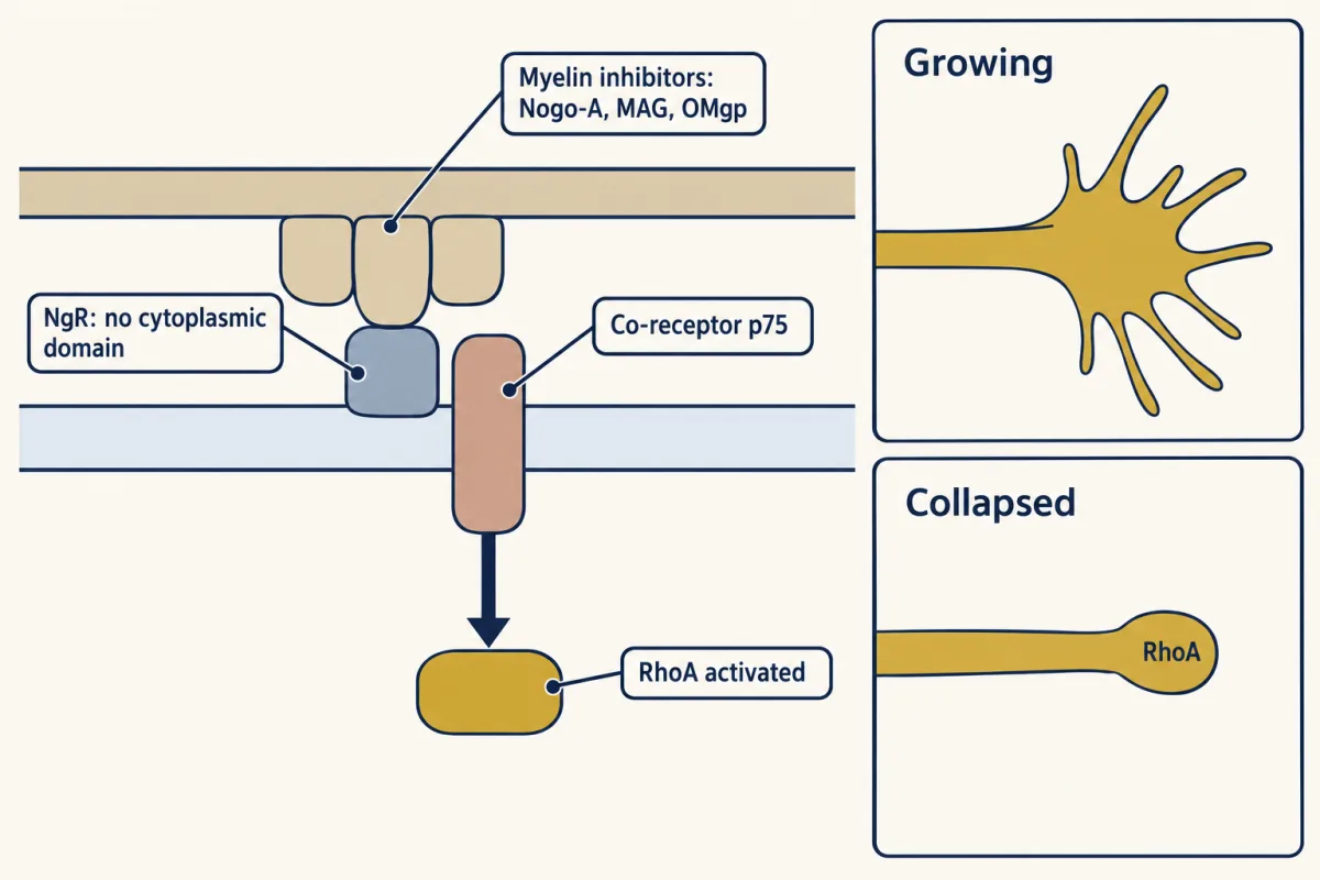
Myelin inhibitors. In 1988 Pico Caroni and Martin Schwab reported membrane proteins from central myelin that block neurite growth, and an antibody, IN-1, that neutralized them. The larger antigen was later cloned as Nogo-A. Nogo-A’s Nogo-66 domain, myelin-associated glycoprotein (MAG) and oligodendrocyte myelin glycoprotein (OMgp) all bind the Nogo receptor, NgR, on the growth cone, which signals through partner proteins such as p75 (p75) to activate RhoA and collapse the growth cone.
The glial scar. Reactive astrocytes form a border around the lesion, and the lesion is rich in chondroitin sulfate proteoglycans, matrix proteins with long sugar chains, made by these astrocytes and by other cells such as NG2 glia. Digesting their sugar chains with the bacterial enzyme chondroitinase ABC improved axon growth and recovery in rats with spinal cord injury. The astrocyte border itself is not the wall it was once thought to be: in mice, preventing or removing it did not let cut axons regrow, and it reduced regrowth that had been stimulated with growth-promoting treatments. The scar also limits the spread of damage, so the aim is to change it, not remove it.
Why each fix is only partial
Predict. Myelin inhibitors really do collapse growth cones. Yet mice lacking all three, Nogo, MAG and OMgp, show little or no extra regeneration of cut axons, though spared fibres may sprout more. How can a real inhibitor matter so little when it is removed?
Check your prediction
One way to picture it is as a threshold. Spread the neurons out by how strongly each drives growth, and let the tissue set a line; only neurons whose drive passes the line regrow. If most adult central neurons sit far below the line, removing one part of the resistance lowers it a little and moves few of them across. Myelin inhibitors are one barrier among several, and the others remain.
The table works the threshold picture with made-up settings: drive spread in a bell curve, with the line far out in its upper tail. Its numbers belong to no experiment; in real peripheral grafts, only some central neurons grew in.
| Change, in the illustrative model | Neurons regrowing, of 100 |
|---|---|
| None: adult central neuron, central tissue | about 1 |
| Lower the resistance a little | about 2 |
| Raise the drive | about 10 |
| Both | about 16 |
| Lower the resistance a lot (an arbitrary large step) | about 50 |
Lowering the resistance a little adds about 1 regrowing neuron and raising the drive adds about 9, so if the gains simply summed, both would add about 10. In this model, both add about 15. That is because the line sits in the thin tail of the bell curve, where each step toward the middle passes more neurons than the last. With a different spread, or with changes large enough to reach most neurons, the gains might only add, or add up to less. So the model is a reason to test combinations, not a promise that they work better. Nor do the gains compare the two kinds of change: here the resistance step is about a quarter of the spread of drive and the drive step about one spread.
Regrowth is not recovery
Useful function returns only when a regrowing axon reaches the right target and forms working connections, and regrowth is slow. Recall that the axon to a toe runs about a metre.
Predict. An axon regrows at around a millimetre a day. Ignoring the delay before growth starts, roughly how long does it take to cover 50 millimetres, 300 millimetres and a metre?
Check your prediction
About 50 days (under two months), about 300 days (roughly ten months) and about 1,000 days (nearly three years).
Even in peripheral nerve, recovery is often incomplete: the repair state of Schwann cells does not last as long as slow regrowth over a long distance needs, and after severe injuries some axons never reinnervate (reconnect to) their targets.
Two problems, two fixes
The two fates need different fixes. The first is to keep axons from degenerating. Small molecules that inhibit SARM1 have protected axons in cultured neurons and in animal models, and raising NAD+ with precursors such as nicotinamide riboside or nicotinamide mononucleotide (NMN) is being explored. The model contains a catch. Both precursors pass through NMN on the way to NAD+ (nicotinamide riboside is first converted to NMN), so in a cut stump a precursor can raise NAD+ only while NMNAT is still there to turn NMN into NAD+. Once the stump has lost nicotinamide mononucleotide adenylyltransferase 2 (NMNAT2), extra NMN could add to the signal that switches SARM1 on. Which precursor reaches the axon, and in what amount, matters. A fully severed stump cannot rejoin its cell body on its own however long it survives, so the strongest case for these drugs is slower damage, such as some peripheral neuropathies, where failing axons are still connected.
The second problem is regrowth after the axon is lost, and no single fix is enough. Blocking Nogo signalling, digesting the scar, raising intrinsic growth and supplying bridges each help in rodents, usually partly, and none yet restores function reliably in people.
For now, acute care for spinal cord injury centres on surgery to relieve pressure on the cord and stabilize the spine, supportive care, and rehabilitation. High-dose methylprednisolone, a steroid, was once widely given in the first hours, but many current guidelines no longer recommend it routinely, because its benefit was small and uncertain and it carries risks.
Putting it together
| Experiment | Animal | Observed | What it supports | What it does not establish |
|---|---|---|---|---|
| Cut nerves of the tongue (Waller, 1850) | frog | fibres beyond the cut break up; attached parts stay normal | the cut-off piece degenerates, the attached piece survives | whether the death is passive or a program |
| Wlds mutation, and the added fusion gene (1989, 2001) | mouse | cut axons intact for two weeks or more; more of the gene, more protection | a longer-lived NMNAT holds off degeneration | that normal axons run a program of their own |
| Loss of dSarm or Sarm1 (2012) | fly, mouse | severed axons intact for weeks in flies and for a long time in mice | an active program, which needs SARM1 | how SARM1 is switched on |
| Forced TIR association; TIR cleaves NAD+ (2015, 2017) | neurons; the TIR domain on its own | NAD+ destroyed, axons degenerate | SARM1’s enzyme is the executioner | what switches it on normally |
| SARM1 that cannot cleave NAD+, in Sarm1-null neurons | cultured mouse sensory neurons | neither NAD+ loss nor fragmentation restored | the NAD+-cleaving activity is required | — |
| NMNAT2 loss, with and without Sarm1 (2013, 2015) | mouse | without NMNAT2, developing axons stall and the mice die around birth; without both, they survive | NMNAT2 loss acts through SARM1 | how long the latent phase lasts in a living nerve |
| Caspases blocked or missing | — | the stump still degenerates | it is not apoptosis | — |
| Peripheral nerve bridge (1981) | adult rat | axons grow about 30 mm along the graft and stop soon after re-entering central tissue | some central neurons can regrow on a peripheral path | that the neuron’s state does not matter |
| PTEN deleted (2008) | mouse retinal ganglion cells | a minority, but far more than normal, regrow through the crushed optic nerve | the neuron’s drive limits regrowth | that the tissue does not |
| Conditioning lesion | sensory neurons | central branch regrows better | the neuron’s state can be raised | — |
| Nogo, MAG and OMgp all deleted | mouse | little or no extra regeneration; spared fibres may sprout more | myelin inhibitors are one barrier among several | that they do not resist at all |
| Chondroitinase ABC | rat, spinal cord injury | better axon growth and recovery | lesion proteoglycans resist | that the recovery came only from regrowth at the lesion |
| Astrocyte border prevented or removed | mouse | no regrowth; stimulated regrowth reduced | the border also supports growth; resistance is what the tissue allows overall | — |
Read down the table and the idea holds: one cut, two fates. A program for the cut-off piece, and a balance of drive against resistance for the attached one.
Going deeper
Who found the slow mouse
Waller’s 1850 paper appeared in the Philosophical Transactions of the Royal Society, and the two nerves he cut were the glossopharyngeal and the hypoglossal. The slow mouse was described by Lunn, Perry, Brown, Rosen and Gordon, and crosses reported in 1990 traced it to a single dominant gene. In 1998 Michael Coleman, Hugh Perry and colleagues found that the gene’s region on mouse chromosome 4 holds an 85-kilobase stretch of DNA in three tandem copies, the rearrangement that makes the fusion gene.
Receptors for myelin inhibitors
NgR has no cytoplasmic domain of its own. Its partners are LINGO-1 with p75, or with TROY in neurons that lack p75. A second receptor, PirB, also binds Nogo-66, myelin-associated glycoprotein (MAG) and OMgp.
Spheroids [Deppmann lab]
Working with sympathetic neurons from newborn mice, the Deppmann laboratory first studied spheroids in axons after withdrawal of nerve growth factor, a setting in which, unlike injury, caspases are needed. The axons stayed intact for about 18 hours (the latent phase) and then came apart over about 3 hours (the catastrophic phase). Toward the end of latency, calcium rose inside the axons, and calcium-rich spheroids grew and ruptured, releasing their contents. Medium from such axons hastened the catastrophic phase in others (Yong et al., J Neurosci 2019). After injury, the same group reported that spheroid growth depends on Rho and actin, that Wlds neurons and added NAD+ suppress it, and that Sarm1-null and DR6-null neurons still form spheroids that rupture (Yong et al., Sci Rep 2020). DR6 is a member of the tumor necrosis factor (TNF) receptor family; the group had found that cut sciatic nerve axons in mice lacking DR6 were preserved up to four weeks after injury (Gamage et al., Curr Biol 2017). The authors propose that spheroid rupture marks the transition from latency to catastrophic degeneration, and they fit it to the model above by suggesting that an early fall in NAD+, not caused by SARM1, drives spheroid formation, and that SARM1 then drives NAD+ down further. This is one laboratory’s culture work and a proposed ordering, and rupture alone does not commit an axon, since Sarm1-null axons rupture spheroids and still resist degeneration. Spheroids and calcium appear in both injury and developmental loss of axons, which raises the question of whether the two share mechanisms.
Your notes
Select any text in the lesson to highlight it or add a note to it. Your highlights and notes are saved in this browser and collected in your notebook.
This browser is not letting the site save anything (a private window or strict privacy settings), so highlights and notes will be lost when you leave the page.
How we measure it
Nerve transection with a time course
A peripheral nerve such as the mouse sciatic nerve is cut, and the distal stump is examined at set times by light and electron microscopy or by testing whether it still conducts. The time to fragmentation is the measure: about one and a half to two days in normal mice, weeks in Wlds or Sarm1-null mice.
Compartmented (microfluidic) neuron culture
Neurons are grown so that their cell bodies sit in one chamber and their axons grow through narrow channels into another. Axons can then be cut, deprived of growth factor or treated with a drug on their own, which shows whether a process runs locally in the axon.
Forward genetic screen in Drosophila
Flies are mutagenized at random, and their offspring are screened for a phenotype, here severed axons that fail to degenerate. Because the screen assumes nothing about which gene is involved, it found dSarm, a protein no one had linked to axon death.
Peripheral nerve bridge graft with tracing
A segment of peripheral nerve is grafted into the central nervous system, and a tracer taken up by axons in the graft is carried back to their cell bodies. Labelled cell bodies show which neurons grew into the graft, which tests whether central neurons can regrow in a peripheral environment.
Practice questions on this lesson (3) → MCAT-style, original, each with an explanation
Where this lesson fits
Before thisReview first
- The Growth Cone: The Tip That Steers the AxonThis course · Lesson 3
You need the growth cone's steering and actin dynamics from there to follow how the attached stump regrows.
- Pruning Without Dying: Axons That Cut Themselves BackThis course · Lesson 34
It shows caspase-dependent axon cutting without cell death, the contrast this lesson draws with injury-induced degeneration.
- Myelination: Wrapping the Axon So the Signal JumpsThis course · Lesson 44
You need the glial wrapping of axons from there to understand myelin debris and myelin inhibitors after a cut.
After thisStudy next
- Two Places the Adult Brain Still Makes NeuronsThis course · Lesson 47
It asks whether the adult brain can replace lost neurons, the other half of repair after the axon repair problem here.
Elsewhere on the siteFilms, simulations and method films in other courses
- Reading annexin V and PISimulation · Cell Biology
It shows how apoptosis is read with annexin V and PI, the programmed death this lesson distinguishes from stump degeneration.
- A Current That Light Can Use UpFilm · Sensory Systems
It shows how a neuron's own signalling loss leads to cell death and degeneration.
Suggested by an AI model from the lessons themselves, then checked. How we use AI
Finish line
Test yourself
8 questions
Practice and MCAT-style, with explained answers.
Next · Lesson 47
Two Places the Adult Brain Still Makes Neurons
Inject an adult rat with a label that marks newly made DNA. A month later a labelled cell sits in its hippocampus. Is it a new neuron, how would you know, and do you make them too?
Continue →