Developmental Neuroscience · Unit 7: Survival and competition
The Signal from the Target Travels Home
Rewritten on : new text, figures, flashcards and film. Your notes and highlights stay, but the text around them may have moved.
A neuron hears its survival signal at the far end of its axon. How does the message get home?
Pause when the reading reaches a film, and offer to play it. Saved on this device.
Watch the film now, or keep listening
Builds on:The Neurotrophic Hypothesis TestedNeurotrophins, Trk Receptors and p75The Death Machinery in NeuronsWhy these?
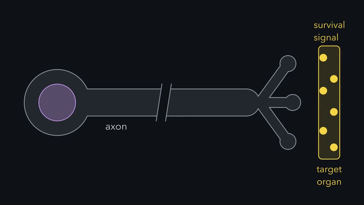 ▶ Watch Still On When It Gets There · 2:45
Read the transcript of “Still On When It Gets There”
Film transcript: Still On When It Gets There
A neuron hears its survival signal at the far end of its axon. How does the message get home? A sympathetic neuron collects NGF at axon terminals that can sit centimetres from its cell body. The terminal hears the factor. But the nucleus decides whether the cell lives. In these young neurons, dying needs new genes switched on in the nucleus. So a survival signal from the tip has to be able to act on the cell body. Here is the idea to carry through. A target keeps a neuron alive only if its signal makes the trip home. And it must be able to signal when it gets there. On when it arrives, not necessarily on every second of the trip. Binding at the tip is not enough.
So could the signal simply drift home, by diffusion? Diffusion time grows with the square of the distance. For a typical soluble protein and an axon one centimetre long, how long would the trip take? Roughly two months. Over a few centimetres, many months.
Worse, many signals are proteins carrying a phosphate. Enzymes called phosphatases keep removing it. Suppose a phosphate lasts about ten seconds. Then the active signal falls to about a third every ten micrometres from the tip. A centimetre is a thousand of those steps. The rates are illustrative, but the conclusion holds. It does not arrive late. It arrives switched off.
So the signal needs a vehicle or a relay. And it must be able to signal when it arrives. One candidate fits both needs. At the tip, NGF binds its receptor, TrkA. Ligand and active receptor are pulled into the cell together, in a vesicle called the signaling endosome. It can keep signaling while carried. The motor protein dynein drags it along microtubules toward the cell body. At roughly a micrometre each second, a centimetre takes a few hours. So what arrives must still be able to signal, after hours on the way. Carrying alone is not enough. This model is favoured. But is it proven? Hold that question. Takeaway: a survival signal must make it home still able to signal.
From the film
- A survival signal received at the axon terminal has to be able to act on a cell body that can sit centimetres away.
- Diffusion alone would take about two months over a centimetre, and an active signal would fade on the way, with illustrative rates.
- In the favoured signaling endosome model, dynein carries the NGF-TrkA complex home, and what arrives must still be able to signal.
A sympathetic neuron’s cell body can sit centimetres away from the axon terminals where it collects nerve growth factor (NGF). The terminals receive the signal. The nucleus decides whether the cell lives.
You met the neurotrophic hypothesis in that lesson: neurons are overproduced, targets make a limited amount of a survival factor, and neurons that do not get enough die. Stated that way, the hypothesis skips a step. NGF is released near the axon terminals, but in these young neurons, the death that follows loss of NGF needs new genes switched on in the nucleus (the lesson on death machinery in neurons), so a survival signal from the terminal has to be able to act on the cell body. Something has to cross the distance between them. This lesson asks what crosses, how we know, and why it matters for which target factor keeps a neuron alive.
The one idea to carry through this lesson. A target keeps a neuron alive only if its signal makes the trip home and is able to signal when it gets there. Binding at the tip is not the test. The sharpest case comes near the end: two factors switch on the same receptor at the tip, and only one keeps the neuron alive.
What the rule covers.
- Which neurons. Target-dependent neurons with long axons, during their death window: sympathetic neurons (autonomic neurons whose axons run from ganglia out to organs), and the sensory neurons that carry TrkA, the receptor NGF binds. Much of the mechanistic evidence comes from rat and mouse sympathetic neurons in compartmented culture.
- Which effect. Survival supported from the axon terminal. Local effects, such as axon growth where the factor is, do not need the trip, and a factor given straight to the cell body bypasses it.
- What it does not say. What carries the signal: whether NGF itself has to travel, and whether a faster signal also contributes, are still open. Nor does it say that every signal that arrives is enough.
Could it just drift?
The simplest idea is that something made at the tip diffuses to the cell body. The typical time to diffuse a distance grows with the square of that distance.
time ≈ (distance)² / (2 × D)
D, the diffusion coefficient, says how fast a molecule spreads. A soluble protein in cytoplasm has a D on the order of 10 square micrometres per second.
Predict. A sympathetic axon is 1 cm long, which is 10,000 micrometres. Roughly how long would a protein take to diffuse from the terminal to the cell body: seconds, hours, or much longer?
Check your prediction
Much longer. 10,000 squared is 10⁸, and 10⁸ / 20 is about 5 million seconds: roughly two months. Over a few centimetres, it would take many months.
Worse, the signal would be switched off on the way. Many signals inside a cell are proteins carrying an added phosphate, and enzymes called phosphatases keep removing those phosphates.
How far does an active signal get? Take a simple model: steady diffusion from the tip, with phosphates removed at a constant rate. Suppose a phosphate lasts about ten seconds on average. The active signal then fades over a distance of about √(D × lifetime) = √(10 × 10) = 10 micrometres: every 10 micrometres from the tip, the active fraction falls to about a third. A centimetre is a thousand such steps, so effectively nothing active arrives. With a phosphatase a hundred times slower, the fade distance is 100 micrometres, and the active fraction at 1 cm is still only about 10⁻⁴⁴. These rates are illustrative, not measurements for TrkA pathways. Protein molecules may still drift in, but inactive: the signal does not arrive late, it arrives switched off.
What a long-distance signal needs
So the signal needs a vehicle or a relay, and it needs two properties.
- It must still be able to signal when it arrives. A carried complex need not be active every second of the trip, but it must reach the cell body intact and able to signal. A relay must switch each stretch on in turn.
- It should keep the target’s message separate from signals the neuron picks up along the way. Keeping the ligand bound to its receptor is one way to do this. Whether the cell body reads which factor started a signal is not shown here. Whether nerve growth factor (NGF) itself must arrive is unsettled: the bead result in Going deeper hints that survival may not need it, but Ye and colleagues found NGF inside the cell bodies was needed (below).
A membrane vesicle holding the ligand still bound to its active receptor fits both. That is why the signaling endosome became the favoured model.
Here is the model. NGF binds TrkA at the axon terminal. TrkA is a receptor tyrosine kinase: an NGF dimer holds two TrkA molecules together, their kinases add phosphates to each other’s tyrosines, and the phosphorylated tails recruit signaling proteins. The ligand and the active receptor are pulled into the cell together in a small membrane vesicle, the signaling endosome. NGF stays bound inside the vesicle, while TrkA’s kinase tails face the cytoplasm outside it, so the complex can keep signaling while it is carried (the cell biology is in the lesson on endocytosis and signaling endosomes). The motor protein dynein drags it along microtubules toward the cell body. Once there, kinase pathways reach transcription factors such as cAMP response element-binding protein (CREB), which binds DNA and switches genes on.
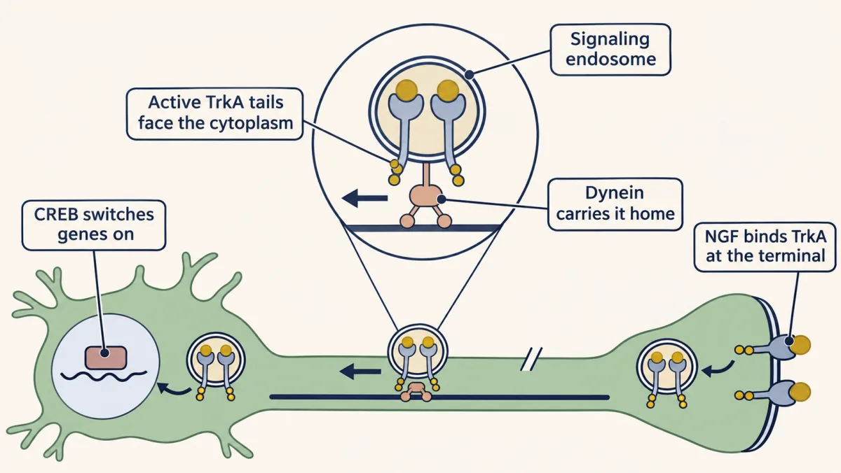
Dynein-driven endosomes move at roughly a micrometre per second, so a centimetre takes about three hours. That makes the first need sharp. Take an illustrative model in which the carried complex loses its ability to signal at a steady rate, for good, with nothing switching it back on. If it halves every hour, a three-hour trip is three halvings: (1/2)³ = 1/8 arrive able to signal.
| If the carried complex’s ability to signal halves every | Fraction still able to signal after a three-hour trip |
|---|---|
| 5 minutes | essentially none (36 halvings) |
| 1 hour | one in eight |
| 10 hours | about four in five |
Carrying alone is not enough. And favoured is not proven: the next two sections ask what the evidence shows.
The evidence that something travels
Read the transcript of “Courier or Relay?”
Film transcript: Courier or Relay?
Does a signal from the axon tip really keep the cell body alive? And what actually makes the trip? First, radioactive NGF placed in a target built up in the matching ganglion on the same side. In the chick, the motor neurons beside it stayed unlabelled. But label shows only where radioactivity went, not whether it mattered for survival. Robert Campenot's dish answered that. Sealed dividers split it into chambers whose fluids do not mix. Cell bodies sit in the centre. Their axons grow under the seal into side chambers. Give the cell bodies no NGF. Let their axons reach a chamber that has it. Do the cell bodies live?
Many survive. A signal from the distal axons, the far ends near the terminals, can keep the cell body alive. With NGF only on distal axons, CREB became phosphorylated in the nuclei. CREB is a transcription factor that switches genes on. That needed TrkA kinase activity in the cell body. But was that the same receptor, carried from the tip? So how did it travel?
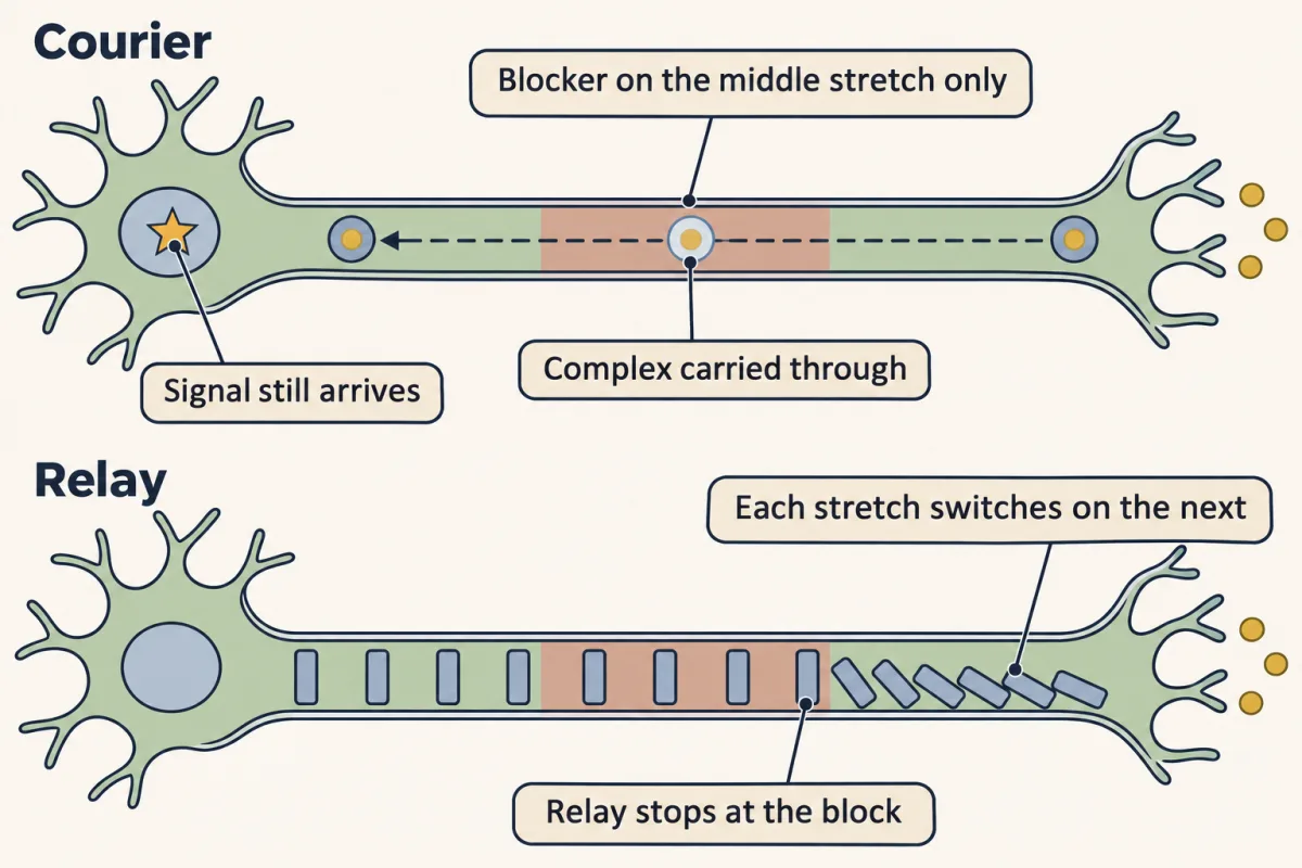
The NGF and TrkA complex itself is carried the whole way. Relay. Each stretch of axon switches on the next, like a row of dominoes. No one molecule makes the whole trip. Blocking dynein stops survival. Does that prove a courier?
No. If each relay step needed dynein, the relay would stop too. This block cannot tell them apart. So block TrkA's kinase only in the middle stretch, with an inhibitor applied only there. What does each model predict? A relay that needs TrkA kinase in each stretch would break at the gap. A carried complex could pass through and signal again. The signal still reached the cell bodies, and the neurons survived. NGF also had to be taken into the cell.
Two results keep the question open. TrkA in the cell bodies was phosphorylated within a minute, far faster than endosomes move. And NGF fixed to beads supported survival with little or no NGF transport. Takeaway: for survival, the evidence favours a courier. Whether a faster signal also helps is still open.
From the film
- In Campenot's compartmented cultures, NGF given only to distal axons can keep many cell bodies alive.
- A dynein block cannot tell a courier from a relay, but a middle-segment TrkA block that left survival intact favours a courier.
- Whether a faster signal also contributes is still open.
The first evidence was anatomical. In 1974, Hendry, Stöckel, Thoenen and Iversen injected radioactive, iodine-labeled nerve growth factor (NGF) into the eye of mice and found the label building up in the superior cervical ganglion on the same side, the sympathetic ganglion whose axons innervate the eye. In 1979, Judy Brunso-Bechtold and Viktor Hamburger did the embryonic version. They implanted a small gel pellet soaked in radioactive NGF under the leg skin of chick embryos at about ten days of incubation. Eight hours later, the lumbar dorsal root ganglia on the implanted side were labeled. The lateral motor columns, ventral roots and dorsal roots were not labeled, though motor axons ran into the same leg.
That selectivity is the point. Label leaking into the blood would reach both sides; uptake by any nerve would label the motor column. Instead it went to the sensory neurons, which respond to NGF. Autoradiography locates radioactivity. It cannot tell you whether NGF arrived intact, whether it was active, or whether its arrival had anything to do with survival.
Robert Campenot’s dish answered the survival question. In 1977 he described a culture in which a divider, sealed to the dish, separated a central chamber from side chambers. Rat sympathetic neurons were plated in the centre, and their axons grew under the seal into the side chambers. The fluids did not mix. Now NGF could be given to the distal axons, the far ends near the terminals, and not to the cell bodies, or the reverse.
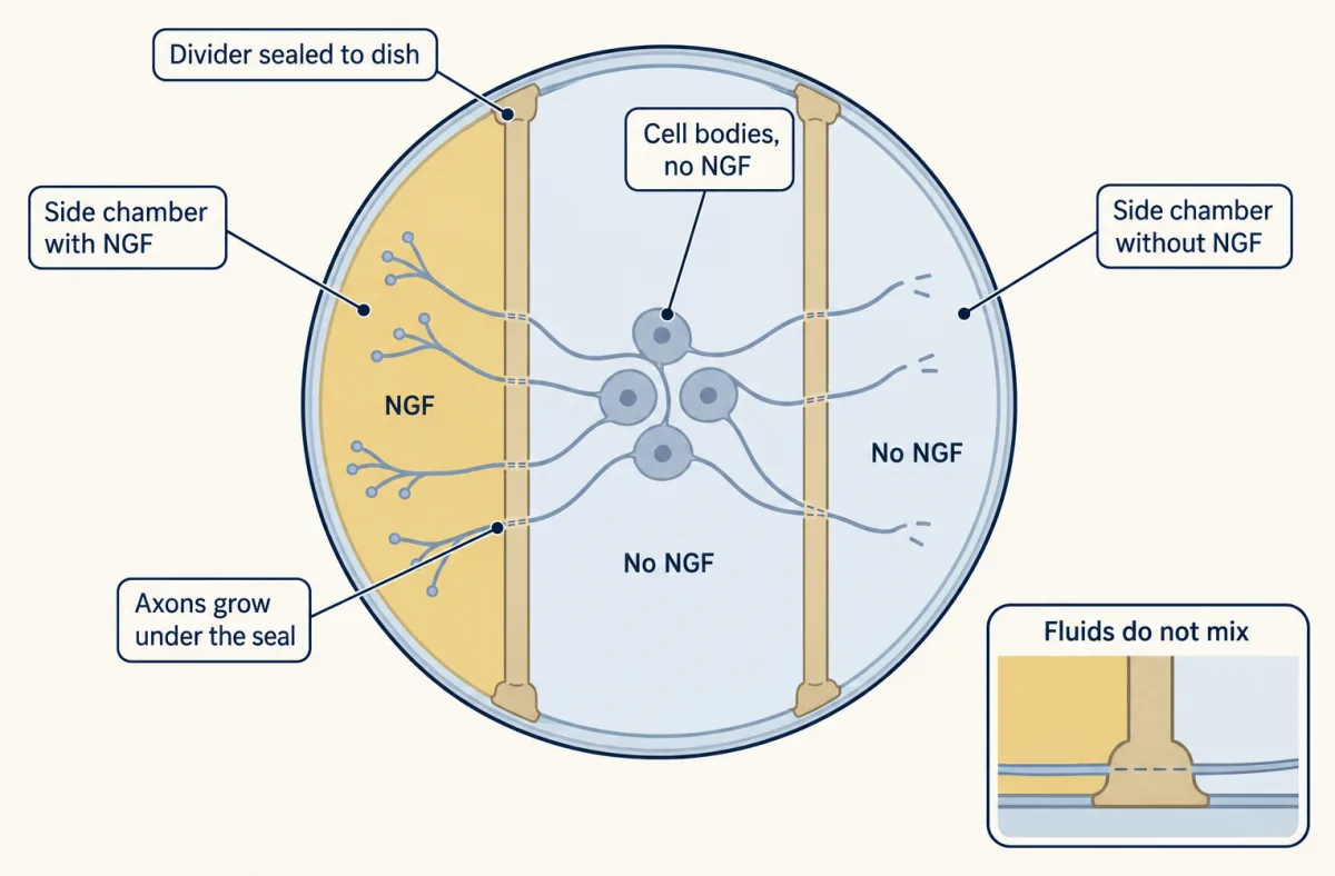
Predict. Two tests. First, take NGF away from one side chamber while the cell bodies stay in NGF. What happens to the axons in that chamber? Second, give the cell bodies no NGF, but let their axons reach a chamber that has it. Do the cell bodies live?
Check your prediction
When NGF was removed from a side chamber, the axons there stopped growing and later degenerated, even though their cell bodies sat in NGF. An axon depends on NGF in its own surroundings. And when cell bodies had no NGF, many still survived, provided their axons reached a chamber that had it. A signal from the distal axon can keep the cell body alive.
Does that signal reach the genome? Antonella Riccio and colleagues in David Ginty’s laboratory gave NGF only to the distal axons of cultured rat sympathetic neurons and found cAMP response element-binding protein (CREB) phosphorylated in the nuclei. The same study found that NGF and TrkA had to be internalized and transported to the cell body for this to happen. And TrkA kinase activity was needed in the cell body, not only at the tip. Was that active TrkA the same receptor, carried from the tip?
Courier or relay?
Two simple models of how an active signal could get from terminal to cell body make different predictions, though they are not the only possibilities.
- Courier. The NGF-TrkA complex itself is carried the whole way and signals when it arrives.
- Relay. Each stretch of axon switches on the next, like a row of dominoes in which each one tips its neighbour. No one molecule makes the whole trip.
Heerssen, Pazyra and Segal reported in 2004 that dynein transports activated Trk receptors and that this transport is needed for the survival of target-dependent neurons. That fits a courier.
Predict. Does it rule out a relay?
Check your prediction
No. Suppose each local step of a relay needed dynein for its own reasons, to bring up a protein the next stretch uses, say. Blocking dynein would stop the relay just as completely. A dynein block, by itself, cannot tell a courier from a relay.
What can tell them apart?
| Test | Courier predicts | Relay predicts |
|---|---|---|
| Watch receptors and NGF in the axon | Labeled complexes move all the way from tip to cell body | Moving complexes may exist, but they would not be what carries the signal |
| Block TrkA’s kinase only in the middle stretch of the axon, with an inhibitor applied only to that stretch (a courier test needs it not to stop transport) | The signal still arrives: the complex is carried through and can signal again once past the inhibitor | A relay that needs TrkA kinase in each stretch stops there |
| Stop NGF from being taken into the cell | Survival fails: there is nothing to carry | A relay could still start from the surface |
Predict. Which test separates the models most cleanly, and which result would favour the courier?
Check your prediction
The middle-segment block. In 2003, Ye, Kuruvilla, Zweifel and Ginty did it: blocking TrkA kinase only in the middle stretch of the axon did not stop the signal reaching the cell bodies or the neurons’ survival. That is evidence against a relay built from TrkA kinase, and for a courier, though not against every kind of relay.
The other tests fit a courier. Heerssen and colleagues imaged fluorescent Trk receptors moving from axon terminals to cell bodies, and in 2007 Cui and colleagues tracked single NGF-containing endosomes moving back along axons. Imaging shows that complexes are carried, not that they are what keeps the neuron alive. Ye and colleagues also found that NGF had to be internalized, and that NGF inside the cell bodies was needed for survival.
For survival, most of the evidence favours the carried endosome. Two results keep the question from being closed: TrkA became phosphorylated in cell bodies within a minute of nerve growth factor (NGF) reaching distal axons, far faster than dynein-driven endosomes move, and NGF fixed to beads supported survival with little or no NGF transport (Going deeper). Whether a faster, propagated signal also contributes is not settled.
NGF versus NT-3: one signal makes the trip
Read the transcript of “Two Factors, One Receptor”
Film transcript: Two Factors, One Receptor
Two factors switch on the same receptor at the axon tip. Why does a neuron need both? In the mouse, sympathetic axons grow out along blood vessels before reaching the organs they innervate. The vessels, an intermediate target, make NT-3. The final targets make NGF. Both factors can activate TrkA. Kuruvilla, Ginty and colleagues gave each factor only to the distal axons of sympathetic neurons in culture. With NT-3 on the axon tips, do the axons grow? Do the cell bodies survive?
NT-3 supported local axon growth through TrkA. But the cell bodies did not survive. NGF supported both growth and survival. Why the difference? Trafficking. NGF and TrkA complexes were taken into the cell and carried back to the cell body. NT-3 and TrkA complexes were not stably carried back. So NT-3 switches the receptor on at the tip. But that signal is not carried home. It is not useless. In mice lacking NT-3, sympathetic axon growth and target innervation were impaired. The paper also proposed a handoff. Retrograde NGF signaling raises a second neurotrophin receptor, p75. As p75 rises, the neuron responds less to NT-3.
A factor that acts only locally can steer an axon. It cannot, by itself, keep the neuron alive. So competition for the final target depends on the trafficking of the receptor, not only on binding. Takeaway: binding at the tip is not enough. Survival needs a signal that makes it home able to signal.
From the film
- NGF and NT-3 both activate TrkA at the axon tip and support local axon growth, but only NGF supports survival of cell bodies in cultured sympathetic neurons.
- Only NGF-TrkA complexes were stably carried back to the cell body.
- A factor that acts only locally can steer an axon but cannot by itself keep the neuron alive.
Development supplies the sharpest test. Sympathetic axons in the mouse grow out along blood vessels before reaching the organs they innervate. The vessels, an intermediate target, make neurotrophin-3 (NT-3). The final targets make nerve growth factor (NGF). Both factors can activate TrkA. So why does a neuron need two?
Predict. Kuruvilla, Ginty and colleagues gave NGF or NT-3 to the distal axons only of sympathetic neurons in compartmented cultures, with no factor at the cell bodies, and measured growth, transport and survival in separate assays. Fill in each cell with yes or no.
| Factor on distal axons only | Local axon growth | Factor-TrkA complexes carried back to the cell body | Cell bodies survive |
|---|---|---|---|
| NGF | ? | ? | ? |
| NT-3 | ? | ? | ? |
Check your prediction
| Factor on distal axons only | Local axon growth | Factor-TrkA complexes carried back to the cell body | Cell bodies survive |
|---|---|---|---|
| NGF | yes | yes | yes |
| NT-3 | yes | no (not stably) | no |
Both factors activate TrkA at the axon, so local signaling for growth works in both cases. The difference was in trafficking. NGF-TrkA complexes were internalized into signaling endosomes and carried back to the cell body by dynein. NT-3-TrkA complexes were not stably carried back. To test that explanation further, look for phosphorylated TrkA arriving in cell bodies, and for nuclear CREB phosphorylation, with each factor on the axons: both with NGF, neither with NT-3.
NT-3 is not useless. In mice lacking NT-3, sympathetic axon growth and target innervation were impaired, so NT-3 matters for the journey to the target.
The paper also proposed a handoff. Retrograde NGF signaling raises the neuron’s level of p75, a second neurotrophin receptor that binds every neurotrophin (the lesson on Trk and p75 receptors). As p75 rises, the neuron’s response to NT-3 falls, so the neuron becomes less responsive to the intermediate target once it reaches the final one.
This is the guiding idea at its sharpest: both factors bind TrkA at the tip, but only NGF’s signal makes the trip, and of the two, only NGF keeps these neurons alive from the axon tip. A factor that acts only locally can steer an axon, and so decide which target it reaches, but it cannot by itself keep the neuron alive. Competition among neurons for the final target, the subject of that lesson, therefore depends on the trafficking of the receptor, not only on binding.
Putting it together
| Experiment | System | Observed result | What it supports | What it does not establish |
|---|---|---|---|---|
| Diffusion arithmetic | a protein with D near 10 µm²/s | about two months for 1 cm (time grows with distance squared); an active signal fades over about 10 µm with illustrative phosphatase rates | a vehicle or relay is needed | what the vehicle is |
| Radiolabeled NGF in the eye (1974) | mouse | label builds up in the same-side superior cervical ganglion | something moves from target to ganglion | that NGF arrived intact or active |
| Radiolabeled NGF under the leg skin (1979) | chick embryo | same-side sensory ganglia labeled; motor column and roots not | selective transport by NGF-responsive neurons | that the transport mattered for survival |
| Compartmented culture (1977) | rat sympathetic neurons | axons degenerate where NGF is removed; many cell bodies survive on distal NGF | NGF acts locally on axons; a distal signal supports the cell body | what the signal is |
| NGF on distal axons only (Riccio and colleagues) | rat sympathetic neurons | nuclear CREB phosphorylated; this needed internalization, transport and TrkA kinase in the cell body | a signal from the axon tip reaches a nuclear transcription factor | whether the receptor that left the tip arrived active, or what carried it |
| Dynein block (2004) | target-dependent neurons | survival lost | transport is needed | courier against relay |
| Imaging (2004, 2007) | live axons | Trk receptors and single NGF endosomes move back along axons | something is carried | that the carried complex is what keeps the neuron alive |
| Middle-segment TrkA block (2003) | sympathetic neurons | signal and survival intact; NGF had to be internalized | a courier over a relay that needs TrkA kinase along the way, for survival | that no faster signal contributes |
| NGF against NT-3 on distal axons (2004) | cultured sympathetic neurons | both support growth; only NGF supports survival; only NGF-TrkA is stably carried back | survival needs a signal that makes the trip | that arrival alone is enough |
| NT-3 knockout | mouse | sympathetic axon growth and target innervation impaired | NT-3 matters for growth toward targets | — |
The experiments trace a shift, though not strictly in date order: from evidence that something moves, to evidence that a signal from the axon keeps the cell body alive, to evidence of what makes the trip and why only one of two factors can use it.
Design problem
A new factor Y activates TrkA at the distal axons of sympathetic neurons in compartmented culture, with no factor at the cell bodies. Labeled Y is found in the cell bodies, yet the neurons die. Name two places the signal could fail, one before it reaches the cell body and one after, and an experiment for each.
Saved in this browser only.
One way to do it
First, remember what label shows. Finding labeled Y in the cell bodies says that Y, or its label, moved. It does not say that Y arrived bound to an active receptor.
- Y never delivers a receptor that can still signal. TrkA might be taken up with Y into endosomes that are not carried home, as with NT-3, or not taken up at all, or it might lose Y or its phosphates on the way. Test: follow TrkA from the distal axons and measure its phosphorylation along the axon and in the cell bodies, compared with NGF. No carried receptor points to uptake or sorting; carried receptor that fades points to switching off on the way.
- Active receptor arrives, but the cell body’s response falls short. Test: measure nuclear CREB phosphorylation, as one readout, with Y on the axons. Active TrkA in the cell bodies without phospho-CREB points here. Normal phospho-CREB alone would not prove the survival response is complete: the rule does not promise that every signal that arrives is enough.
A useful control for both: give Y directly to the cell bodies at a matched dose. If the neurons then survive, they can use Y, so the failure lies somewhere between the axon tip and a sufficient response in the cell body. The measurements above say where.
Going deeper
Where the decision is made
The nucleus is not the only place where death machinery sits. The Bcl-2 family proteins act at mitochondria, and mitochondria run along the axon too; the machinery that dismantles an axon locally is the subject of the lesson on axon pruning. What is confined to the cell body is the nucleus, whose new transcription the death program in these neurons needs.
A faster signal?
Senger and Campenot (1997) saw the cell-body TrkA phosphorylation before any labeled nerve growth factor (NGF) was detected there. Even across one millimetre, arriving within a minute would need about 17 micrometres per second, far above the roughly one micrometre per second measured for dynein transport.
In the bead experiment, MacInnis and Campenot (2002) supplied the beads to the distal axons of rat sympathetic neurons, and they kept over 80% of the neurons alive for 30 hours. Compare Riccio and colleagues, who found that internalization and transport were needed for nuclear cAMP response element-binding protein (CREB) phosphorylation. The two studies measured different things, a nuclear transcription factor against survival, and how they fit together is part of the open question.
The cell biology of the trip
How dynein moves cargo along an axon is covered in the lesson on long-distance signaling in neurons, and how kinase signals reach transcription factors in the lesson on signaling to the nucleus.
Does a single flavor of endosome mediate all of these functions?
NGF arriving at the cell body does more than prevent death. It regulates dendrite growth, transmitter phenotype, cell size and, through the dendrites, synapse formation. One carrier, or several?
Work from David Ginty’s laboratory, where Deppmann was a postdoctoral fellow, and later from Deppmann’s own laboratory addresses this. Sharma, Deppmann, Harrington and colleagues in the Ginty laboratory (Neuron, 2010) found that in sympathetic neurons, NGF-TrkA signaling endosomes travel from distal axons to cell bodies and dendrites and promote assembly of the postsynaptic specialization, a long-distance relationship between target NGF and the synapses a neuron receives.
Suo and colleagues (Nature Neuroscience, 2014) identified Coronin-1, a protein whose expression NGF induces, as an endosomal effector that keeps NGF-TrkA endosomes from fusing with lysosomes, and found that it is required for developmental competition for survival. Together these results suggest that biasing an endosome’s maturation changes which function it serves, so that distinct endosome pools may govern distinct developmental processes. The review by Barford, Deppmann and Winckler (Developmental Neurobiology, 2017) sets out this view. Whether every message rides in its own kind of endosome is still open.
Retrograde transport in disease models
In disease models, dynein and dynactin mutations that impair retrograde transport are linked to motor neuron degeneration, and a mouse model of Down syndrome shows failed NGF transport with degeneration of cholinergic neurons.
Your notes
Select any text in the lesson to highlight it or add a note to it. Your highlights and notes are saved in this browser and collected in your notebook.
This browser is not letting the site save anything (a private window or strict privacy settings), so highlights and notes will be lost when you leave the page.
How we measure it
Compartmented (Campenot) culture
Neurons are plated in one chamber and their axons grow under a sealed barrier into others, so cell bodies and distal axons sit in separate fluids. You can give a factor to one part of the neuron and measure survival, growth or gene expression in another.
Retrograde transport of a radiolabeled ligand
A radioactive form of a factor such as iodinated NGF is placed in a target, and after some hours the ganglia are sectioned and the label located by autoradiography or counting. It shows that the label moved toward cell bodies along a specific route, not that the molecule arrived intact or active.
Knockout mice
A knockout is an animal in which a gene has been deliberately inactivated. Comparing NT-3 or NGF knockouts, often on a Bax-null background that prevents neuron death, separates effects on axon growth from effects on survival.
Practice questions on this lesson (3) → MCAT-style, original, each with an explanation
Where this lesson fits
Before thisReview first
- The Neurotrophic Hypothesis TestedThis course · Lesson 29
This lesson starts from the idea that targets supply a limited survival factor, and asks how that signal reaches the cell body.
- Neurotrophins, Trk Receptors and p75This course · Lesson 30
You need TrkA activation by NGF and the role of p75 from there to follow the NGF versus NT-3 comparison.
- The Death Machinery in NeuronsThis course · Lesson 31
The claim that the survival signal must reach the nucleus rests on death needing new gene expression in the cell body.
Elsewhere on the siteFilms, simulations and method films in other courses
- A neuron can take its survival signal in at the far end of its axonFilm · Cell Signaling
It shows how a survival signal taken in at the axon tip is carried to the nucleus.
- Sealed Inside, a Receptor Goes SilentFilm · Cell Signaling
It covers the signaling endosome that carries a receptor and its signal along the axon.
- Grip, Swing, Let GoFilm · Cell Biology
It shows dynein walking toward the minus end, the motor that carries NGF-TrkA back to the cell body.
- Cell-free extracts and reconstitutionMethod film
It shares the idea of isolating parts of a process in a tube to test which are needed.
Suggested by an AI model from the lessons themselves, then checked. How we use AI
Finish line
Test yourself
8 questions
Practice and MCAT-style, with explained answers.
Next · Lesson 34
Pruning Without Dying: Axons That Cut Themselves Back
How does a neuron cut off one branch with caspases, the enzymes of cell death, and still live?
Continue →