Developmental Neuroscience · Unit 8: Building synapses
Synapse Elimination: One Muscle Fiber, One Axon
Rewritten on : new text, figures, flashcards and film. Your notes and highlights stay, but the text around them may have moved.
A newborn rat muscle fiber has two or more motor axons on one synapse, and every one of them works. Within weeks all but one are gone. What decides which one stays?
Pause when the reading reaches a film, and offer to play it. Saved on this device.
Watch the film now, or keep listening
Builds on:Building the Neuromuscular Junction: Agrin, Lrp4 and MuSKNeurotrophins, Trk Receptors and p75Why these?
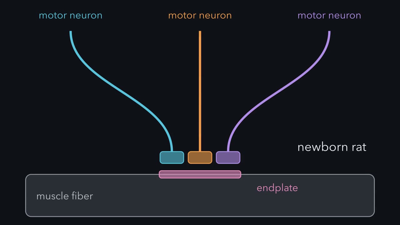 ▶ Watch Quieter Than Its Neighbour · 3:12
Read the transcript of “Quieter Than Its Neighbour”
Film transcript: Quieter Than Its Neighbour
A newborn rat muscle fiber is wired to two or more motor neurons. Every one works. Within weeks, all but one are gone. What decides which stays? Each extra input releases acetylcholine and evokes an electrical response in the fiber. Nothing is wrong with them. Then they are removed. How do you count them? Put an electrode inside one fiber, and slowly turn up the stimulus to its nerve. Each axon in the nerve has its own threshold. It fires only once the stimulus passes that level. Predict. If three axons reach this fiber, what does the response look like as the stimulus rises?
A staircase. Each threshold crossed adds one step. Three distinct steps mean three axons. One axon gives one step, at its own threshold. One action potential ordinarily reaches all its release sites at once. In 1970, Redfern did this in newborn rat diaphragm. Most responses at birth had two to four steps. By 16 to 18 days, single steps. So did the extra axons' motor neurons die? Two measures tell these apart. Count the motor neurons, and measure one motor unit's share of the twitch. A motor unit is one motor neuron and all the fibers it supplies. A twitch is the muscle's brief contraction to one impulse. Predict. If every fiber gives similar force, what happens to one unit's share if neurons die? And if they live?
If neurons died, and the survivors still reach every fiber, each keeps its fibers, so its share stays the same. If neurons live and trim branches, the count holds. Each unit's share falls. This assumes the muscle does not gain many fibers. In 1976, Brown, Jansen and Van Essen measured both, in rat soleus muscle. Each unit's share of the twitch fell sharply. The number of motor neurons did not change significantly. The neurons lived, and their branches went. Synapse elimination is branch loss, not neuron death. Here is the idea we will test through this lesson. A synapse is not removed just for being quiet. What puts it at risk is being quieter than its neighbours at the same junction.
The idea says which input goes. Overall activity sets how fast. Silence slows it, and some patterns of stimulation speed it. Quieter seems to mean weaker transmission, as the muscle sees it. And it is a bias, not a verdict. So the neurons stay, and each fiber holds a contest.
From the film
- A newborn rat muscle fiber is contacted by two or more motor axons, and within weeks all but one are gone.
- Counting the steps in a fiber's response as nerve stimulation rises counts its inputs.
- The lost synapses are branch loss: the motor neurons do not die.
- A synapse is not removed just for being quiet; being quieter than its neighbours at the same junction puts it at risk.
A rat is born with most of its muscle fibers wired to two or more motor neurons. Within two to three weeks, each fiber listens to one.
Nothing is wrong with the extra inputs. Each releases acetylcholine and evokes an electrical response in the fiber, and they work. Then all but one are removed. Earlier lessons removed neurons: those that fail to get enough target-derived factor die, as you saw in competition for targets. This lesson removes synapses from neurons that stay alive. It asks three questions in order. How did anyone count the inputs? Where do the lost ones go? And what decides which axon keeps the fiber?
Counting the inputs
The place to study this is the neuromuscular junction, the synapse between a motor axon and a skeletal muscle fiber. Its assembly, with the receptor patch that agrin and its partners build, is covered in the neuromuscular junction. Here what matters is that a typical adult mammalian muscle fiber has one junction and one axon. The postsynaptic site is called the endplate. When the nerve releases acetylcholine, the fiber makes a small depolarization there, the endplate potential.
Anatomists had drawn several axons on one young endplate long before, starting with Tello, a student of Cajal. But in a simple stain, tangled terminals are hard to tell apart or count. The first functional count was electrical.
Predict. Put an electrode inside one newborn muscle fiber and stimulate its nerve, starting weak and turning the strength up in small steps. Each axon in the nerve has its own threshold: it fires only once the stimulus passes that level. If three axons reach this fiber, what does the endplate potential look like as you turn the stimulus up? And if only one axon reaches it?
Check your prediction
With three axons, a plot of response size against stimulus strength is a staircase, one step for each threshold crossed:
| Stimulus strength | Fiber with three axons |
|---|---|
| Below every threshold | no response |
| Past the first axon’s threshold | first step |
| Past the second | second step added |
| Past the third | third step added |
| Stronger still | no change |
The number of steps counts the axons, provided the muscle’s own action potential is kept from firing (with curare, for example) and each axon’s step can be told apart. With one axon you get a single step, at that axon’s own threshold: one action potential reaches all of its release sites, so its terminals are ordinarily recruited together.
In 1970 P. A. Redfern did this in fibers of newborn rat diaphragm, stimulating the phrenic nerve. Most responses at birth grew in discrete jumps, each with its own threshold and latency (the delay from stimulus to response): two to four units summed together. By 16 to 18 days they were single steps, like an adult’s. The method is called stepwise recruitment, and it is still a standard way to count the inputs to one cell, at muscle and in the brain.
Did the neurons die?
Each fiber lost its extra axons. Did their motor neurons die, or did surviving neurons pull back some of their branches?
Two measures tell these apart. A motor unit is one motor neuron and all the fibers it supplies. Stimulate one motor unit and you can measure its force as a share of the whole muscle’s twitch, the brief contraction to a single nerve impulse. Separately, you can count the motor neurons that supply the muscle.
Predict. Here is the bookkeeping for a made-up muscle. Each synapse here is one neuron–fiber connection, so fibers × inputs per fiber = motor neurons × fibers per neuron. If every fiber gives about the same force, a unit’s share of the twitch is the fraction of fibers it reaches: a neuron that supplies fewer fibers gives a smaller slice of the force. Which day-14 row would show that branches, not neurons, were lost?
| Case | Fibers | Inputs per fiber | Motor neurons | Fibers per neuron | One unit’s share of the twitch |
|---|---|---|---|---|---|
| Newborn | 2,400 | 3 | 24 | 300 | 12.5% |
| Day 14, if neurons were kept | 2,400 | 1 | 24 | 100 | about 4% |
| Day 14, if neurons had died | 2,400 | 1 | 8 | 300 | 12.5% |
Check your prediction
The “if neurons were kept” row: the count stays put and each unit’s share falls, because each neuron now supplies fewer fibers. If neurons die, each survivor keeps its fibers, so its share stays the same while the count falls. (That row is the tidiest case, in which the 8 survivors between them cover every fiber once.)
In 1976 Michael Brown, Jan Jansen and David Van Essen measured both in rat soleus muscle. Fibers lost their extra inputs by the end of the second week. Each motor unit’s share of the twitch fell sharply, yet the number of motor neurons supplying the soleus did not change significantly: the “if neurons were kept” row.
One condition holds the argument together. Each unit’s share would also fall if the muscle tripled its fibers to 7,200 and the same 7,200 connections spread out one per fiber: 300 of 7,200 is again about 4%. So the inference needs fiber number not to have risen anywhere near that much. Tracing single axons later closed the gap, as the next section shows: each axon was losing branches. Synapse elimination is branch loss, the subject of axon pruning, not neuron death.
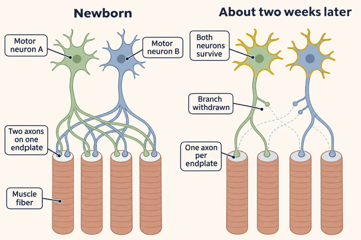
That leaves the third question: at each fiber, what picks the branch that stays? Here is the answer the rest of the lesson has to earn.
The one idea to carry through this lesson. A synapse is not removed for being quiet. What puts it at risk is being quieter than its neighbours at the same junction.
What the rule covers.
- Where. Inputs that share one neonatal mammalian endplate, or that sit close together on one fiber. In an experiment with a foreign nerve implanted into muscle, two endplates on one fiber more than about 1 mm apart could both persist, so the contest seems to act only between nearby synapses. For two whole axons the evidence is the Buffelli experiment, below.
- Which input, not how fast. The idea is about which input goes. Overall activity sets the pace, as the section on activity shows.
- What “quieter” means. The evidence points to less activation of the acetylcholine receptors at that site, as the muscle sees it, not the axon’s firing alone.
- A bias, not a verdict. The less active input usually loses, but the outcome is not fixed in advance.
A contest at every junction
Read the transcript of “A Contest at Every Junction”
Film transcript: A Contest at Every Junction
One motor neuron loses many branches. Does it pull them all back together, on one schedule? If the neuron ran one program for its whole arbor, its branches would retreat together. In 2001, Keller-Peck, Lichtman and colleagues made a small, random subset of motor axons glow. They traced every branch of single axons. Each axon was losing branches. Its branches withdrew at different times, at different junctions, without an obvious spatial pattern. They did not go together. That points to a separate contest at every junction.
In 2003, Mark Walsh and Lichtman watched one of those contests. Two motor axons carried different colours. The same living junction was imaged again and again, over days. A fluorescent snake toxin marked the receptors. The dose was small, so transmission still worked. Predict. Axon A starts with more of the receptor map. Does the bigger territory win?
Not reliably. The starting territory did not reliably predict the winner. The share could shift one way, then back. Typically, one axon withdrew from the receptor sites. The other extended into the sites it left. So starting size alone does not reliably decide. Each junction runs its own contest, and what tips it is the next question.
From the film
- One motor neuron's branches withdrew at different times and at different junctions, which points to a separate contest at every junction.
- In the mouse time-lapse, the starting territory did not reliably predict the winner.
- Typically one axon withdrew from the receptor sites while the other extended into the sites it left.
If each neuron ran one program to shrink its whole arbor, its branches would retreat together. Fluorescent proteins let later work check. In 2001 Cynthia Keller-Peck, Jeff Lichtman and colleagues used transgenic mice in which only a small, random subset of motor axons glowed, and traced every branch of single axons. Each axon was losing branches, but they withdrew at different times, at different junctions, without an obvious spatial pattern. That points to a separate contest at every junction.
Watching the contest
In 2003 Mark Walsh and Lichtman watched one. They used mice in which two motor axons carried different colours of fluorescent protein, and imaged the same living junction again and again over days. To see the receptors they labelled them with fluorescent alpha-bungarotoxin, a snake toxin that binds acetylcholine receptors almost irreversibly and blocks each one it binds. The imaging dose bound only some of the receptors, so transmission still worked. Later in this lesson a larger dose blocks a patch.
Predict. At the first image, axon A covers more of the receptor map than axon B. Does the bigger territory win?
Check your prediction
Not reliably: the outcome could not be predicted from how much territory each axon held at the start. The relative areas could also shift in favour of one axon and then the other, so the contest was not always a steady retreat.
Typically one axon withdrew from the receptor sites while the other extended into the sites it left: a takeover. In rare cases the winner did not move into a vacated site, and the receptors there quickly disappeared.
Activity biases the contest
Read the transcript of “The Focal Block”
Film transcript: The Focal Block
If starting size alone does not decide, what does? Is it silence, or being quieter than a neighbour? In newborn rat muscle, blocking nerve impulses with tetrodotoxin slows elimination. Chronic electrical stimulation, with some patterns, speeds it up. So activity sets the pace. But these tests changed every input at once. They could not say whether a quiet input is lost because of its neighbours. Two rules were left to separate. Rule one. A silent synapse is removed. Rule two. A synapse is removed when it is less active than nearby synapses, if enough of them are active. To tell them apart, silence part of one junction, and leave the rest working.
Alpha-bungarotoxin, the snake toxin from the imaging, binds acetylcholine receptors almost irreversibly. It blocks each one it binds. Where it sits, the muscle stops responding. The axon above is untouched. So quieter here means the muscle responds less. In a living mouse, put toxin on one small patch of one endplate. At a second endplate, cover every receptor. Predict. Over the following weeks, what does each rule say will happen at each junction?
Rule one predicts loss at both junctions. Both have silent sites. Rule two predicts loss only at a partial block, where a small silent patch sits beside a larger working area. In 1994, Rita Balice-Gordon and Lichtman reported the result. At the partial block, the patch lost its receptors, and the nerve terminal withdrew from it. At the full block, nothing was eliminated.
At the full block, every site is equally silent. No site is quieter than its neighbour. Rule two wins. the loss stayed inside the patch. The signal behind it, and how far it travels, were not identified. The amount of active territory mattered. A large active area removed a small silent one. A small active area could not. The loss happened where the toxin was put. So a local difference in activity directs which sites go. Lo and Poo built the smallest contest, in cultures from the frog Xenopus. Two spinal neurons shared one muscle cell. A burst to one neuron immediately weakened the other's synapse. Predict. What if both neurons get the burst together?
The suppression was largely absent. Neither input was left quiet while the other was driven. This is heterosynaptic depression, a weakening within minutes. It fits rule two, but it is not removal of a synapse. For two whole axons, mice had one of two inputs made weaker. The more powerful inputs were strongly favoured. Still, it is a bias, not a verdict.
What carries the message is still open. In one model, the muscle punishes inactive sites and protects the active one. Without one form of PKC, elimination in mice is delayed, but it still completes. Mice lacking BDNF or p75 eliminate synapses on schedule. So none of these is yet shown to be the indispensable signal. So silence alone does not remove a synapse. Being quieter than its neighbours puts it at risk.
From the film
- Overall activity sets the pace of elimination: blocking impulses slows it, and some patterns of stimulation speed it.
- In mice, a small blocked patch within a working junction was lost, while a fully blocked junction was not, so being quieter than neighbours is what puts a site at risk.
- A difference in activity is a bias, not a verdict, and no candidate molecular signal is yet established as the one that picks the winner.
If the starting territory does not reliably decide, what does? The first clue came from newborn rat muscle. Block nerve impulses with tetrodotoxin, a toxin that stops action potentials, and elimination slows. Apply chronic electrical stimulation and, with some patterns, elimination speeds up; other patterns did not. So activity sets the pace. But each of these experiments changed every input at once. Slowing under whole-nerve block is already awkward for rule one below, which predicts that silent inputs go, not that elimination slows. What these experiments could not show is whether a quiet input is lost because of what its neighbours are doing. Two rules frame that question.
- Rule one. A silent synapse is removed.
- Rule two. A synapse is removed when it is less active than the synapses near it, provided enough of its neighbourhood is active.
To tell them apart, you have to silence part of one junction and leave the rest working.
Predict. Use alpha-bungarotoxin, the toxin from the time-lapse, at a dose that blocks every receptor it reaches. Wherever it sits, the muscle no longer responds to transmitter; the axon above is untouched. In a living mouse, put the toxin on one small patch of a single endplate. At a second endplate, put it on every receptor. Image both junctions over the following weeks. What does each rule predict for the receptors, and for the nerve terminal above them?
Check your prediction
Rule one predicts loss at both junctions, because both have silent sites. Rule two predicts loss only at the partly blocked junction, if enough of the junction stays active: there a silent patch sits beside working patches. At the fully blocked junction every site is equally silent, so no site is less active than its neighbour.
Rita Balice-Gordon and Lichtman did this experiment and reported it in 1994. Blocking a small patch caused long-lasting synapse elimination at that site: the receptors in the patch were lost, and the nerve terminal withdrew from it. Blocking the whole junction did not cause elimination. Rule two wins.
Three points are worth slowing down for. First, what the blocked site lacked was the muscle’s response, not the nerve’s firing. That is why “quieter” seems to mean weaker transmission as the muscle sees it. Second, what lost was a patch of one junction, not a whole axon. Sites compete, and this experiment does not by itself pit two axons against each other. Third, how much stayed active mattered. Barber and Lichtman (1999), from the same laboratory, describe the result as depending on active area: a large active area could eliminate a small blocked one, but a small active area could not eliminate a large blocked one. The loss stayed inside the patch, but the signal behind it, and how far it travels, were not identified.
This is the guiding idea at work: silence alone did not remove a synapse, but being quieter than active neighbours did. And because the loss happened where the toxin was put, the difference in activity directs where loss happens rather than merely allowing it: in the language of the neurotrophic hypothesis, it is instructive.
Can you watch the comparison happen? In 1991 Yi-Jun Lo and Mu-ming Poo built the smallest contest possible: in cultures from embryos of the frog Xenopus, one embryonic muscle cell innervated by two spinal neurons. A brief burst of high-frequency stimulation, a tetanus, to one neuron immediately suppressed the synapse made by the other. This is heterosynaptic depression: activity at one synapse weakens an inactive neighbour on the same cell.
Predict. Now give the burst to both neurons together. What does rule two predict for each synapse?
Check your prediction
Little or no suppression, because neither input is left quiet while the other is driven. That is what Lo and Poo found: when both neurons were stimulated together, the suppression was largely absent. Activity at the same time seems to protect both inputs, though matching the stimulus trains does not prove the two synapses are equally strong.
This is a weakening of transmission within minutes, not the removal of a synapse. It fits rule two, but it does not show that the same step removes synapses in an animal. Nor can the culture alone separate the two rules for removal: its readout is weakening, and an unstimulated input is not a silenced one. The focal block did that.
Does it hold for two whole axons in a living animal? In 2003 Buffelli, Sanes, Lichtman and colleagues used a genetic method in mice to inhibit transmission from a subset of motor axons, so that one of two inputs to the same fibers was weaker. The more powerful inputs were strongly favoured.
Strongly favoured does not mean always, and one experiment in newborn rabbit found the reverse of the activity rule (Going deeper). Relative activity is not the only factor either: firing patterns, muscle fiber type and the terminal Schwann cells also matter. Terminal Schwann cells, the glial cells that cap the junction, engulf the tips of retreating branches. Whether they take part in choosing the winner is not known.
Rule to keep: a synapse is removed when it is less active than synapses near it, not simply when it is silent, so silencing part of a junction removes that part and silencing all of it removes nothing. This is a sketch of the logic in relative units, not to scale.
What carries the signal
Several candidate signals have good evidence, from culture and from developing rodents, but how they fit together is unclear. In one working model, a muscle activated by an input’s transmitter makes two kinds of signal: one that punishes nearby inactive sites and one that protects the active site. Two candidate pairs fit this picture.
The first pair is two kinases. In compartmented nerve-muscle cultures from Phillip Nelson’s laboratory, protein kinase C (PKC) mediated heterosynaptic loss of the less active input and protein kinase A (PKA) stabilized the stimulated input, both through the number of acetylcholine receptors at the synapse.
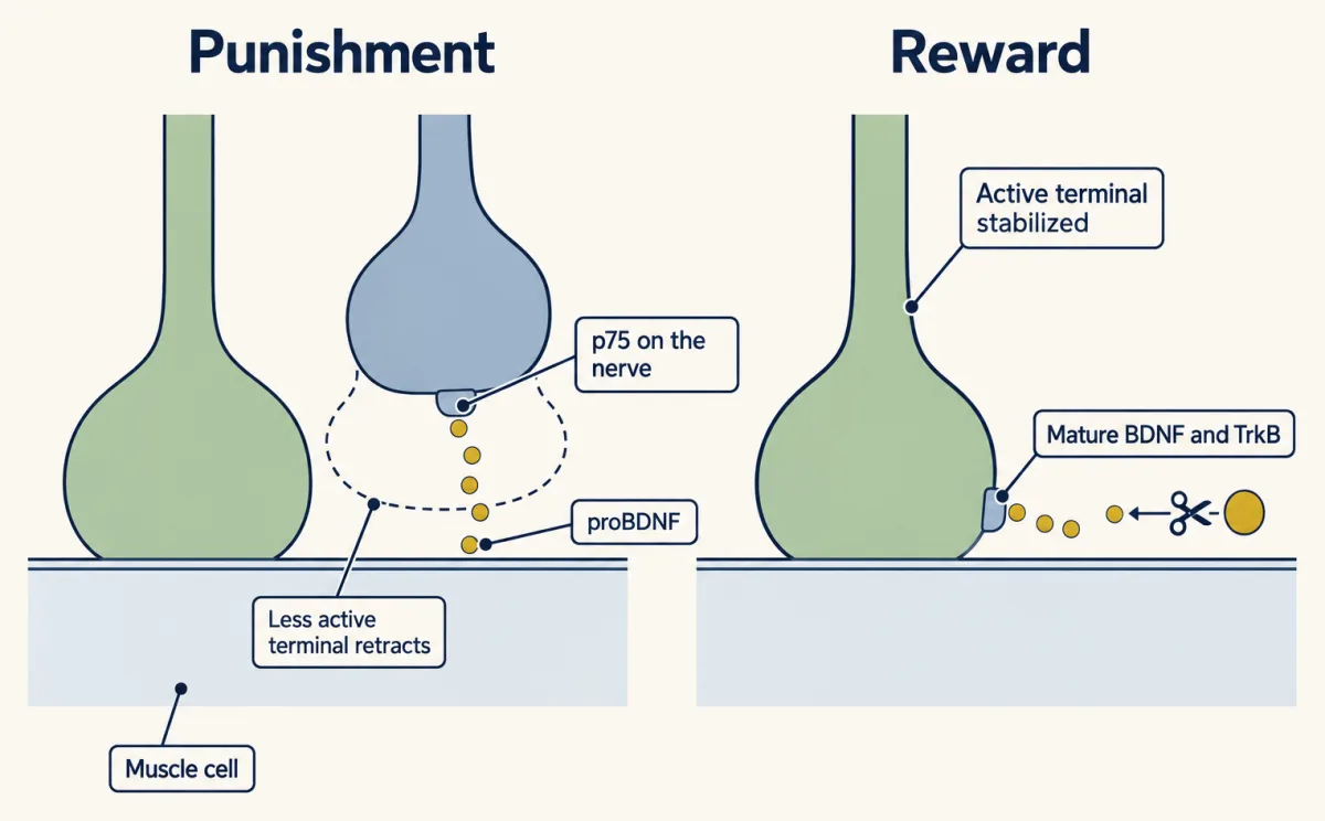
The second pair is two forms of the neurotrophin brain-derived neurotrophic factor (BDNF), which act mainly on different receptors, as Trk and p75 explains. In Xenopus nerve-muscle cultures, proBDNF acting through p75 promoted retraction of the less active terminal, while mature BDNF acting through TrkB stabilized the active one. A related punish-and-reward logic, acting on whole neurons rather than synapses, appears among sympathetic neurons in the neurotrophic hypothesis.
In living rodents, PKC activity has been linked to the normal pace of elimination in rat muscle, and in mice lacking the theta form of PKC elimination is delayed but eventually completes (Li, Nelson and colleagues, 2004). Mice lacking BDNF or p75 eliminate synapses on a normal schedule. So none of the candidates is established as the indispensable signal that selects the winner. What remains unknown is how these signals are confined to one patch, and how they relate to the cytoskeletal steps inside a retreating branch and to the Schwann cells that engulf its tips.
A similar contest in the brain
In the rodent cerebellum, each Purkinje cell, the large output neuron of the cerebellar cortex, is innervated by several climbing fibers, the axons from the inferior olive in the brainstem, in the first postnatal week, and by about the end of the third week most have one. Stepwise climbing-fiber responses, Redfern’s counting method, track the change; that literature, from groups such as Masanobu Kano’s, is separate from the muscle work. The detailed mechanisms differ, but glia take part in both places: terminal Schwann cells at the muscle, and astrocytes and microglia, which remove synapses, in the brain, as microglia and complement describes. Starting with several working inputs and removing some recurs in many developing circuits, though the number kept varies: a Purkinje cell usually keeps one climbing fiber, while a thalamic relay neuron keeps a few retinal inputs.
Putting it together
| Experiment | Preparation | Observed result | What it supports | What it does not establish |
|---|---|---|---|---|
| Stepwise recruitment (Redfern 1970) | newborn rat diaphragm | two to four steps at birth; single steps by 16 to 18 days | most fibers start with several axons and end with one | where the lost inputs go |
| Motor-unit force and motor neuron counts (Brown, Jansen and Van Essen 1976) | newborn rat soleus | each unit’s share of the twitch fell sharply; neuron number unchanged | branch loss, not neuron death | holds only if fiber number did not rise greatly |
| Single axons traced (Keller-Peck and colleagues 2001) | transgenic mice | branches withdrew at different times, without an obvious spatial pattern | a separate contest at each junction | what decides each contest |
| Two-colour time-lapse (Walsh and Lichtman 2003) | living mouse junction, imaged over days | takeover; the lead could shift; the starting area did not reliably predict the winner | inputs compete for the same receptor sites | which input will win |
| Tetrodotoxin; chronic stimulation | neonatal rat muscle | tetrodotoxin slows elimination; some stimulation patterns speed it | overall activity sets the pace | which input goes |
| Focal versus whole-junction block (Balice-Gordon and Lichtman 1994) | living mice | small blocked patch lost; fully blocked junction kept | a local difference in transmission biases which sites are lost, if enough of the junction stays active | the signal, its range, or a contest between two whole axons |
| Tetanus to one neuron or both (Lo and Poo 1991) | Xenopus culture, two neurons on one muscle cell | one: the other synapse suppressed at once; both: largely absent | activity at one input can weaken another within minutes | that this weakening is the step that removes synapses in an animal |
| One input made weaker (Buffelli and colleagues 2003) | mice | the more powerful inputs strongly favoured | the activity bias for two whole axons in vivo | that the stronger input always wins |
| Silenced motor neurons (Callaway, Soha and Van Essen 1987) | neonatal rabbit soleus | the silenced neurons had a competitive advantage | an exception the rule does not explain | — |
| Candidate signals (PKC and PKA; proBDNF and mature BDNF) | cultures; developing mouse junctions | punish-and-protect effects; PKC theta loss delays elimination in vivo | a working model of how the comparison is carried out | that any of them is the indispensable signal that picks the winner |
The table answers the first two questions: the inputs were counted in steps, and the lost ones belong to neurons that live. The third is answered in part, because the rows answer different questions. The focal block and the weakened-input mice support the guiding idea directly, the rabbit result goes against it, and the signal that carries it is still open.
Design problem
A Purkinje cell starts with several climbing fibers and usually ends with one. Design an experiment that would tell rule one from rule two in the cerebellum. Say how you would count the inputs, what you would manipulate, and what each rule predicts.
Saved in this browser only.
One way to do it
- Count, and label. Stepwise climbing-fiber responses count only inputs that transmit, so a silenced fiber will not show up as a step. Label the climbing fibers before you silence any, and follow them and their contacts over time. Use stepwise counts for transmitting inputs in controls, plus an untreated age series across the first three postnatal weeks to measure normal formation and removal.
- Make one input quieter. Silence a few climbing fibers, so that a silenced fiber shares its Purkinje cell with active ones. At the muscle, “quieter” meant weaker transmission as the target sees it, so a fiber that fires but releases no transmitter counts as quieter.
- The control that separates the rules. Silence every climbing fiber onto some Purkinje cells. This silences the climbing-fiber contest, not the whole cell: parallel fibers still drive it, so measure the cell’s activity.
- Predictions. Rule one: silenced fibers are lost wherever they are. Rule two: silenced fibers are lost where they share a cell with active fibers, and where every climbing fiber is silent none is singled out for loss. Score the outcome anatomically.
- Pace. Silencing many inputs may slow elimination overall, as tetrodotoxin does at muscle. Compare timing across cells and do not mistake a slower contest for a different rule.
This lesson reports no such cerebellar result, and the mechanisms there differ, so treat your answer as a prediction to test, not a fact to recall.
Going deeper
Words for the change
Wiring a mature circuit, sometimes called neural circuit refinement, changes three quantities: the number of neurons, the number of synapses, and the strength of synapses. This lesson is about the number of synapses; heterosynaptic depression is where it touches strength. A useful measure is convergence, the number of axons that innervate one postsynaptic cell. In many developing circuits where it has been measured, convergence falls during a particular period, a change called refinement of convergence. At the neonatal rodent junction it falls from two or more to one. In their review of synapse elimination at the mammalian neuromuscular junction (NMJ), Lichtman and Colman point out why muscle allows it: the fibers of one muscle are nearly interchangeable targets, so a motor neuron can first diverge onto many equally suitable fibers and then sort out which it keeps. The diffuse, redundant early wiring becomes a specific one.
The imaging preparation
Walsh and Lichtman imaged the junction in the sternomastoid muscle of an anaesthetised pup, let it recover, and found the junction again days later.
Kinases in the muscle
In Nelson’s cultures at the NIH, two groups of neurons innervated the same myotubes, and stimulating one group weakened the other’s input. Protein kinase C (PKC) is the kinase family whose conventional members are switched on by diacylglycerol and calcium, which you met in the PLC lesson; protein kinase A (PKA) is the cAMP-driven kinase of cAMP and PKA, and its action in stabilizing the stimulated input was activity-dependent. Heterosynaptic depression itself, the effect described in the Xenopus co-cultures, is reported in culture to need a rise of calcium in the postsynaptic cell, whichever signal carries it.
A neurotrophin with two faces
Brain-derived neurotrophic factor (BDNF) is made as a pro-form, proBDNF, which can be cut to mature BDNF. As Trk and p75 explains, the pro-form signals mainly through p75, with its partner sortilin, and the mature form mainly through TrkB. Bai Lu’s group, working at the NIH, found in Xenopus nerve-muscle cultures that muscle stimulation releases proBDNF, which depresses synapses and then causes the presynaptic terminal to retract, through p75 on the nerve. Cutting proBDNF to mature BDNF at the synapse turned the same molecule into a signal that potentiates; the two-neuron result in the main text comes from the same work. In developing mouse junctions, adding proBDNF promoted elimination, infusing mature BDNF delayed it, and blocking the cutting step sped it up through p75. The group called the two forms “punishment” and “reward” signals. Given the knockout mice that eliminate on schedule, whether the pathway is required is still open.
A rabbit result that went the other way
The activity rule was not always accepted. In 1987 Callaway, Soha and Van Essen silenced a small fraction of the motor neurons to neonatal rabbit soleus for four days and found that the inactive neurons had a competitive advantage, the reverse of the prediction. The later genetic test in mice (Buffelli and colleagues, in the main text) favoured the activity rule. The rule in this lesson does not explain the rabbit result.
Your notes
Select any text in the lesson to highlight it or add a note to it. Your highlights and notes are saved in this browser and collected in your notebook.
This browser is not letting the site save anything (a private window or strict privacy settings), so highlights and notes will be lost when you leave the page.
How we measure it
Stepwise recruitment of endplate potentials
You record from one muscle fiber and raise the stimulus to its nerve in small increments. Each new axon that reaches threshold adds a discrete step to the response, so the number of steps counts the axons innervating that fiber.
Multicolour transgenic labelling
Mice are engineered so that fluorescent proteins of different colours appear in small, random subsets of motor axons. Two axons sharing one junction can then be told apart, and a single axon's whole arbor can be traced.
In vivo time-lapse imaging
The same identified junction in an anaesthetised pup is imaged, the animal recovers, and the junction is found and imaged again days later. Fluorescent alpha-bungarotoxin marks the receptors, so you see which axon covers which part of the receptor map at each time.
Focal receptor blockade
Alpha-bungarotoxin, a snake toxin that binds acetylcholine receptors almost irreversibly, is applied to part or all of one junction. Comparing the two cases tests whether inactivity itself, or a difference in activity, removes a synapse.
Practice questions on this lesson (3) → MCAT-style, original, each with an explanation
Where this lesson fits
Before thisReview first
- Building the Neuromuscular Junction: Agrin, Lrp4 and MuSKThis course · Lesson 35
You need the endplate and its receptor patch from there, since the contest happens at that junction.
- Neurotrophins, Trk Receptors and p75This course · Lesson 30
You need proBDNF acting through p75 and mature BDNF through TrkB to weigh the candidate signals.
After thisStudy next
- Microglia and Complement Prune SynapsesThis course · Lesson 41
It shows how glia in the brain remove synapses, extending the glial clearance of retreating branches seen here.
- Ocular Dominance and the Critical PeriodThis course · Lesson 39
Competition between inputs from the two eyes, biased by activity, is a similar contest in the cortex.
Suggested by an AI model from the lessons themselves, then checked. How we use AI
Finish line
Test yourself
8 questions
Practice and MCAT-style, with explained answers.
Next · Lesson 38
Activity Before Experience: Spontaneous Retinal Waves
Weeks before vision begins, the retina already fires in waves. What are they for?
Continue →← Lesson 36 · Synapses in the Brain: Adhesion, Scaffolds and Astrocytes