Biochemistry · Protein Three-Dimensional Structure
Protein Three-Dimensional Structure: The Sequence Sets the Shape
Rewritten on : new text, figures, flashcards and film. Your notes and highlights stay, but the text around them may have moved.
One sequence, one working shape. Change a single letter and the shape, and the job, can break.
Pause when the reading reaches a film, and offer to play it. Saved on this device.
Watch the film now, or keep listening
Builds on:Amino Acids: How One Side Chain Decides EverythingWater, Weak Bonds, and the Generation of OrderWhy these?
 ▶ Watch One String, One Shape · 2:60
Read the transcript of “One String, One Shape”
Film transcript: One String, One Shape
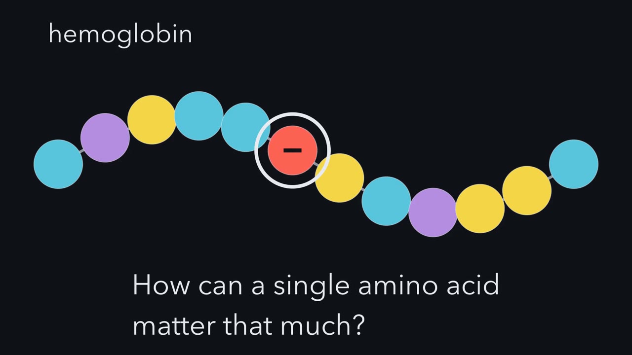
One changed letter can break a protein. How can a single amino acid matter that much? In sickle cell anemia, one amino acid in hemoglobin is swapped. Glutamate at position 6 of beta-globin becomes valine. A charged residue on the surface becomes an oily one. Once the hemoglobin releases its oxygen, it sticks together into long fibers. One letter changed the shape, and the shape changed the job. So the shape must be stored somewhere, and stored precisely. But where? Start with the chain. Its primary structure is the order of its amino acids, linked end to end by peptide bonds. That order is exact, not random. Insulin, for example, is 51 residues in one defined order.
Christian Anfinsen tested where the shape lives. He used ribonuclease, an enzyme with four disulfide bonds. Tube one holds the working enzyme. Its activity is full. In tube two, he added urea and a reducing agent. The reducing agent broke the disulfide bonds. Urea pulled the chain apart. Activity fell to zero. The enzyme was now a loose, random coil. In tube three, he washed the chemicals away. No other proteins were there. Just the chain, a buffer, and air. Here is the test. If the cell's machinery stored the shape, what would tube three show?
The enzyme refolded on its own, back to full activity. No helper guided it, only its own sequence, in that buffer. Look at the disulfides. Eight cysteines can pair up in 105 different ways. Random pairing would get them right less than one time in a hundred. Yet the chain found its correct partners. Anfinsen's conclusion was this. The fold is set by the amino acid sequence, in a given environment. For most small proteins, the fold is the lowest free energy state the chain can reach. Free energy here is a trade. The chain gives up freedom. In return, it gains favorable contacts and freer water. Keep those words in mind: in a given environment.
They will matter later. Three questions remain. Which shapes are possible? What picks one? And how is it found so fast? One rule will answer the first two. Geometry allows the shapes. Water drives the choice. The third question needs one more idea. The shape of a protein is written in its sequence.
Swap one amino acid in hemoglobin, glutamate for valine at position 6 of beta-globin, and the protein sticks together into long fibers once it releases its oxygen.
That is sickle cell anemia. One wrong letter in a chain of amino acids breaks a machine that carries oxygen. So the shape of a protein must be stored somewhere, and stored precisely. Where is it stored, and how does a floppy chain find that one shape?
One sequence, one shape
Start with the chain itself. The primary structure of a protein is its amino acid sequence: amino acids linked by peptide bonds into a chain, from its free amino end (the N-terminus) to its free carboxyl end (the C-terminus). Each amino acid in the chain is called a residue. The order is exact, not random. Insulin, for example, is 51 residues in one defined order. That single fact carries the whole chapter:
Sequence dictates structure. Structure dictates function. One changed residue can break both.
The fold decides what a protein can do: carry oxygen, cut another protein, hold a tendon together. Sickle cell shows the whole chain of cause in one residue.
The sequence raises four questions, and this lesson answers them in order.
- Where is the shape stored? In the sequence, in helper proteins, or in the cell?
- Which shapes are even possible? A chain of a hundred residues looks as if it could bend any way at all.
- What picks one shape out of the possible ones?
- How does the chain find it fast? There are far too many shapes to try one at a time.
Two terms first. The backbone is the repeating chain of atoms that every residue shares: N–Cα–C, over and over, where Cα is the central carbon of each amino acid. The side chains (R groups) hang off each Cα. Some are nonpolar (oily, like leucine), and some are polar or charged (like lysine and aspartate).
The one idea to carry through this lesson. Geometry allows; water chooses. The backbone can bend only a few ways, so only a few local shapes are possible: helices, strands and the turns that link them. Among the ways to pack those pieces, the winner tucks the oily side chains inside, away from water, and packs them well. Contacts like these tilt the chain’s landscape toward that fold, so it does not have to search for it at random. The rule is for water-soluble proteins in water, with small single-domain proteins as the clean case.
The proof: Anfinsen’s refolding experiment
So where does the folding information actually live? Christian Anfinsen settled it with ribonuclease, an enzyme held together in part by four disulfide bonds: covalent –S–S– bridges between the side chains of two cysteines. He unfolded it completely with urea and a reducing agent: zero activity, a random coil. The reducing agent broke the disulfide bonds, and urea pulled the chain apart. Then he washed the chemicals away and waited.
Predict. The third tube holds the same unfolded chain, with the chemicals removed. No other proteins are in the tube, just the enzyme in a buffered solution, with air to re-form its disulfide bonds. If the cell’s machinery held the shape, what would the tube show? If the sequence held it, what would it show?
Check your prediction
The enzyme refolded on its own to full activity. With nothing else in the tube, the information had to come from the chain itself. Its four disulfide bonds re-formed with their correct partners, even though eight cysteines can pair in 7 × 5 × 3 × 1 = 105 different ways. Random pairing would get the native set less than 1% of the time.
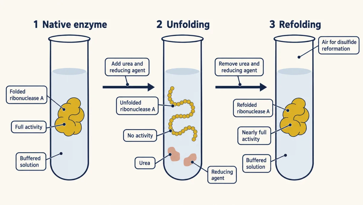
The conclusion, in his words: “The native conformation is determined by the totality of interatomic interactions and hence by the amino acid sequence, in a given environment.” For most small proteins, the fold is the lowest free-energy state the sequence can reach. Free energy here is a balance. Folding costs the chain freedom: an unfolded chain can wiggle through a huge number of shapes, and a folded one keeps very few. In return it gains favorable contacts and, as you will see, freedom for the water around it.
The words “in a given environment” matter, and the disulfides show why. A disulfide forms only when two cysteine thiols (–SH groups) are oxidized; the bridged pair is called cystine, and it can link sites within a chain or between chains. The cytosol is reducing and keeps cysteines as free thiols. The inside of the endoplasmic reticulum (the ER lumen) and the outside of the cell are oxidizing. Secreted proteins make their disulfides in the ER, which is why disulfides are found mostly in extracellular, secreted proteins. Anfinsen’s tube, open to air, was oxidizing too.
The flat peptide bond: two hinges per residue
Read the transcript of “Two Hinges per Residue”
Film transcript: Two Hinges per Residue
A chain of a hundred residues looks as if it could bend any way at all. Can it? The answer starts with the bond that joins one residue to the next. It is the peptide bond. It joins the carbon of one residue's C=O group to the nitrogen of the next residue's N–H group. Chemists can draw this bond two ways. In one drawing, carbon and oxygen share a double bond. The bond from carbon to nitrogen is single. In the other, oxygen carries a negative charge. Carbon and nitrogen share a double bond, and nitrogen carries a positive charge. The real bond is a blend of both. So the bond from carbon to nitrogen is partly double. That makes it short, about 1.32 Å.
A double bond cannot twist without losing its overlap. So the peptide bond does not rotate. Six atoms are locked in one flat plane. The Cα of the first residue. Its C and O. The N and H of the next residue. And that residue's Cα. Almost always, the two alpha carbons sit on opposite sides of the bond. That is the trans form. The cis form, with both on the same side, is rare. It is more common when the next residue is proline. So picture the backbone as a string of flat, rigid cards. One card for each peptide bond. The cards are joined at each Cα. Watch what can move. Each residue has three backbone bonds. The peptide bond is frozen. Only two can turn, one on each side of the Cα.
Watch one hinge turn. It is the rotation about the bond from nitrogen to the Cα. It is called phi. Now the other. It is the rotation about the bond from the Cα to the next carbon. It is called psi. Two angles per residue, repeated down the chain. Together they set the path of the whole backbone. Two free angles sound like freedom. Next, we see how much freedom they really give. The peptide bond is flat and rigid. Each residue turns on just two hinges.
The shape is in the sequence. Next question: which shapes can a chain take at all? The answer starts with the bond that links one residue to the next.
Look at the peptide bond. It joins the carbon of one residue’s C=O group to the nitrogen of the next residue’s N–H group. It can be drawn two ways. In one drawing, C=O is a double bond and C–N is single. In the other, the oxygen carries a negative charge (C–O⁻) and the C–N bond is double, with a positive charge on the nitrogen (C=N⁺). The real bond is a blend of the two. So the C–N bond has partial double-bond character, and it is short, about 1.32 Å.
A double bond cannot twist without breaking its overlap, so the peptide bond does not rotate. Six atoms are locked in one flat plane: the Cα of the first residue, its C and O, the N and H of the next residue, and that residue’s Cα. Almost always the two Cα atoms sit on opposite sides of the bond, the trans form. The cis form, with both on the same side, is rare, though markedly more common when the next residue is proline (an X-Pro bond).
So picture the backbone as a string of flat, rigid cards, one card per peptide bond. The cards are joined at each Cα. Of the three backbone bonds per residue, one (the peptide bond) is frozen. Only two can turn, one on each side of the Cα. Read along the backbone, N–Cα–C, and phi comes first, psi second:
- phi (φ), the rotation about the N–Cα bond;
- psi (ψ), the rotation about the Cα–C bond.
The path of a protein backbone is set by these two angles, repeated down the chain.
The map of allowed shapes: the Ramachandran plot
Read the transcript of “The Map of Allowed Shapes”
Film transcript: The Map of Allowed Shapes
Two hinges per residue sounds like a lot of freedom. How many of those settings actually work? Take two linked cards. Hold one hinge still, and turn the other. At many settings, an atom on one card runs into an atom on the next. A C=O group comes too close to an N–H group. Or a side chain runs into the backbone. The atoms would have to sit in the same space. Those settings are strongly disfavored. Now try every pair of angles. Put phi along the bottom and psi up the side. Each runs from minus −180° degrees to plus +180°. degrees. Shade every pair that does not clash. G. N. Ramachandran did this calculation. The survivors cluster into a few compact islands.
The rest of the map is empty. Only a small fraction of the map is open to the backbone. This is the Ramachandran plot. Here is the test. Put both angles at zero, right in the center. Will that pair survive? No. The atoms of neighbouring peptide planes run into each other. The center of the map is empty. Now read the islands. The broad one at the upper left is for extended strands, like the beta sheet. The compact one at the lower left is the right-handed alpha helix. A small island at the upper right is left-handed. It is used mostly by glycine. Glycine roams more widely than any other residue. Its side chain is a single hydrogen, so it bumps into less.
Each island matches a real shape. The helix island builds a coil. The strand island builds a stretched chain. The map is also a check on new structures. Plot every residue's two angles. If the points land on the islands, the structure is plausible. If many stray, something is probably wrong. Forbidden means strongly disfavored, not impossible. A few residues sit in strained spots, held by their surroundings. Those outliers are often the ones that matter for catalysis. Most hinge settings crash. The backbone is left with only a few local shapes.
Two free angles sounds like freedom. It is not.
Take two linked cards and turn one hinge while holding the other still. At some settings, nothing happens. But at many settings an atom on one card runs into an atom on the next: a C=O comes too close to an N–H, or a side chain runs into the backbone. Those settings are sterically disfavored. The atoms would have to sit in the same space.
Now do this for every pair of angles. Put φ on the horizontal axis and ψ on the vertical, each running from −180° to +180°, and shade every pair that does not clash. When G. N. Ramachandran calculated which pairs were sterically allowed, the survivors clustered into three islands. Only a small fraction of φ/ψ space is accessible. That map is the Ramachandran plot:
- a broad region for extended strands, the beta sheet and other stretched chains (upper left);
- a compact region for the right-handed alpha helix, around φ ≈ −57°, ψ ≈ −47° (lower left);
- a small left-handed region (upper right), used mostly by glycine.
Glycine roams more widely than any other residue. Its side chain is a single hydrogen. Every other residue has a Cβ, the first carbon of its side chain, which can bump into the backbone; glycine has none.
Predict. Drag the point below to φ = 0°, ψ = 0°. Will that pair survive?
Check your prediction
No. With both angles at zero, the atoms of the neighbouring peptide planes run into each other, and the readout shows a steric clash. Most of the plane is like that.
The panel beside the plot draws what each φ/ψ pair does to the backbone: a helix, a stretched strand, or atoms crashing together.
Glycine has no Cβ to bump into the backbone, so much more of the map is allowed, including the left-handed region.
What this φ/ψ looks like
Geometry allows these few shapes. Which ones a protein uses, and how they pack, is chosen later by water.
The plot is also the cheapest first check on any new structure, measured or predicted. If every residue’s φ/ψ falls in the allowed islands, the structure is plausible; if many stray into the forbidden zone, something is probably wrong. “Forbidden” means strongly disfavored, not impossible. A few residues, held by their surroundings, do sit in strained spots, and these outliers are often the ones that matter for catalysis.
Helices and sheets: the backbone bonds to itself
Read the transcript of “Helices, Sheets and the Rule-Breakers”
Film transcript: Helices, Sheets and the Rule-Breakers
The map has islands. Why do they become helices and sheets? Each island is the geometry of a regular shape. That shape lets the backbone hydrogen bond to itself. The bonds run between backbone C=O groups and N–H groups. Shapes built this way are called secondary structure. Secondary structure is local. It describes a stretch of chain, not the whole protein. First, the alpha helix. It is a right-handed coil. It has a little, over three and a half residues per turn. Inside the helix, each C=O group bonds to the N–H group four residues ahead. In the first turn, the N–H groups have no partner four residues back. The side chains stick outward. In a helix, the partners are near each other in the sequence.
Second, the beta sheet. Its strands are stretched nearly flat. Side chains alternate, above and below the sheet. Each strand bonds to its neighbouring strands. They can run in the same direction, or in opposite directions. Those strands can lie far apart in the sequence. Now meet a rule-breaker, proline. Its side chain loops back and bonds to its own backbone nitrogen. That makes a ring. So proline has no N–H group to donate, and its ring is bulky. Here is the test. Where in a helix could a proline sit without breaking it?
In the first turn. There, the N–H groups have no partners anyway. Losing one costs nothing. In the middle, proline breaks the helix. It cannot donate its hydrogen bond. And its ring bumps into the backbone of the turn before it. Its fixed angle is not the problem. That angle is a helical one. The same ring builds something else, collagen. It pre-shapes each strand into an extended helix. Collagen repeats a pattern: Gly-X-Y. X and Y are often proline and hydroxyproline. Three chains wind together into a triple helix. Every third residue sits at the crowded center. Only glycine is small enough to fit there. Put a larger residue in place of that glycine, in type I collagen.
That is a common cause of osteogenesis imperfecta, brittle-bone disease. What kills one structure builds another. One residue, again, decides the shape. Geometry allows these local shapes. But it does not choose which ones a protein makes. Helices and sheets are the backbone bonding to itself.
The islands are not random. Each one is the geometry of a regular shape that lets the backbone hydrogen-bond to itself. Shapes built this way, from hydrogen bonds between backbone C=O and N–H groups, are secondary structure. It is local: it describes a stretch of chain, not the whole protein.
- The alpha helix is a right-handed coil with 3.6 residues per turn. In the interior of the helix, every backbone C=O is hydrogen-bonded to the N–H four residues ahead (residue i to residue i+4). At the ends, some groups have no partner: in the first turn, the N–H groups have no C=O four residues back, and in the last turn, the C=O groups have no N–H four ahead. The side chains bristle outward. Hydrogen-bond partners in a helix are near each other in sequence.
- The beta sheet is built from strands stretched nearly flat, with side chains alternating above and below. Each strand is hydrogen-bonded to its neighbouring strands, running parallel or antiparallel. Those strands can lie far apart in sequence.
Proline shows the rule has teeth. Its side chain loops back and bonds to its own backbone nitrogen, forming a ring.
Predict. Proline has no N–H to donate, and its ring is bulky. Where in a helix could a proline sit without breaking it?
Check your prediction
In the first turn. There, the N–H groups have no partners four residues back anyway, so losing one costs nothing. In the middle of a helix, proline breaks it for two reasons. It has no N–H to donate to the C=O four residues back, and its ring bumps into the backbone of the turn before it. (The ring also fixes proline’s φ near −65°. That is a helical angle, so the fixed φ is not what breaks the helix.)
Collagen: the same ring, the opposite effect. In collagen, the rigid ring is an asset. It pre-shapes each strand into an extended helix. A repeating Gly-X-Y sequence, where X and Y are often proline and hydroxyproline, lets three chains wind together into a triple helix. Glycine, the only residue small enough, sits at the crowded center every third position. What kills one structure builds another. Substitute a larger residue for glycine at that center of type I collagen and you get one of the most common causes of osteogenesis imperfecta, brittle-bone disease.
Geometry has now done its job. It allows a few local shapes. It does not say which helices and strands a protein makes, or how they pack together. Something else chooses.
The hydrophobic core: water pushes oil inward
Read the transcript of “Water Chooses”
Film transcript: Water Chooses
Geometry allows a few shapes. So what chooses among them? Mostly, water does. Picture one oily side chain, like leucine's, sitting in water. Water molecules hydrogen bond to each other all the time, in a shifting network. Next to the oily surface, they cannot hydrogen bond to it. So the water in contact forms a tighter, more ordered network around it, like a cage. Ordered water has lost freedom. That is lost entropy. Now bring two oily side chains together. Less oily surface faces the water. Less water has to be caged. The freed water tumbles away and gains entropy. That gain drives the oily groups together. It is called the hydrophobic effect.
A common mistake is that oil is pulled to oil. But oily groups attract water about as well. Oily groups are pushed together by water, not pulled together by each other. So in a water-soluble protein, the nonpolar side chains end up buried in a core. Polar and charged side chains sit on the surface, in the water. This packs the whole chain into a compact shape. That is tertiary structure. Burial also helps explain helices and sheets. Buried backbone groups lose their water partners. A buried polar group with no partner costs free energy. The cheapest fix is for the backbone to bond to itself. Helices and sheets are the regular ways to do that.
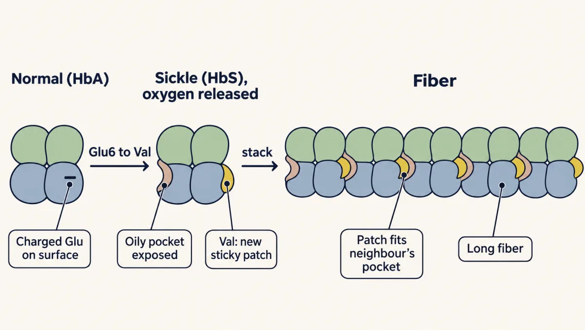
Burial does not stop at one chain. Folded chains can bury oily surface against each other. That assembly is quaternary structure. Hemoglobin is two alpha and two beta subunits, working as one. Now return to sickle cell. Valine puts an oily residue on the surface, where a charged one was. That makes a new sticky patch. Once oxygen is released, the patch fits an oily pocket on a neighbouring hemoglobin. Sickle cell is an unwanted contact between whole hemoglobins, built by the same rule. One more surprise. A folded protein is only marginally more stable than the unfolded coil. The chain gives up a lot of freedom to fold. Its contacts and freed water only just pay for it.
So heat can undo a fold. The melting temperature is where half the molecules are unfolded. Here is the test. A buried leucine becomes aspartate. Or a surface lysine becomes arginine. Which one melts lower? The buried aspartate. It is charged, with no water to bond with, and it breaks up the oily packing. The surface swap trades one positive charge for another, still in water. Stability barely changes. Water pushes oil inward. That burial builds the core and the interfaces.
What holds a fold together? Mostly water’s push.
Picture one oily side chain, such as leucine’s, sitting in water. Water molecules hydrogen-bond to each other all the time, in a shifting network. Next to the oily surface they cannot hydrogen-bond to it. So the water molecules in contact with it arrange themselves into a tighter, more ordered network around it, like a cage. Ordered water has lost freedom, which is lost entropy.
Now bring two oily side chains together. Less oily surface faces the water, so less water has to be caged. The freed water molecules tumble away and gain entropy. That gain is what drives the oily groups together. It is called the hydrophobic effect.
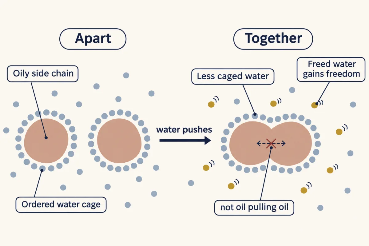
A common mistake is to think oil attracts oil and pulls itself together. Oily groups do attract each other, but they attract water about as well, so that attraction gives them no net preference. They are pushed together by water, not pulled together by each other. Once they are together, tight packing adds to the gain.
So in a water-soluble protein, nonpolar side chains are buried in a core and polar and charged side chains sit on the surface, where they hydrogen-bond with water. The hydrophobic effect is the main driving force of tertiary structure, the whole chain packed into a compact 3D shape by interactions between side chains far apart in sequence. Myoglobin is the classic example: eight helices cradle a heme group around an oily core.
Burial adds a strong push toward secondary structure. The backbone’s C=O and N–H groups want hydrogen-bond partners, and in an unfolded chain, water supplies them. When part of the backbone is buried in the core, those partners are gone. A buried polar group with no partner costs free energy. The cheapest fix is for the backbone to hydrogen-bond to itself, and helices and sheets are the regular ways to do that. So geometry offers helices and sheets, and burial makes them more worth forming.
Water drives the collapse; tight packing and hydrogen bonding, from the backbone and from side chains, make the result specific. Disulfide bonds and salt bridges add more.
Between chains: quaternary structure
Burial does not stop at the edge of one chain. Several folded chains can bury nonpolar surface against each other and hold together as one machine. That assembly is quaternary structure. It uses the same non-covalent forces as tertiary structure, now acting between chains. Hemoglobin is two alpha and two beta subunits, each myoglobin-like, working as one.
Now return to sickle cell. Swapping glutamate for valine at position 6 puts a nonpolar residue on the surface of beta-globin, where a charged one used to be. That makes a new sticky patch. Releasing oxygen changes hemoglobin’s shape and exposes an oily pocket. The patch on one hemoglobin fits into that pocket on a neighbouring one, and the molecules stack into long fibers. Sickle cell is an interface between whole hemoglobin molecules that should not exist, built by the same rule that builds the core and holds the subunits together.
The four levels, each with the force that builds it:
| Level | What it is | Main force | Example |
|---|---|---|---|
| Primary | The sequence of residues | Covalent peptide bonds | Insulin’s 51 residues in one exact order |
| Secondary | Local helices and sheets | Hydrogen bonds between backbone C=O and N–H | The alpha helix; the beta sheet |
| Tertiary | The whole chain’s fold | The hydrophobic effect, plus hydrogen bonds, salt bridges and disulfides | Myoglobin: eight helices around a heme |
| Quaternary | Several chains as one unit | The same non-covalent forces, between chains | Hemoglobin: two alpha and two beta subunits |
Only just stable: the melt
Here is the surprise. The favorable contacts and freed water only just pay for the chain’s lost freedom, so a folded protein is only marginally more stable than the coil. That is why heat can undo it, and why one bad residue can tip it.
Two terms describe a melt. The melting temperature (Tm) is the temperature at which half the molecules are unfolded. Cooperativity means the structure unfolds as one unit: the contacts support each other like the cards in a house of cards, so when enough of them fail, the rest let go together. A cooperative protein gives a steep curve. When a small, single-domain protein melts, it often goes from mostly folded to mostly unfolded over roughly ten degrees.
Predict. Two point mutants of one small enzyme. In one, a leucine buried in the core becomes aspartate. In the other, a lysine on the surface becomes arginine. Which one melts at a lower temperature?
Check your prediction
The core leucine to aspartate. Aspartate is charged. Buried in the core, it has no water to hydrogen-bond with, and it breaks up the oily packing. That costs much of the small margin, so Tm falls. Lysine to arginine on the surface swaps one positive charge for another, still in water, so stability barely changes.
Slide the melting temperature and the cooperativity below, and watch how sharp the collapse is.
- Folded at 37 °C
- ~100%
- Melting temperature
- 62 °C
A protein does not loosen gradually; it holds its shape, then unravels over roughly ten degrees. That sharp transition is the signature of cooperative folding.
Favorable contacts and freed water only just pay for the chain's lost freedom, so the fold is only marginally stable; one buried charged residue can tip it. A single disulfide or a thermophile's extra salt bridge can push Tm well up.
Finding the fold fast: Levinthal’s paradox and the funnel
Read the transcript of “Downhill, Not Random”
Film transcript: Downhill, Not Random
Water picks the fold. But how does the chain find it, without trying every shape? Count the options. Give each residue of a 100-residue chain just three backbone shapes. One comes from each island of the map. Helix-like, strand-like, or left-handed. That gives about 5 × 10⁴⁷ shapes. with 47 zeros. Suppose the chain tried a new shape every tenth of a trillionth of a second. That is about as fast as a bond can turn. Trying them all would take far longer than the universe has existed. Yet small proteins fold in microseconds to seconds. That gap is Levinthal's paradox. You know the Ramachandran plot forbids most backbone angles. Does that solve the paradox?
No. The count already used it. Three shapes per residue is what the filter left behind. Now give each residue only two shapes. The search still takes billions of years. A filter on each residue makes each factor smaller. But there are still a hundred factors. So the answer is not fewer shapes. It is that the residues do not choose blindly. Picture a flat golf course with one hole. Every wrong shape is as good as every other. The ball rolls at random and almost never drops in. That is a random search. Now tilt the ground toward the hole from every side. Each roll tends to carry the ball downhill. This sloping landscape is the folding funnel. Its width stands for the many unfolded shapes.
Its depth stands for how favorable the contacts are. The native fold sits at the bottom. What makes the slope? The same forces that make the fold stable. Two oily side chains meet in the core. Some water is freed, and the energy drops a little. A stretch of backbone makes its first helical hydrogen bonds. It drops a little more. Partly correct shapes tend to last. Wrong contacts tend to break apart again. The chain needs only to keep more of what is right. A small preference at each step is enough. For small proteins, the first few contacts climb a small hill. Once a core forms, the rest runs downhill. Real funnels have dips, called kinetic traps.
Chaperones stop half-folded chains from clumping while they search. Chaperones do not choose the destination. The sequence, in its environment, still does. The rule bends in places. Disordered proteins have too few oily residues to pay for a core. Prions take one sequence to a second fold, rich in beta sheet, that aggregates. And membrane proteins sit in lipid, not water, so their surface flips. Greasy residues face the lipid. Folding is downhill, not random. Native-like contacts tilt the landscape toward the fold.
One puzzle is left. How does the chain find its fold?
Count the options. Give each residue of a 100-residue chain just three backbone shapes, one from each island of the Ramachandran map: helix-like, strand-like or left-handed. That is a generous count, since the left-handed island is rarely used outside glycine. The chain then has 3 × 3 × 3 … one hundred times over, which is 3¹⁰⁰, about 5 × 10⁴⁷ shapes. Suppose it could try a new shape every 10⁻¹³ seconds, about as fast as a bond can turn. Trying them all would take about 10²⁷ years. The universe is about 10¹⁰ years old. Yet small proteins fold in microseconds to seconds. That gap is Levinthal’s paradox, after Cyrus Levinthal, who pointed it out in the late 1960s.
Predict. You have just learned that the Ramachandran plot forbids most backbone angles. Is that what solves the paradox?
Check your prediction
No. The count already used the Ramachandran filter: three shapes per residue is what survives it, generously counted. Push the filter further and give each residue only two shapes. You still have 2¹⁰⁰, about 10³⁰ shapes, and billions of years of searching. A filter that works one residue at a time makes each factor smaller, but there are still a hundred factors.
So the answer is not fewer shapes. It is that the residues do not choose blindly.
Picture two landscapes. On a flat golf course with a single hole, every wrong shape is as good as every other. The ball rolls at random and almost never drops in. That is the random search Levinthal described. Now let the ground slope toward the hole from every side. Wherever the ball starts, each roll tends to carry it downhill. This sloping landscape is the folding funnel. Its width at the top stands for the huge number of unfolded shapes. Its depth stands for how favorable the chain’s contacts are, with the water they free counted in. The native fold sits at the bottom.
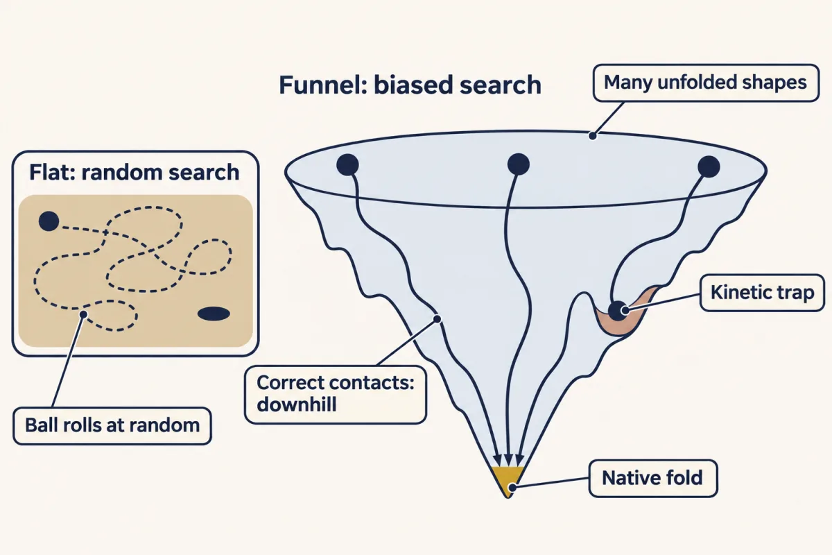
What makes the slope? The same forces that make the fold stable. When two oily side chains that belong together in the core meet, they push out some ordered water and the energy drops a little. When a stretch of backbone makes its first helical hydrogen bonds, it drops a little more. But for small proteins, the first few contacts cost more freedom than they pay back, so folding starts with a small uphill step, and partly folded shapes are less stable than either end: the same cooperativity you saw in the melt. Once a core is in place, each further correct contact makes that shape more stable than its neighbours, so it tends to last. Wrong contacts tend to break apart again. The chain does not need to know the answer in advance. It needs only to keep more of what is right than of what is wrong, and a small preference at each step is enough to make the search fast once the first contacts are in place. The interactions that make the fold stable also guide the chain to it.
Real funnels are not smooth. They have dips where a partly wrong shape gets stuck for a while: kinetic traps. That is one reason cells make chaperones, proteins that stop half-folded chains from clumping together while they search. Chaperones prevent accidents and raise the yield of folded protein. They do not choose the destination. The sequence, in its environment, still does.
Where the rule bends
The rule was stated for water-soluble proteins in water. Each case below either breaks one of those conditions or shows where the idea of one stable, lowest-energy working fold fails. The rule itself still helps explain each one.
- Intrinsically disordered proteins have no stable fold on their own. Their sequences have too few nonpolar residues and too much net charge to pay for a core. Many fold only when they meet a partner, and disordered regions are common in many proteins.
- Prions take the same sequence to a second stable fold, rich in beta sheet, that aggregates and kills. The normal prion protein is helix-rich. So a sequence sets a shape, or a set of shapes, for a given environment.
- Serpins, a family of protease inhibitors that includes alpha-1-antitrypsin, work in a fold that is not their most stable one. The working fold is a kinetic trap; the lowest free-energy state is a more stable, inactive form.
- Membrane proteins sit in lipid, not water, so the solvent is flipped and so is their surface: greasy residues face the lipid. Porins even run a polar channel through the middle.
For folded domains, the rule holds very widely. Today we see folds by X-ray crystallography, nuclear magnetic resonance (NMR) and cryo-electron microscopy, and predict them from sequence.
Going deeper
None of these details changes the main line above.
How small a preference is enough?
In a simple model of a 100-residue chain (after Zwanzig, Szabo and Bagchi, 1992), each residue makes a local move about once a nanosecond, and each residue in its correct state is favored by a small energy U. With no preference, folding takes about 10²⁹ years, longer than the 10²⁷ years above because of the slower move rate. A preference of about 2 kT per correct residue brings it to about a second, and about 3 kT brings it to microseconds. (kT is the thermal energy per molecule.) The model gives each residue three states, one right and two wrong. Its per-residue preference, summed over the chain, is far larger than any real protein’s net stability. In a real protein most of the contact energy is cancelled by the chain’s lost freedom, a balance this simple model leaves out.
How marginal is marginal?
A typical net free energy of unfolding of 20 to 60 kJ/mol at 37 °C means folded molecules outnumber unfolded ones by roughly a thousand to ten billion to one. That is stable. But a single buried polar or charged substitution can cost 10 to 25 kJ/mol or more, enough to wipe out much of that margin.
The hydrophobic effect and temperature
The picture of freed, ordered water is right near room temperature, where the hydrophobic effect is mostly entropic. At higher temperatures it becomes increasingly enthalpic, because burying nonpolar surface comes with a large change in heat capacity.
Seeing a fold: crystallography and cryo-EM
X-ray crystallography turns a protein crystal’s diffraction pattern into an electron-density map, atom by atom at high resolution. It gave Kendrew myoglobin’s fold in 1958 and Perutz hemoglobin in 1960, after more than twenty years of work, and together the two structures founded structural biology. Cryo-electron microscopy now does the same for large assemblies without crystals, by averaging many thousands, often hundreds of thousands, of images of frozen-hydrated particles.
Predicting a fold: AlphaFold
Fifty years after Anfinsen, AlphaFold gave striking computational support for the idea that the sequence holds the fold. Given a sequence and alignments of its evolutionary relatives, the neural network predicts many structures to near-experimental accuracy. Its creators, Demis Hassabis and John Jumper, shared the 2024 Chemistry Nobel with David Baker, who was honored for protein design.
History: how the shapes were found
Sanger read insulin’s sequence in 1955. Pauling worked out the alpha helix from a sickbed in 1948 — paper, a slide rule, and the chemistry — and, with Robert Corey and Herman Branson (on the helix), published both helix and sheet in 1951. They predicted both structures from chemical first principles before anyone had seen either. The helix even seemed at first to clash with keratin’s X-ray pattern. Perutz soon found the helix’s predicted 1.5 Å repeat in hair and hemoglobin, and X-ray work confirmed the helix. In Madras in 1963, Ramachandran’s plot showed why the predictions worked: both structures sit where the chemistry allows the backbone to go.
Prions: a debated energy picture
Why can a prion’s sequence reach a second fold at all? One influential view is that the native helix-rich fold is kinetically trapped, and the deadly aggregate is a lower-energy state the sequence could always have reached. This is still debated. Other researchers argue that Anfinsen’s idea still fits once you count the environment and all the states a sequence can reach. A prion also spreads by templating its shape onto normal copies of the same protein, with no DNA or RNA required.
Alpha-1-antitrypsin: one mutation, two organs
Alpha-1-antitrypsin is another one-letter disease. A single substitution, Glu342 to Lys, changes how the protein folds: it folds slowly, and its molecules can link into long chains (polymers) instead of staying single. In the liver, the misfolded protein is held inside the cells that make it instead of being secreted, and damages them. In the lungs, too little of the protein circulates, so the enzyme elastase is poorly controlled and lung tissue breaks down, causing emphysema.
A rule of thumb: protein mass
An average residue adds about 110 Da to a protein’s mass, after the loss of the elements of water when each peptide bond forms. Multiply residues by 110 for daltons and divide by 1,000 for kilodaltons; most proteins are 5 to 200 kDa.
Your notes
Select any text in the lesson to highlight it or add a note to it. Your highlights and notes are saved in this browser and collected in your notebook.
This browser is not letting the site save anything (a private window or strict privacy settings), so highlights and notes will be lost when you leave the page.
How we measure it
X-ray crystallography
Fire X-rays at a protein crystal and the diffraction pattern, once its missing phases are recovered, yields an electron-density map; at high resolution it shows where individual atoms sit. Kendrew solved myoglobin this way in 1958 — the first three-dimensional protein fold ever solved, at low resolution — by stacking hand-drawn electron-density contours on sheets of plastic until the fold rose into three dimensions.
Reading a Ramachandran plot
Plot a residue's two rotatable backbone angles, phi against psi, and the allowed combinations cluster into a few islands — mainly the alpha-helix and the broad beta/extended region, plus a small left-handed region — with the rest of the map disfavored by atoms colliding. It is a standard sanity check on new structures.
Denaturation and refolding (Anfinsen's assay)
Strip a protein to a random coil with urea and a reducing agent, watch activity vanish, then wash the chemicals away. If the protein refolds to full activity on its own, the folding instructions had to be in the sequence — nowhere else.
Practice questions on this lesson (6) → MCAT-style, original, each with an explanation
Where this lesson fits
Before thisReview first
- Amino Acids: How One Side Chain Decides EverythingThis course · Lesson 4
You need the side chains and the peptide-linked chain from there to see why sequence sets the fold.
- Water, Weak Bonds, and the Generation of OrderThis course · Lesson 3
The hydrophobic effect and weak bonds from there are the forces that choose and hold the fold here.
After thisStudy next
- Techniques in Protein Biochemistry: Purify It, Then Prove ItThis course · Lesson 6
Purifying and proving a protein relies on its folded shape, size and surface, which this lesson explains.
- Protein Binding and Allostery: How Hemoglobin Loads and Delivers OxygenThis course · Lesson 9
Hemoglobin's shape change on binding oxygen builds directly on quaternary structure and the fold-function link.
- Basic Concepts of Enzyme ActionThis course · Lesson 7
An enzyme's folded active site, set by the sequence, is what makes catalysis possible.
Elsewhere on the siteFilms, simulations and method films in other courses
- From Gene Copy to Protein ChainFilm · Biology
It shows how one changed letter in a gene alters the amino acid chain, as in sickle cell.
- Take It Apart, Build It AgainFilm · Biology
It shows how amino acids link into a protein chain, the starting point for folding.
- Count the Working SitesFilm · Biology
It shows an enzyme's shape holding an active site that fits its substrate.
Suggested by an AI model from the lessons themselves, then checked. How we use AI
Finish line
Test yourself
9 questions
Practice and MCAT-style, with explained answers.
Next · Lesson 6
Techniques in Protein Biochemistry: Purify It, Then Prove It
Pull one protein out of a soup of ten thousand others — then prove you got it.
Continue →← Lesson 4 · Amino Acids: How One Side Chain Decides Everything